Curcumin Attenuates Acrolein-induced COX-2 Expression and Prostaglandin Production in Human Umbilical Vein Endothelial Cells
- Affiliations
-
- 1Department of Microbiology, College of Medicine, Kyung Hee University, Seoul, Korea. yongseek@khu.ac.kr
- 2Department of Biomedical Science, Graduate School, Kyung Hee University, Seoul, Korea.
- KMID: 2470810
- DOI: http://doi.org/10.12997/jla.2020.9.1.184
Abstract
OBJECTIVE
Inflammation is crucial to limiting vascular disease. Previously we reported that acrolein, a known toxin in tobacco smoke, might play an important role in the progression of atherosclerosis via an inflammatory response involving cyclooxygenase-2 (COX-2) and prostaglandin production in human umbilical vein endothelial cells (HUVECs). Curcumin has been known to improve vascular function and have anti-inflammatory properties. In this study, we investigated whether curcumin prevents the induction of inflammatory response caused by acrolein.
METHODS
Anti-inflammatory effects of curcumin were examined in acrolein-stimulated HUVECs. Induction of proteins, mRNA, prostaglandin and reactive oxygen species (ROS) were measured using immunoblot analysis, real-time reverse-transcription polymerase chain reaction, enzyme-linked immunosorbent assay and flow cytometry, respectively.
RESULTS
Curcumin attenuates inflammatory response via inhibition of COX-2 expression and prostaglandin production in acrolein-induced human endothelial cells. This inhibition by curcumin results in the abolition of phosphorylation of protein kinase C, p38 mitogen-activated protein kinase, and cAMP response element-binding protein. Furthermore, curcumin suppresses the production of ROS and endoplasmic reticulum stress via phosphorylation of eukaryotic initiation factor-2α caused by acrolein.
CONCLUSION
These results suggest that curcumin might be a useful agent against endothelial dysfunction caused by acrolein-induced inflammatory response.
MeSH Terms
-
Acrolein
Atherosclerosis
Curcumin*
Cyclic AMP Response Element-Binding Protein
Cyclooxygenase 2
Endoplasmic Reticulum Stress
Endothelial Cells
Enzyme-Linked Immunosorbent Assay
Flow Cytometry
Human Umbilical Vein Endothelial Cells*
Humans*
Inflammation
Phosphorylation
Polymerase Chain Reaction
Protein Kinase C
Protein Kinases
Reactive Oxygen Species
RNA, Messenger
Smoke
Tobacco
Vascular Diseases
Acrolein
Curcumin
Cyclic AMP Response Element-Binding Protein
Cyclooxygenase 2
Protein Kinase C
Protein Kinases
RNA, Messenger
Reactive Oxygen Species
Smoke
Figure
-

Fig. 1 Curcumin suppresses acrolein-induced COX-2 and prostaglandin production in HUVECs. (A) HUVECs were preincubated with 25 µM curcumin before treatment with 10 µM acrolein and COX-2 protein levels were assessed by western blot. (B) Relative expression of COX-2 was measured by real-time quantitative PCR as described in the methods section. (C) The production of PGE2 was measured in the supernatants as described in the methods section. (D) Inhibitory effect of curcumin on acrolein-induced activation of COX-2 promoter. HUVECs were transfected with COX-2 promoter construct (−1,432 to +59) for 48 hours. After transfection, cells were preincubated with 25 µM curcumin for 30 minutes before exposure to 10 µM acrolein for 6 hours. Data represents luciferase activity that has been normalized to co-transfected β-galactosidase activity. Data were analyzed by the Student's t-test. Data represent the mean±standard deviation of results from three independent experiments. HUVEC, human umbilical vein endothelial cell; COX-2, cyclooxygenase-2; PGE2, prostaglandin E2. *p<0.05 vs control group.
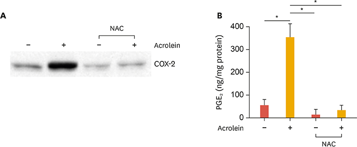
Fig. 2 Effect of NAC on acrolein-induced COX-2 and PGE2 production in HUVECs. (A) HUVECs were preincubated with 10 mM NAC for 12 hours. The cells were treated with 10 µM acrolein for 16 hours and western blotting was performed for COX-2 expression. (B) HUVECs were preincubated with 10 mM NAC for 12 hours. The cells were then treated with 10 µM acrolein for 16 hours and then release of PGE2 was measured from supernatants as described in the methods section. The values shown for PGE2 production are the mean±standard deviation of 3 independent experiments. Data were analyzed by the Student's t-test. Results are from 3 independent experiments. NAC, N-acetyl-cysteine; COX-2, cyclooxygenase-2; HUVEC, human umbilical vein endothelial cell; PGE2, prostaglandin E2. *p<0.05 compared to untreated control cells.
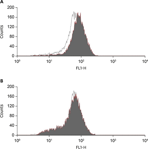
Fig. 3 Effect of curcumin on acrolein-induced intracellular peroxide production. (A) Cells were incubated with (black area) or without (white area) 10 µM acrolein for 30 minutes, and then treated with a peroxide sensitive dye, H2DCF-DA (20 µM) during the final 30 minutes of each treatment. The relative peroxide concentrations in cells were then measured by flow cytometry. (B) After preincubation with 10 mM NAC for 4 hours, the cells were treated with (black area) or without (white area) 10 µM acrolein and flow cytometric analysis was performed. X-axis represents intensity of fluorescence. NAC, N-acetyl-cysteine.

Fig. 4 Effects of curcumin on acrolein-induced phosphorylation of PKCδ, p38, and CREB in HUVECs. Cells were pre-treated with curcumin (10 and 25 µM) for 30 minutes prior to acrolein treatment (10 µM). After 30 minutes of incubation, the cell lysates (20 µg) were prepared and western blot analysis was performed with Abs against phosphorylated PKCδ, p38, and CREB or total PKCδ, p38, and CREB. Quantitative data were obtained using an imaging densitometer (ImageJ version 1.52a software, NIH, Bethesda, MD, USA). Data were analyzed by the Student's t-test. Data represent the mean±standard deviation of results from 3 independent experiments. PKC, protein kinase C; CREB, cAMP response element-binding protein; HUVEC, human umbilical vein endothelial cell; Ab, antibody; p, phosphorylated. *p<0.05 vs. control group.

Fig. 5 Effects of curcumin on acrolein-induced phosphorylation of eIF-2α in HUVECs. HUVECs were treated with curcumin (10 and 25 µM) for 30 minutes before acrolein (10 µM) treatment. After 30 minutes, cell lysates were prepared, and 20 µg of protein was used to perform western blotting using the anti-phosphorylated eIF-2α, and total eIF-2α antibody was used as a loading control. Quantitative data were obtained using an imaging densitometer (ImageJ version 1.52a software, NIH). Data were analyzed by the Student's t-test. Data represent the mean±standard deviation of results from three independent experiments. eIF, eukaryotic initiation factor; HUVEC, human umbilical vein endothelial cell; p, phosphorylated. *p<0.05 vs. control group.
Reference
-
1. Geovanini GR, Libby P. Atherosclerosis and inflammation: overview and updates. Clin Sci (Lond). 2018; 132:1243–1252.
Article2. Libby P. Inflammation in atherosclerosis. Arterioscler Thromb Vasc Biol. 2012; 32:2045–2051.
Article3. Ambrose JA, Barua RS. The pathophysiology of cigarette smoking and cardiovascular disease: an update. J Am Coll Cardiol. 2004; 43:1731–1737.4. Stevens JF, Maier CS. Acrolein: sources, metabolism, and biomolecular interactions relevant to human health and disease. Mol Nutr Food Res. 2008; 52:7–25.
Article5. Thweatt WD, Harward CN Sr, Parrish ME. Measurement of acrolein and 1,3-butadiene in a single puff of cigarette smoke using lead-salt tunable diode laser infrared spectroscopy. Spectrochim Acta A Mol Biomol Spectrosc. 2007; 67:16–24.
Article6. Zirak MR, Mehri S, Karimani A, Zeinali M, Hayes AW, Karimi G. Mechanisms behind the atherothrombotic effects of acrolein, a review. Food Chem Toxicol. 2019; 129:38–53.
Article7. Park YS, Kim J, Misonou Y, Takamiya R, Takahashi M, Freeman MR, et al. Acrolein induces cyclooxygenase-2 and prostaglandin production in human umbilical vein endothelial cells: roles of p38 MAP kinase. Arterioscler Thromb Vasc Biol. 2007; 27:1319–1325.
Article8. Park YS, Taniguchi N. Acrolein induces inflammatory response underlying endothelial dysfunction: a risk factor for atherosclerosis. Ann N Y Acad Sci. 2008; 1126:185–189.
Article9. Rumzhum NN, Ammit AJ. Cyclooxygenase 2: its regulation, role and impact in airway inflammation. Clin Exp Allergy. 2016; 46:397–410.
Article10. Gupta SC, Patchva S, Aggarwal BB. Therapeutic roles of curcumin: lessons learned from clinical trials. AAPS J. 2013; 15:195–218.
Article11. Kunnumakkara AB, Bordoloi D, Padmavathi G, Monisha J, Roy NK, Prasad S, et al. Curcumin, the golden nutraceutical: multitargeting for multiple chronic diseases. Br J Pharmacol. 2017; 174:1325–1348.
Article12. Binion DG, Otterson MF, Rafiee P. Curcumin inhibits VEGF-mediated angiogenesis in human intestinal microvascular endothelial cells through COX-2 and MAPK inhibition. Gut. 2008; 57:1509–1517.
Article13. Devassy JG, Nwachukwu ID, Jones PJ. Curcumin and cancer: barriers to obtaining a health claim. Nutr Rev. 2015; 73:155–165.
Article14. Gardener SL, Rainey-Smith SR, Martins RN. Diet and inflammation in Alzheimer's disease and related chronic diseases: a review. J Alzheimers Dis. 2016; 50:301–334.
Article15. Karimian MS, Pirro M, Johnston TP, Majeed M, Sahebkar A. Curcumin and endothelial function: evidence and mechanisms of protective effects. Curr Pharm Des. 2017; 23:2462–2473.
Article16. Midura-Kiela MT, Radhakrishnan VM, Larmonier CB, Laubitz D, Ghishan FK, Kiela PR. Curcumin inhibits interferon-γ signaling in colonic epithelial cells. Am J Physiol Gastrointest Liver Physiol. 2012; 302:G85–G96.
Article17. Cho JW, Park K, Kweon GR, Jang BC, Baek WK, Suh MH, et al. Curcumin inhibits the expression of COX-2 in UVB-irradiated human keratinocytes (HaCaT) by inhibiting activation of AP-1: p38 MAP kinase and JNK as potential upstream targets. Exp Mol Med. 2005; 37:186–192.
Article18. He Y, Yue Y, Zheng X, Zhang K, Chen S, Du Z. Curcumin, inflammation, and chronic diseases: how are they linked? Molecules. 2015; 20:9183–9213.
Article19. Lee SE, Park HR, Park CS, Ahn HJ, Cho JJ, Lee J, et al. Autophagy in crotonaldehyde-induced endothelial toxicity. Molecules. 2019; 24:E1137.
Article20. Lee SE, Park HR, Kim H, Choi Y, Jin YH, Park CS, et al. Effect of crotonaldehyde on the induction of COX-2 expression in human endothelial cells. Mol Cell Toxicol. 2017; 13:345–350.
Article21. Lee SE, Park YS. Korean Red Ginseng water extract inhibits COX-2 expression by suppressing p38 in acrolein-treated human endothelial cells. J Ginseng Res. 2014; 38:34–39.
Article22. Haberzettl P, Vladykovskaya E, Srivastava S, Bhatnagar A. Role of endoplasmic reticulum stress in acrolein-induced endothelial activation. Toxicol Appl Pharmacol. 2009; 234:14–24.
Article23. Gómez-Hernández A, Martín-Ventura JL, Sánchez-Galán E, Vidal C, Ortego M, Blanco-Colio LM, et al. Overexpression of COX-2, Prostaglandin E synthase-1 and prostaglandin E receptors in blood mononuclear cells and plaque of patients with carotid atherosclerosis: regulation by nuclear factor-κB. Atherosclerosis. 2006; 187:139–149.
Article24. Hatcher H, Planalp R, Cho J, Torti FM, Torti SV. Curcumin: from ancient medicine to current clinical trials. Cell Mol Life Sci. 2008; 65:1631–1652.
Article25. Burge K, Gunasekaran A, Eckert J, Chaaban H. Curcumin and intestinal inflammatory diseases: molecular mechanisms of protection. Int J Mol Sci. 2019; 20:E1912.
Article26. Sharma C, Kaur J, Shishodia S, Aggarwal BB, Ralhan R. Curcumin down regulates smokeless tobacco-induced NF-κB activation and COX-2 expression in human oral premalignant and cancer cells. Toxicology. 2006; 228:1–15.
Article27. Lelli D, Sahebkar A, Johnston TP, Pedone C. Curcumin use in pulmonary diseases: state of the art and future perspectives. Pharmacol Res. 2017; 115:133–148.
Article28. Ghosh S, Banerjee S, Sil PC. The beneficial role of curcumin on inflammation, diabetes and neurodegenerative disease: a recent update. Food Chem Toxicol. 2015; 83:111–124.
Article29. Shehzad A, Ha T, Subhan F, Lee YS. New mechanisms and the anti-inflammatory role of curcumin in obesity and obesity-related metabolic diseases. Eur J Nutr. 2011; 50:151–161.
Article30. Sikora E, Scapagnini G, Barbagallo M. Curcumin, inflammation, ageing and age-related diseases. Immun Ageing. 2010; 7:1.
Article31. Camacho-Barquero L, Villegas I, Sánchez-Calvo JM, Talero E, Sánchez-Fidalgo S, Motilva V, et al. Curcumin, a Curcuma longa constituent, acts on MAPK p38 pathway modulating COX-2 and iNOS expression in chronic experimental colitis. Int Immunopharmacol. 2007; 7:333–342.
Article32. Ramírez-Tortosa MC, Mesa MD, Aguilera MC, Quiles JL, Baró L, Ramirez-Tortosa CL, et al. Oral administration of a turmeric extract inhibits LDL oxidation and has hypocholesterolemic effects in rabbits with experimental atherosclerosis. Atherosclerosis. 1999; 147:371–378.
Article33. Haybar H, Shahrabi S, Rezaeeyan H, Shirzad R, Saki N. Endothelial cells: from dysfunction mechanism to pharmacological effect in cardiovascular disease. Cardiovasc Toxicol. 2019; 19:13–22.
Article34. Incalza MA, D'Oria R, Natalicchio A, Perrini S, Laviola L, Giorgino F. Oxidative stress and reactive oxygen species in endothelial dysfunction associated with cardiovascular and metabolic diseases. Vascul Pharmacol. 2018; 100:1–19.
Article
- Full Text Links
-
- Actions
-
Cited
- CITED
-
- Close
- Share
-
- Similar articles
-
- Epigallocatechin-3-gallate Regulates Inducible Nitric Oxide Synthase Expression in Human Umbilical Vein Endothelial Cells
- The Effect of Erythropoietin on the Production of Endothelin in Human Glomerular and Umbilical Endothelial Cells
- DHA and EPA Down-regulate COX-2 Expression through Suppression of NF-kappa B Activity in LPS-treated Human Umbilical Vein Endothelial Cells
- Propofol attenuates hydrogenperoxide-induced apoptosis in human umbilical vein endothelial cells via multiple signaling pathways
- Curcumin Attenuates Nuclear Factor-kappaB, c-Jun N-Terminal Kinase and p38 in Tumor Necrosis Factor-alpha-Stimulated Endothelial Cells
