Inhibition of miRNA-222-3p Relieves Staphylococcal Enterotoxin B-Induced Liver Inflammatory Injury by Upregulating Suppressors of Cytokine Signaling 1
- Affiliations
-
- 1Department of Clinical Laboratory, the Third People's Hospital of Dalian, Dalian, China.
- 2Department of Clinical Laboratory, the Baotou Medical College of Inner Mongolia University of Science and Technology, Inner Mongolia, China.
- 3Department of Clinical Laboratory, the Hongqi Hospital Affiliated to Mudanjiang Medical University, Mudanjiang, China.
- 4Department of Clinical Laboratory, the Second Hospital of Dalian Medical University, Dalian, China. zdgkua@163.com
- KMID: 2460227
- DOI: http://doi.org/10.3349/ymj.2019.60.11.1093
Abstract
- PURPOSE
Staphylococcal enterotoxin B (SEB) has been well-documented to induce liver injury. miRNA-222-3p (miR-222-3p) was implicated in SEB-induced lung injury and several liver injuries. This study aimed to explore the role of miR-222-3p in SEB-induced liver injury.
MATERIALS AND METHODS
Expression of miR-222-3p and suppressors of cytokine signaling 1 (SOCS1) was detected using real-time quantitative PCR and western blot. Liver injury was determined by levels of aspartate aminotransferase (AST), alanine aminotransferase (ALT), and inflammatory cytokines, numbers of infiltrating mononuclear cells using AST/ALT assay kit, enzyme-linked immunosorbent assay (ELISA), and hematoxylin-eosin staining, respectively. Target binding between miR-222-3p and SOCS1 was predicted on targetScan software, and confirmed by luciferase reporter assay.
RESULTS
SEB induced liver injury in D-galactosamine (D-gal)-sensitized mice, as demonstrated by increased serum levels of AST and ALT, elevated release of interferon-gamma (INF-γ), tumor necrosis factor-alpha (TNF-α), interleukin-6 (IL-6), and IL-2, and promoted infiltrating immune cells into liver. Expression of miR-222-3p was dramatically upregulated, and SOCS1 was downregulated in SEB-induced liver injury both in mice and splenocytes. Moreover, miR-222-3p knockout (KO) mice exhibited alleviated liver injury accompanied with SOCS1 upregulation. Besides, splenocytes under SEB challenge released less INF-γ, TNF-α, IL-6, and IL-2 during miR-222-3p knockdown. Mechanically, SOCS1 was targeted and downregulated by miR-222-3p. Upregulation of SOCS1 attenuated INF-γ, TNF-α, IL-6, and IL-2 release in SEB-induced splenocytes; downregulation of SOCS1 could block the suppressive role of miR-222-3p knockdown in SEB-induced splenocytes.
CONCLUSION
Inhibition of miR-222-3p relieves SEB-induced liver inflammatory injury by upregulating SOCS1, thereby providing the first evidence of miR-222-3p in SEB-induced liver injury.
Keyword
MeSH Terms
-
Alanine Transaminase
Animals
Aspartate Aminotransferases
Blotting, Western
Cytokines
Down-Regulation
Enterotoxins*
Enzyme-Linked Immunosorbent Assay
Interferon-gamma
Interleukin-2
Interleukin-6
Liver*
Luciferases
Lung Injury
Mice
Polymerase Chain Reaction
Tumor Necrosis Factor-alpha
Up-Regulation
Alanine Transaminase
Aspartate Aminotransferases
Cytokines
Enterotoxins
Interferon-gamma
Interleukin-2
Interleukin-6
Luciferases
Tumor Necrosis Factor-alpha
Figure
-

Fig. 1 The role of miR-222-3p in staphylococcal enterotoxin B (SEB)-mediated liver injury in mice. Wild type (WT) and miR-222 knockout (KO) mice were treated with 20 mg of D-galactosamine (D-gal) for 1 h, followed by 40 µg of SEB injection for 24 h or not. (A) Levels of miR-222 serum were confirmed by real-time quantitative PCR. Data were presented by 2−ΔΔCt value and normalized to WT control. (B and C) Serum levels of aspartate aminotransferase (AST) and alanine aminotransferase (ALT) were determined using special assay kits. (D) Histopathological examination of liver tissues was conducted using hematoxylin-eosin (HE) staining. Magnification: ×100. (E) Liver-infiltrating mononuclear cells were obtained by density gradient centrifugation, and total number of viable cells were counted using a hemocytometer. (F-I) Serum interferon-gamma (INF-γ), tumor necrosis factor-alpha (TNF-α), interleukin-6 (IL-6), and IL-2 were detected by enzyme-linked immunosorbent assay. *p<0.05. KO control group (without SEB challenge) versus WT control group (without SEB challenge); KO SEB (with standard error of mean challenge) group versus KO control group and WT SEB group, respectively; WT SEB group versus WT control group.

Fig. 2 Knockdown of miR-222-3p inhibited staphylococcal enterotoxin B (SEB)-induced inflammatory injury in splenocytes. Splenocytes from C57BL/6 mice were transfected with miR-222-3p/NC inhibitor (miR-222-3p/NC-in), and then challenged with 1 µg/mL of SEB for 24 h. (A) Expression levels of miR-222-3p were measured using real-time quantitative PCR. Data was presented by 2−ΔΔCt value and normalized to miR-NC-in. (B) Treated splenocytes were collected, and total cell number was determined by a hemocytometer. (C-F) Interferon-gamma (INF-γ), tumor necrosis factor-alpha (TNF-α), interleukin-6 (IL-6), and IL-2 in culture supernatant were detected by enzyme-linked immunosorbent assay. *p<0.05. miR-222-3p-in group (without SEB challenge) versus miR-NC-in group; SEB+miR-222-3p-in group versus miR-222-3p-in group and SEB+miR-NC-in group, respectively; SEB+miR-NC-in group versus miR-NC-in group.

Fig. 3 Suppressors of cytokine signaling 1 (SOCS1) was negatively regulated by miR-222-3p in splenocytes. (A and B) Real-time quantitative PCR (RT-qPCR) detected expression of SOCS1 in staphylococcal enterotoxin B (SEB)-induced mice and splenocytes. Data was presented by 2−ΔΔCt value and normalized to control. (C) The predicted miR-222-3p binding sites in mouse SOCS1 gene wild type (SOCS1-Wt) according to targetScan software. Corresponding sequence in the mutated version (SOCS1-Mut) was also shown. (D) Levels of miR-222-3p were confirmed in splenocytes when transfected with miR-222-3p mimic (miR-222-3p), inhibitor, or its corresponding control. (E and F) Luciferase activity of SOCS1 wild type (SOCS1-Wt) or SOCS1-Mut in splenocyte cells transfected with miR-222-3p/NC mimic (miR-222-3p/NC) or miR-222-3p/NC-in. (G and H) Expression levels of SOCS1 were confirmed by RT-qPCR and western blot in splenocytes when transfected with miR-222-3p, miR-222-3p-in, or its corresponding control. *p<0.05 compared to controls. GAPDH, glyceraldehyde 3-phosphate dehydrogenase.
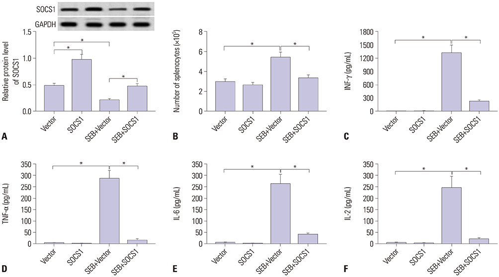
Fig. 4 Overexpression of suppressors of cytokine signaling 1 (SOCS1) suppressed inflammatory cytokines release in staphylococcal enterotoxin B (SEB)-induced splenocytes ex-vivo. Splenocytes from C57BL/6 mice were transfected with pcDNA-SOCS1/Vector (SOCS1/Vector), and then challenged with 1 µg/mL of SEB for 24 h. (A) Expression of SOCS1 was measured by western blot. GAPDH expression was used as internal reference. (B) Treated splenocytes were collected, and total cell number was determined by a hemocytometer. (C–F) Interferon-gamma (INF-γ), tumor necrosis factor-alpha (TNF-α), interleukin-6 (IL-6), and IL-2 in culture supernatant were detected by enzyme-linked immunosorbent assay. *p<0.05. SOCS1 group (without SEB challenge) versus Vector group; SEB+SOCS1 group versus SOCS1 group and SEB+Vector group, respectively; SEB+ SOCS1 group versus Vector group.

Fig. 5 Suppressors of cytokine signaling 1 (SOCS1) downregulation blocked the suppressive effect of miR-222-3p knockdown in staphylococcal enterotoxin B (SEB)-induced splenocytes. Splenocytes were transfected with miR-222-3p/NC, or co-transfected with miR-222-3p-in and si-SOCS1/NC. (A) Western blot showed SOCS1 levels after transfection. GAPDH level was used as internal reference. (B) Transfected splenocytes were collected, and total cell number was determined by a hemocytometer. (C-F) Interferon-gamma (INF-γ), tumor necrosis factor-alpha (TNF-α), interleukin-6 (IL-6), and IL-2 in culture supernatant were detected by enzyme-linked immunosorbent assay. *p<0.05 compared to miR-NC-in group or miR-222-3p+si-NC group.
Reference
-
1. Pinchuk IV, Beswick EJ, Reyes VE. Staphylococcal enterotoxins. Toxins (Basel). 2010; 2:2177–2197.
Article2. Chambers HF, Deleo FR. Waves of resistance: Staphylococcus aureus in the antibiotic era. Nat Rev Microbiol. 2009; 7:629–641.
Article3. Henghold WB 2nd. Other biologic toxin bioweapons: ricin, staphylococcal enterotoxin B, and trichothecene mycotoxins. Dermatol Clin. 2004; 22:257–262. v
Article4. Nagaki M, Tanaka M, Sugiyama A, Ohnishi H, Moriwaki H. Interleukin-10 inhibits hepatic injury and tumor necrosis factor-alpha and interferon-gamma mRNA expression induced by staphylococcal enterotoxin B or lipopolysaccharide in galactosamine-sensitized mice. J Hepatol. 1999; 31:815–824.
Article5. Rieder SA, Nagarkatti P, Nagarkatti M. CD1d-independent activation of invariant natural killer T cells by staphylococcal enterotoxin B through major histocompatibility complex class II/T cell receptor interaction results in acute lung injury. Infect Immun. 2011; 79:3141–3148.
Article6. Krakauer T. Staphylococcal superantigens: pyrogenic toxins induce toxic shock. Toxins (Basel). 2019; 11:E178.
Article7. Ler SG, Lee FK, Gopalakrishnakone P. Trends in detection of warfare agents. Detection methods for ricin, staphylococcal enterotoxin B and T-2 toxin. J Chromatogr A. 2006; 1133:1–12.8. Dilda F, Gioia G, Pisani L, Restelli L, Lecchi C, Albonico F, et al. Escherichia coli lipopolysaccharides and Staphylococcus aureus enterotoxin B differentially modulate inflammatory microRNAs in bovine monocytes. Vet J. 2012; 192:514–516.
Article9. Amini S, Abak A, Sakhinia E, Abhari A. MicroRNA-221 and microrna-222 in common human cancers: expression, function, and triggering of tumor progression as a key modulator. Lab Med. 2019; 05. 02. [Epub]. Available at: https://doi.org/10.1093/labmed/lmz002.
Article10. Fiorino S, Bacchi-Reggiani ML, Visani M, Acquaviva G, Fornelli A, Masetti M, et al. MicroRNAs as possible biomarkers for diagnosis and prognosis of hepatitis B- and C-related-hepatocellular-carcinoma. World J Gastroenterol. 2016; 22:3907–3936.
Article11. Mirzaei HR, Sahebkar A, Mohammadi M, Yari R, Salehi H, Jafari MH, et al. Circulating microRNAs in hepatocellular carcinoma: potential diagnostic and prognostic biomarkers. Curr Pharm Des. 2016; 22:5257–5269.
Article12. Qi P, Cheng SQ, Wang H, Li N, Chen YF, Gao CF. Serum microRNAs as biomarkers for hepatocellular carcinoma in Chinese patients with chronic hepatitis B virus infection. PLoS One. 2011; 6:e28486.
Article13. Motawi TM, Sadik NA, Shaker OG, Ghaleb MH. Elevated serum microRNA-122/222 levels are potential diagnostic biomarkers in Egyptian patients with chronic hepatitis C but not hepatic cancer. Tumour Biol. 2016; 37:9865–9874.
Article14. Higashi M, Yoneda M, Nakagawa T, Ikeda M, Ito T. miR-222 regulates proliferation of primary mouse hepatocytes in vitro. Biochem Biophys Res Commun. 2019; 511:644–649.15. Selten JW, Verhoeven CJ, Heedfeld V, Roest HP, de Jonge J, Pirenne J, et al. The release of microRNA-122 during liver preservation is associated with early allograft dysfunction and graft survival after transplantation. Liver Transpl. 2017; 23:946–956.
Article16. Elliott DM, Nagarkatti M, Nagarkatti PS. 3,39-diindolylmethane ameliorates staphylococcal enterotoxin B-induced acute lung injury through alterations in the expression of microRNA that target apoptosis and cell-cycle arrest in activated T cells. J Pharmacol Exp Ther. 2016; 357:177–187.
Article17. Rao R, Nagarkatti P, Nagarkatti M. Role of miRNA in the regulation of inflammatory genes in staphylococcal enterotoxin B-induced acute inflammatory lung injury and mortality. Toxicol Sci. 2015; 144:284–297.
Article18. Ying J, Qiu X, Lu Y, Zhang M. SOCS1 and its potential clinical role in tumor. Pathol Oncol Res. 2019; 02. 13. [Epub]. Available at: https://doi.org/10.1007/s12253-019-00612-5.
Article19. Inagaki-Ohara K, Kondo T, Ito M, Yoshimura A. SOCS, inflammation, and cancer. JAKSTAT. 2013; 2:e24053.
Article20. Mann M, Mehta A, Zhao JL, Lee K, Marinov GK, Garcia-Flores Y, et al. An NF-κB-microRNA regulatory network tunes macrophage inflammatory responses. Nat Commun. 2017; 8:851.
Article21. Yu J, Zhang W, Qian H, Tang H, Lin W, Lu B. SOCS1 regulates hepatic regenerative response and provides prognostic makers for acute obstructive cholangitis. Sci Rep. 2017; 7:9482.
Article22. Tan L, Jiang W, Lu A, Cai H, Kong L. miR-155 aggravates liver ischemia/ reperfusion injury by suppressing SOCS1 in mice. Transplant Proc. 2018; 50:3831–3839.
Article23. Li SS, Yang M, Chen YP, Tang XY, Zhang SG, Ni SL, et al. Dendritic cells with increased expression of suppressor of cytokine signaling 1(SOCS1) gene ameliorate lipopolysaccharide/d-galactosamineinduced acute liver failure. Mol Immunol. 2018; 101:10–18.
Article24. Araújo Júnior RF, Garcia VB, Leitão RF, Brito GA, Miguel Ede C, Guedes PM, et al. Carvedilol improves inflammatory response, oxidative stress and fibrosis in the alcohol-induced liver injury in rats by regulating kuppfer cells and hepatic stellate cells. PLoS One. 2016; 11:e0148868.
Article25. Rao R, Rieder SA, Nagarkatti P, Nagarkatti M. Staphylococcal enterotoxin B-induced microRNA-155 targets SOCS1 to promote acute inflammatory lung injury. Infect Immun. 2014; 82:2971–2979.
Article26. Plaza R, Vidal S, Rodriguez-Sanchez JL, Juarez C. Implication of STAT1 and STAT3 transcription factors in the response to superantigens. Cytokine. 2004; 25:1–10.
Article27. Kadhim S, Singh NP, Zumbrun EE, Cui T, Chatterjee S, Hofseth L, et al. Resveratrol-mediated attenuation of Staphylococcus aureus enterotoxin b-induced acute liver injury is associated with regulation of microrna and induction of myeloid-derived suppressor cells. Front Microbiol. 2018; 9:2910.
Article28. Szabo PA, Goswami A, Memarnejadian A, Mallett CL, Foster PJ, McCormick JK, et al. Swift intrahepatic accumulation of granulocytic myeloid-derived suppressor cells in a humanized mouse model of toxic shock syndrome. J Infect Dis. 2016; 213:1990–1995.
Article29. Busbee PB, Nagarkatti M, Nagarkatti PS. Natural indoles, indole-3-carbinol (I3C) and 3,3’-diindolylmethane (DIM), attenuate staphylococcal enterotoxin B-mediated liver injury by downregulating miR-31 expression and promoting caspase-2-mediated apoptosis. PLoS One. 2015; 10:e0118506.
Article30. McKallip RJ, Fisher M, Gunthert U, Szakal AK, Nagarkatti PS, Nagarkatti M. Role of CD44 and its v7 isoform in staphylococcal enterotoxin B-induced toxic shock: CD44 deficiency on hepatic mononuclear cells leads to reduced activation-induced apoptosis that results in increased liver damage. Infect Immun. 2005; 73:50–61.
Article31. Yin T, Tong SQ, Xie YC, Lu DY. Cyclosporin A protects Balb/c mice from liver damage induced by superan tigen SEB and D-GalN. World J Gastroenterol. 1999; 5:209–212.
Article32. Ogawa T, Enomoto M, Fujii H, Sekiya Y, Yoshizato K, Ikeda K, et al. MicroRNA-221/222 upregulation indicates the activation of stellate cells and the progression of liver fibrosis. Gut. 2012; 61:1600–1609.
Article33. Pineau P, Volinia S, McJunkin K, Marchio A, Battiston C, Terris B, et al. miR-221 overexpression contributes to liver tumorigenesis. Proc Natl Acad Sci U S A. 2010; 107:264–269.
Article34. Li Y, Liang C, Ma H, Zhao Q, Lu Y, Xiang Z, et al. miR-221/222 promotes S-phase entry and cellular migration in control of basal-like breast cancer. Molecules. 2014; 19:7122–7137.
Article35. Lv X, Zhang Y, Cui Y, Ren Y, Li R, Rong Q. Inhibition of microRNA-155 relieves sepsis-induced liver injury through inactivating the JAK/ STAT pathway. Mol Med Rep. 2015; 12:6013–6018.
Article36. Ren JP, Ying RS, Cheng YQ, Wang L, El Gazzar M, Li GY, et al. HCV-induced miR146a controls SOCS1/STAT3 and cytokine expression in monocytes to promote regulatory T-cell development. J Viral Hepat. 2016; 23:755–766.
Article37. Zhang H, Zhao Z, Pang X, Yang J, Yu H, Zhang Y, et al. Genistein protects against Ox-LDL-induced inflammation through microRNA-155/SOCS1-mediated repression of NF-kB signaling pathway in HUVECs. Inflammation. 2017; 40:1450–1459.
Article
- Full Text Links
-
- Actions
-
Cited
- CITED
-
- Close
- Share
-
- Similar articles
-
- Hsa-miRNA-143-3p Reverses Multidrug Resistance of Triple-Negative Breast Cancer by Inhibiting the Expression of Its Target Protein Cytokine-Induced Apoptosis Inhibitor 1 In Vivo
- Improved Differentiation Ability and Therapeutic Effect of miR-23a-3p Expressing Bone Marrow-Derived Mesenchymal Stem Cells in Mice Model with Acute Lung Injury
- Increase of Rhinovirus Replication in Airway Epithelial Cells by Staphylococcal Enterotoxin A and B
- SOCS3 Attenuates DexamethasoneInduced M2 Polarization by DownRegulation of GILZ via ROS- and p38 MAPK-Dependent Pathways
- Cytokine Inductions and Intracellular Signal Profiles by Stimulation of dsRNA and SEB in the Macrophages and Epithelial Cells
