MiR-590 Inhibits Endothelial Cell Apoptosis by Inactivating the TLR4/NF-κB Pathway in Atherosclerosis
- Affiliations
-
- 1Department of Emergency, Zhengzhou University People's Hospital, Zhengzhou, China.
- 2Department of Coronary Heart Disease, Zhengzhou University People's Hospital, Fuwai Central China Cardiovascular Hospital, Zhengzhou, China. alonewoof@126.com
- KMID: 2441308
- DOI: http://doi.org/10.3349/ymj.2019.60.3.298
Abstract
- PURPOSE
Previous study has well documented the anti-apoptotic effects of miR-590 on oxidized low-density lipoprotein (ox-LDL)-treated endothelial cells (ECs). However, the mechanism underlying the anti-apoptotic effects of miR-590 in ox-LDL-treated ECs remains to be further addressed.
MATERIALS AND METHODS
ApoE(−/−) mice fed with a high-fat diet (HFD) and human aortic endothelial cells (HAECs) treated with ox-LDL were used as in vivo and in vitro models of atherosclerosis. The expressions of miR-590 and toll-like receptor 4 (TLR4) were detected by quantitative real-time PCR and Western blot, respectively. Atherosclerotic lesion analysis was performed using Evans blue and hematoxylin-eosin staining. Cell proliferation was assessed by MTT assay. Apoptosis was examined using flow cytometry analysis and Western blot analysis of Cleaved poly (ADP-ribose) polymerase (PARP) and Cleaved Caspase-3 levels. The effect of miR-590 on TLR4/nuclear factor kappa B (NF-κB) pathway was evaluated by Western blot. Binding between miR-590 and TLR4 was confirmed by luciferase reporter assay and Western blot.
RESULTS
miR-590 was downregulated in the aorta tissues from HFD-fed apoE(−/−) mice and ox-LDL-treated HAECs. miR-590 overexpression inhibited atherosclerotic lesion in HFD-induced apoE(−/−) mice and promoted proliferation and inhibited apoptosis of ox-LDL-treated HAECs. Additionally, TLR4 was identified as a direct target of miR-590 in ox-LDL-treated HAECs. Moreover, anti-miR-590 reversed TLR4 knockdown-mediated promotion of cell proliferation and suppression of apoptosis in ox-LDL-treated HAECs. miR-590 overexpression suppressed the TLR4/NF-κB pathway, and inhibition of the TLR4/NF-κB pathway promoted cell proliferation and impeded apoptosis in ox-LDL-treated HAECs.
CONCLUSION
miR-590 promoted proliferation and blocked ox-LDL-induced apoptosis in HAECs through inhibition of the TLR4/NF-κB pathway.
MeSH Terms
-
Animals
Aorta
Apoptosis*
Atherosclerosis*
Blotting, Western
Caspase 3
Cell Proliferation
Diet, High-Fat
Endothelial Cells*
Evans Blue
Flow Cytometry
Humans
In Vitro Techniques
Lipoproteins
Luciferases
Mice
Real-Time Polymerase Chain Reaction
Toll-Like Receptor 4
Caspase 3
Evans Blue
Lipoproteins
Luciferases
Toll-Like Receptor 4
Figure
-
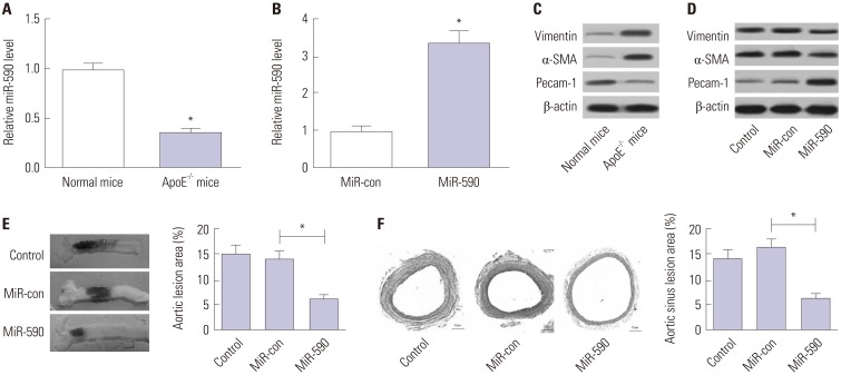
Fig. 1 The effects of miR-590 on atherosclerotic lesion in HFD-induced apoE−/− mice. ApoE−/− mice were fed on a HFD for 12 weeks and injected with miR-590 or miR-control via tail vein once every 4 weeks after starting HFD. (A) qRT-PCR analysis of miR-590 expression in the aorta derived from apoE−/− mice fed on a HFD or wild-type C57BL/6 J controls fed on a normal diet. U6 was used as the normalization. (B) qRT-PCR analysis of miR-590 expression in HFD-fed apoE−/− mice injected with miR-590 or miR-control. U6 was used as the normalization. (C) Western blot analysis of the protein levels of Pecam-1, α-SMA and Vimentin in apoE−/− mice fed on a HFD or wild-type C57BL/6 J controls fed on a normal diet. (D) Western blot analysis of the protein levels of Pecam-1, α-SMA and Vimentin HFD-fed apoE−/− mice injected with miR-590 or miR-control. (E) Atherosclerotic plaque formation in the resected aortic sinuses was assessed by Evans blue staining. (F) The atherosclerotic lesion in aortic sinuses was examined by hematoxylin and eosin staining (×40). *p<0.05. HFD, high-fat diet; α-SMA, α-smooth muscle actin.
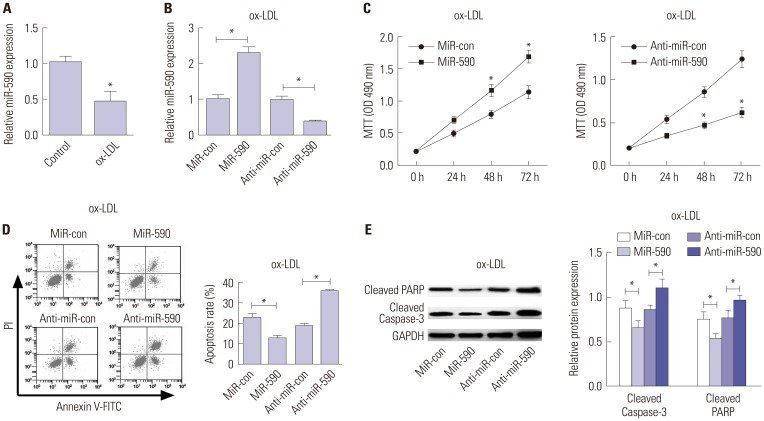
Fig. 2 The effects of miR-590 on the proliferation and apoptosis in ox-LDL-treated HAECs. (A) qRT-PCR analysis of miR-590 expression in HAECs following ox-LDL challenge. U6 was used as the normalization. (B) qRT-PCR analysis of miR-590 expression in HAECs transfected with miR-590, anti-miR-590, or matched controls, followed by ox-LDL treatment. U6 was used as the normalization. (C) MTT assay was performed to evaluate cell proliferation at 24, 48, and 72 h in HAECs transfected with miR-590, anti-miR-590, or matched controls, followed by ox-LDL stimulation. (D) Flow cytometry analysis was conducted to determine the percentage of HAEC apoptosis after transfection with miR-590, anti-miR-590, or matched controls, followed by ox-LDL administration. (E) Western blot was employed to detect the expression levels of Cleaved PARP and Cleaved-Caspase-3 in HAECs transfected with miR-590, anti-miR-590, or matched controls, followed by ox-LDL stimulation. *p<0.05. ox-LDL, oxidized low-density lipoprotein; HAECs, human aortic endothelial cells.

Fig. 3 MiR-590 directly targets TLR4 in ox-LDL-treated HAECs. (A) Bioinformatics analysis of the predicted interaction of miR-590 in the 3′UTR of TLR4. (B) Luciferase activity was determined by luciferase reporter assay in HAECs cells co-transfected with TLR4-WT or TLR4-MUT and miR-590, anti-miR-590, or respective controls. (C) Western blot (left) was performed to detect the cellular protein level of TLR4 in HAECs with or without ox-LDL treatment, while TLR4 protein level on cell surface was evaluated by flow cytometry (right). (D) The cellular protein level of TLR4 in ox-LDL-treated HAECs transfected with miR-590, anti-miR-590, or matched controls was detected by Western blot (left), while TLR4 protein level on cell surface was evaluated by flow cytometry (right). *p<0.05. TLR4, toll-like receptor 4; ox-LDL, oxidized low-density lipoprotein; HAECs, human aortic endothelial cells.
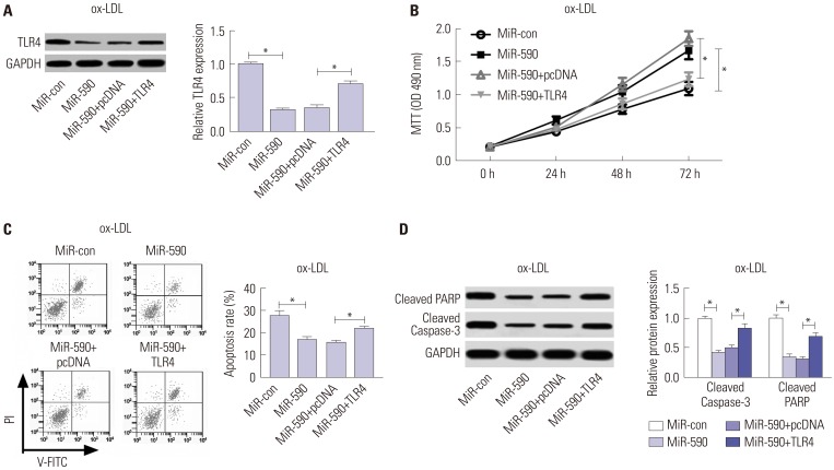
Fig. 4 Anti-miR-590 reverses TLR4 knockdown-mediated promotion of cell proliferation and suppression of apoptosis in ox-LDL-treated HAECs. HAECs were transfected with si-TLR4, si-con, or cotransfected with si-TLR4 and anti-miR-590 or anti-miR-con, following ox-LDL stimulation. (A) Western blot analysis of TLR4 protein level in the treated HAECs. (B) Cell proliferation at 24, 48, and 72 h in the treated HAECs was evaluated by MTT assay. (C) Flow cytometry analysis was performed to detect the apoptotic rates in the treated HAECs. (D) Western blot was applied to analyze the protein levels of Cleaved PARP and Cleaved Caspase-3 in the treated HAECs. *p<0.05. ox-LDL, oxidized low-density lipoprotein; TLR4, toll-like receptor 4; HAECs, human aortic endothelial cells.
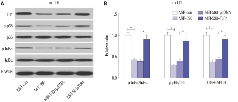
Fig. 5 The effects of miR-590 on the TLR4/NF-κB pathway in ox-LDL-treated HAECs. (A) Western blot analysis of TLR4, p-IκBα, IκBα, p-p65, and p65 in HAECs after transfection with miR-590, miR-con, miR-590+TLR4, or miR-590+pcDNA, following ox-LDL challenge. (B) Quantification analysis of the protein level of TLR4, p-IκBα/IκBα ratio and p-p65/p65 ratio in the treated HAECs. *p<0.05. ox-LDL, oxidized low-density lipoprotein; TLR4, toll-like receptor 4; NF-κB, nuclear factor kappa B; HAECs, human aortic endothelial cells.
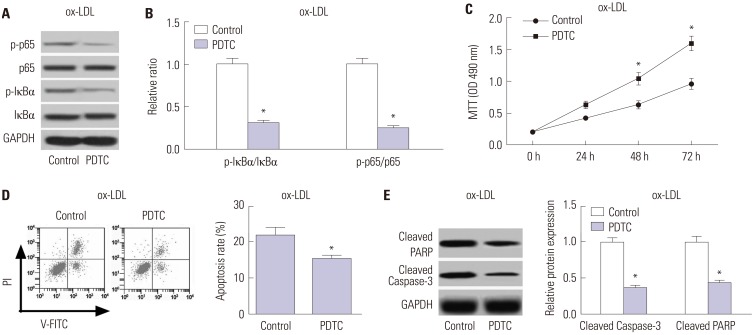
Fig. 6 TInhibition of the TLR4/NF-κB pathway facilitates cell proliferation and restrained apoptosis in ox-LDL-treated HAECs. HAECs were exposed to 50 µM PDTC for 24 h, followed by treatment with 100 µg/mL ox-LDL for 24 h. (A) The protein levels of p-p65, p65, p-IκBα and IκBα in the treated HAECs were detected by Western blot. (B) Quantification analysis of the ratio of p-IκBα/IκBα and p-p65/p65 in the treated HAECs. (C) MTT assay was performed to assess cell proliferation at 24, 48, and 72 h in the treated HAECs. (D) Flow cytometry analysis was conducted to examine the apoptosis of treated HAECs. (E) Western blot was employed to determine the protein levels of Cleaved PARP and Cleaved Caspase-3 in the treated HAECs. *p<0.05. ox-LDL, oxidized low-density lipoprotein; TLR4, toll-like receptor 4; NF-κB, nuclear factor kappa B; HAECs, human aortic endothelial cells.
Reference
-
1. Rafieian-Kopaei M, Setorki M, Doudi M, Baradaran A, Nasri H. Atherosclerosis: process, indicators, risk factors and new hopes. Int J Prev Med. 2014; 5:927–946. PMID: 25489440.2. Tabas I, García-Cardeña G, Owens GK. Recent insights into the cellular biology of atherosclerosis. J Cell Biol. 2015; 209:13–22. PMID: 25869663.
Article3. Trpkovic A, Resanovic I, Stanimirovic J, Radak D, Mousa SA, Cenic-Milosevic D, et al. Oxidized low-density lipoprotein as a biomarker of cardiovascular diseases. Crit Rev Clin Lab Sci. 2015; 52:70–85. PMID: 25537066.
Article4. Najafpour Boushehri S, Yusof RM, Nasir Mohammad Taib M, Mirzaei K, Yazdekhasti N, Akbarzadeh S. Effect of vitamin supplementation on serum oxidized low-density lipoprotein levels in male subjects with cardiovascular disease risk factors. Iran J Basic Med Sci. 2012; 15:958–964. PMID: 23493764.5. Bartel DP. MicroRNAs: target recognition and regulatory functions. Cell. 2009; 136:215–233. PMID: 19167326.
Article6. Natarelli L, Schober A. MicroRNAs and the response to injury in atherosclerosis. Hamostaseologie. 2015; 35:142–150. PMID: 25612846.
Article7. Shan Z, Yao C, Li ZL, Teng Y, Li W, Wang JS, et al. Differentially expressed microRNAs at different stages of atherosclerosis in ApoE-deficient mice. Chin Med J (Engl). 2013; 126:515–520. PMID: 23422117.8. Menghini R, Stöhr R, Federici M. MicroRNAs in vascular aging and atherosclerosis. Ageing Res Rev. 2014; 17:68–78. PMID: 24681293.
Article9. Eulalio A, Mano M, Dal Ferro M, Zentilin L, Sinagra G, Zacchigna S, et al. Functional screening identifies miRNAs inducing cardiac regeneration. Nature. 2012; 492:376–381. PMID: 23222520.
Article10. Bao MH, Li JM, Zhou QL, Li GY, Zeng J, Zhao J, et al. Effects of miR-590 on oxLDL-induced endothelial cell apoptosis: roles of p53 and NF-κB. Mol Med Rep. 2016; 13:867–873. PMID: 26648441.
Article11. den Dekker WK, Cheng C, Pasterkamp G, Duckers HJ. Toll like receptor 4 in atherosclerosis and plaque destabilization. Atherosclerosis. 2010; 209:314–320. PMID: 19900676.
Article12. Xing S, Zheng F, Zhang W, Wang D, Xing Q. Relationship between toll-like receptor 4 levels in aorta and severity of atherosclerosis. J Int Med Res. 2014; 42:958–965. PMID: 24925583.
Article13. Corbi G, Bianco A, Turchiarelli V, Cellurale M, Fatica F, Daniele A, et al. Potential mechanisms linking atherosclerosis and increased cardiovascular risk in COPD: focus on Sirtuins. Int J Mol Sci. 2013; 14:12696–12713. PMID: 23774840.
Article14. Gimbrone MA Jr, Topper JN, Nagel T, Anderson KR, Garcia-Cardeña G. Endothelial dysfunction, hemodynamic forces, and atherogenesis. Ann N Y Acad Sci. 2000; 902:230–239. PMID: 10865843.15. Sun X, Belkin N, Feinberg MW. Endothelial microRNAs and atherosclerosis. Curr Atheroscler Rep. 2013; 15:372. PMID: 24158362.
Article16. Chen Z, Wang M, He Q, Li Z, Zhao Y, Wang W, et al. MicroRNA-98 rescues proliferation and alleviates ox-LDL-induced apoptosis in HUVECs by targeting LOX-1. Exp Ther Med. 2017; 13:1702–1710. PMID: 28565756.
Article17. Tang F, Yang TL. MicroRNA-126 alleviates endothelial cells injury in atherosclerosis by restoring autophagic flux via inhibiting of PI3K/Akt/mTOR pathway. Biochem Biophys Res Commun. 2018; 495:1482–1489. PMID: 29203244.
Article18. Cui J, Ren Z, Zou W, Jiang Y. miR-497 accelerates oxidized low-density lipoprotein-induced lipid accumulation in macrophages by repressing the expression of apelin. Cell Biol Int. 2017; 41:1012–1019. PMID: 28653788.
Article19. Luo P, Zhang WF, Qian ZX, Xiao LF, Wang H, Zhu TT, et al. MiR-590-5p-meidated LOX-1 upregulation promotes Angiotensin II-induced endothelial cell apoptosis. Biochem Biophys Res Commun. 2016; 471:402–408. PMID: 26906623.
Article20. He PP, OuYang XP, Li Y, Lv YC, Wang ZB, Yao F, et al. MicroRNA-590 inhibits lipoprotein lipase expression and prevents atherosclerosis in apoE knockout mice. PLoS One. 2015; 10:e0138788. PMID: 26397958.
Article21. He PP, Ouyang XP, Tang YY, Liao L, Wang ZB, Lv YC, et al. MicroRNA-590 attenuates lipid accumulation and pro-inflammatory cytokine secretion by targeting lipoprotein lipase gene in human THP-1 macrophages. Biochimie. 2014; 106:81–90. PMID: 25149060.
Article22. Thompson MR, Kaminski JJ, Kurt-Jones EA, Fitzgerald KA. Pattern recognition receptors and the innate immune response to viral infection. Viruses. 2011; 3:920–940. PMID: 21994762.
Article23. Yang K, Zhang XJ, Cao LJ, Liu XH, Liu ZH, Wang XQ, et al. Toll-like receptor 4 mediates inflammatory cytokine secretion in smooth muscle cells induced by oxidized low-density lipoprotein. PLoS One. 2014; 9:e95935. PMID: 24755612.
Article24. Stoll LL, Denning GM, Li WG, Rice JB, Harrelson AL, Romig SA, et al. Regulation of endotoxin-induced proinflammatory activation in human coronary artery cells: expression of functional membrane-bound CD14 by human coronary artery smooth muscle cells. J Immunol. 2004; 173:1336–1343. PMID: 15240728.
Article25. Michelsen KS, Wong MH, Shah PK, Zhang W, Yano J, Doherty TM, et al. Lack of Toll-like receptor 4 or myeloid differentiation factor 88 reduces atherosclerosis and alters plaque phenotype in mice deficient in apolipoprotein E. Proc Natl Acad Sci U S A. 2004; 101:10679–10684. PMID: 15249654.
Article26. Pasterkamp G, Van Keulen JK, De Kleijn DP. Role of Toll-like receptor 4 in the initiation and progression of atherosclerotic disease. Eur J Clin Invest. 2004; 34:328–334. PMID: 15147329.
Article27. Baker RG, Hayden MS, Ghosh S. NF-κB, inflammation, and metabolic disease. Cell Metab. 2011; 13:11–22. PMID: 21195345.
Article28. Tang YL, Jiang JH, Wang S, Liu Z, Tang XQ, Peng J, et al. TLR4/NF-κB signaling contributes to chronic unpredictable mild stress-induced atherosclerosis in ApoE-/- mice. PLoS One. 2015; 10:e0123685. PMID: 25860573.
Article29. Hu ZP, Fang XL, Fang N, Wang XB, Qian HY, Cao Z, et al. Melatonin ameliorates vascular endothelial dysfunction, inflammation, and atherosclerosis by suppressing the TLR4/NF-κB system in high-fat-fed rabbits. J Pineal Res. 2013; 55:388–398. PMID: 24006943.
Article30. Lu Z, Zhang X, Li Y, Jin J, Huang Y. TLR4 antagonist reduces early-stage atherosclerosis in diabetic apolipoprotein E-deficient mice. J Endocrinol. 2013; 216:61–71. PMID: 23060524.
Article31. Lu Z, Zhang X, Li Y, Lopes-Virella MF, Huang Y. TLR4 antagonist attenuates atherogenesis in LDL receptor-deficient mice with diet-induced type 2 diabetes. Immunobiology. 2015; 220:1246–1254. PMID: 26162692.
Article32. Zhou Q, Zhu Z, Hu X, Shu C. HMGB1: a critical mediator for oxidized-low density lipoproteins induced atherosclerosis. Int J Cardiol. 2016; 202:956–957. PMID: 26549559.
Article33. Chen M, Li W, Zhang Y, Yang J. MicroRNA-20a protects human aortic endothelial cells from Ox-LDL-induced inflammation through targeting TLR4 and TXNIP signaling. Biomed Pharmacother. 2018; 103:191–197. PMID: 29653364.
Article34. Du F, Yu F, Wang Y, Hui Y, Carnevale K, Fu M, et al. MicroRNA-155 deficiency results in decreased macrophage inflammation and attenuated atherogenesis in apolipoprotein E-deficient mice. Arterioscler Thromb Vasc Biol. 2014; 34:759–767. PMID: 24504735.
Article35. Wang J, Bai X, Song Q, Fan F, Hu Z, Cheng G, et al. miR-223 inhibits lipid deposition and inflammation by suppressing toll-like receptor 4 signaling in macrophages. Int J Mol Sci. 2015; 16:24965–24982. PMID: 26492242.
Article36. Du XJ, Lu JM, Sha Y. MiR-181a inhibits vascular inflammation induced by ox-LDL via targeting TLR4 in human macrophages. J Cell Physiol. 2018; 233:6996–7003.
Article
- Full Text Links
-
- Actions
-
Cited
- CITED
-
- Close
- Share
-
- Similar articles
-
- miR-215 Enhances HCV Replication by Targeting TRIM22 and Inactivating NF-κB Signaling
- Upregulation of miR-27b Facilitates Apoptosis of TNF-α-Stimulated Fibroblast-Like Synoviocytes
- Adipose-Derived Stem Cell Transplantation Inhibits Vascular Inflammatory Responses and Endothelial Dysfunction in Rats with Atherosclerosis
- MiR-182-5p Mediated by Exosomes Derived From Bone Marrow Mesenchymal Stem Cell Attenuates Inflammatory Responses by Targeting TLR4 in a Mouse Model of Myocardial Infraction
- Exosome-mediated lnc-ABCA12-3 promotes proliferation and glycolysis but inhibits apoptosis by regulating the tolllike receptor 4/nuclear factor kappa-B signaling pathway in esophageal squamous cell carcinoma
