Therapeutic Effects and Immunomodulation of Suanbo Mineral Water Therapy in a Murine Model of Atopic Dermatitis
- Affiliations
-
- 1Department of Dermatology, The Catholic University of Korea Uijeongbu St. Mary's Hospital, Uijeongbu, Korea. jwkim52@catholic.ac.kr
- 2Department of Microbiology, Ewha Womans University School of Medicine, Seoul, Korea.
- 3Department of Dermatology, The Catholic University of Korea Bucheon St. Mary's Hospital, Bucheon, Korea.
- 4Department of Earth and Environmental Sciences, Korea University, Seoul, Korea.
- 5Department of Physical Medicine and Rehabilitation, Konkuk University Chungju Hospital, Chungju, Korea.
- 6The Korean Academy of Hot Spring, Seoul, Korea.
- KMID: 2265057
- DOI: http://doi.org/10.5021/ad.2013.25.4.462
Abstract
- BACKGROUND
Balneotherapy is widely used as an alternative treatment modality for AD. Although the clinical benefit of some mineral waters has been established, their mechanisms of action in alleviating AD are only partly understood.
OBJECTIVE
The clinical modification and immunomodulatory or anti-inflammatory effects of mineral water from the Suanbo hot springs on the differentiation and cytokine production of Th1, Th2, and regulatory T cells (Treg) were investigated using spleen, skin tissue, and serum from NC/Nga mice.
METHODS
The therapeutic effects of bathing in mineral water in a Dermatophagoides farinae body extract ointment (Dfb ointment)-induced AD mouse model were assessed by measuring the modified Scoring atopic dermatitis (SCORAD) index scores, transepidermal water loss (TEWL), histological and immunohistochemical changes of the skin lesion, serum levels of interferon (IFN)-gamma, interleukin (IL)-4, IL-5 and immunoglobulin E, mRNA expression of IFN-gamma, IL-4 and IL-5 of dorsal skin, and helper T cell differentiation in the spleen.
RESULTS
Bathing in mineral water significantly reduced the modified SCORAD index scores, TEWL, epidermal hyperplasia, and inflammatory cell infiltration. IL-4 production and Th2 cell differentiation showed a decreasing tendency with mineral water bathing, but the Th1 cells did not. On the contrary, differentiation to Treg cells was promoted with mineral water bathing.
CONCLUSION
Balneotherapy not only has anti-inflammatory activity, but also shows positive effects on cutaneous barrier homeostasis. These results suggest that the favorable effects of balneotherapy may be mediated by modifying the Th2 response, and possibly in part by inducing Treg cell differentiation.
Keyword
MeSH Terms
-
Animals
Balneology
Baths
Cell Differentiation
Dermatitis, Atopic*
Dermatophagoides farinae
Homeostasis
Hot Springs
Hyperplasia
Immunoglobulin E
Immunoglobulins
Immunomodulation*
Interferons
Interleukin-4
Interleukin-5
Interleukins
Mice
Mineral Waters*
RNA, Messenger
Skin
Spleen
T-Lymphocytes, Regulatory
Th1 Cells
Th2 Cells
Mineral Waters
Immunoglobulin E
Immunoglobulins
Interferons
Interleukin-4
Interleukin-5
Interleukins
Mineral Waters
RNA, Messenger
Mineral Waterss
Figure
-
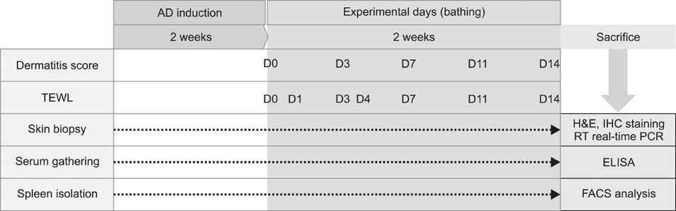
Fig. 1 Schematic diagram of experimental design. AD: atopic dermatitis, TEWL: transepidermal water loss, RT: reverse transcription, PCR: polymerase chain reaction, FACS: fluorescence-activated cell sorting.
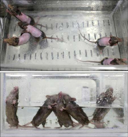
Fig. 2 Bathing was performed daily for 5 minutes during 2 weeks with a regular water temperature.

Fig. 3 (A) Photographs of the clinical features of NC/Nga mice (a) in the untreated negative control (CONT), (b, c, d) Dermatophagoides farinae body (Dfb) ointment applied experimental groups on day 14 following the first challenge, (e) Dfb ointment positive control (AD), (f) Dfb ointment+distilled water bathing (AD+DW), and (g) Dfb ointment+mineral water bathing (AD+MW), on day 14 following first bathing. (B) Clinical skin severity scores. (C) Transepidermal water loss (TEWL). Data are presented as the mean±standard deviation. SCORAD: Scoring atopic dermatitis. *p<0.05 vs. AD+DW.
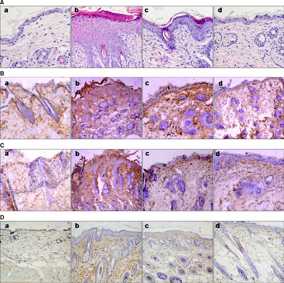
Fig. 4 (A) Histological features of dorsal skin (H&E, ×200). Effects of bathing in mineral water on Dermatophagoides farinae body (Dfb) ointment-induced infiltration of CD1+, CD4+, and CD8+ cells in the dorsal skin of NC/Nga mice (B: anti-CD1, C: anti-CD4 and D: anti-CD8; ×100). (a) The untreated negative control, (b) Dfb ointment positive control (AD), (c) Dfb ointment+distilled water bathing (AD+DW), and (d) Dfb ointment+mineral water bathing (AD+MW) on day 14 following the first bathing.
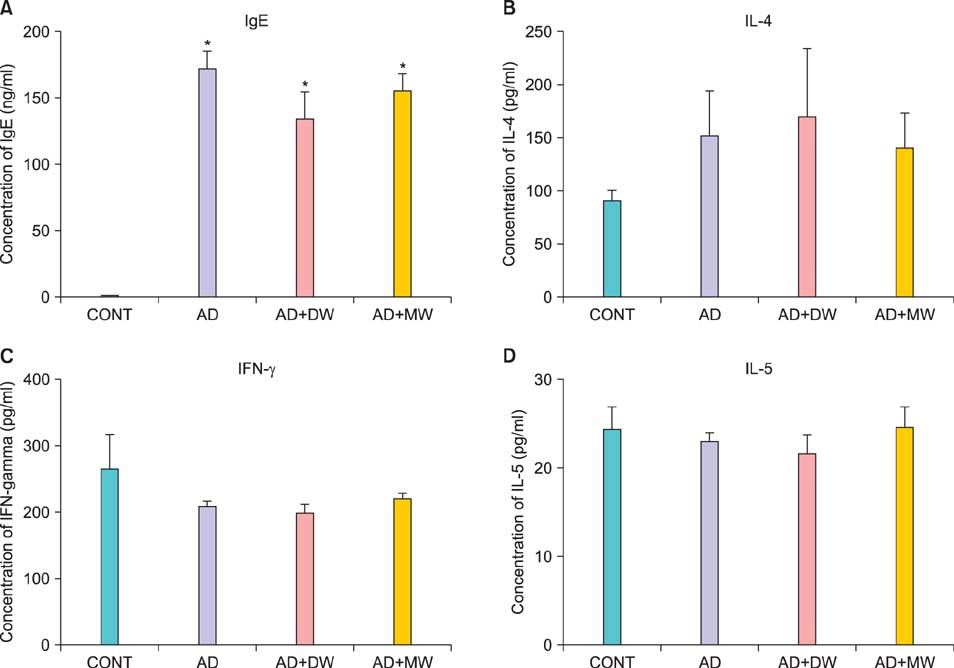
Fig. 5 Serum levels of (A) immunoglobulin E (IgE), (B) interleukin (IL)-4, (C) interferon (IFN)-γ, and (D) IL-5 on experimental day 14. Data are presented as the mean±standard deviation. CONT: the untreated negative control, AD: Dfb ointment positive control, AD+DW: Dfb ointment+distilled water bathing, AD+MW: Dfb ointment+mineral water bathing. *p<0.05 vs. CONT.
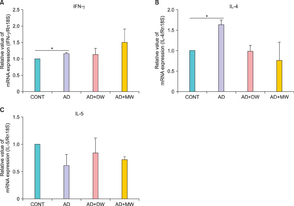
Fig. 6 Effects of bathing in mineral water on Dermatophagoides farinae body (Dfb) ointment-induced mRNA expression of (A) interferon (IFN)-γ, (B) interleukin (IL)-4 and (C) IL-5 of dorsal skin. Data are presented as the mean±standard deviation. CONT: the untreated negative control, AD: Dfb ointment positive control, AD+DW: Dfb ointment+distilled water bathing, AD+MW: Dfb ointment+mineral water bathing. *p<0.05.

Fig. 7 Effects of bathing in the mineral water on the Dermatophagoides farinae body (Dfb) ointment-induced T cell profile of spleen cells. Data are presented as the mean±standard deviation. CONT: the untreated negative control, AD: Dfb ointment positive control, AD+DW: Dfb ointment+distilled water bathing, AD+MW: Dfb ointment+mineral water bathing. *p<0.05.
Cited by 3 articles
-
Immunomodulatory Effects of Deokgu Thermomineral Water Balneotherapy on Oxazolone-Induced Atopic Dermatitis Murine Model
Young Bok Lee, Su Jin Kim, Sae Mi Park, Kyung Ho Lee, Hyung Jin Han, Dong Soo Yu, So Youn Woo, Seong Taek Yun, Se-Yeong Hamm, Hong Jig Kim, Jin-Wou Kim
Ann Dermatol. 2016;28(2):192-198. doi: 10.5021/ad.2016.28.2.192.Basement Membrane Status Is Intact in Urticarial Dermatitis vs. Adult-Onset Atopic Dermatitis
Tae In Kim, Hyung-Jin Park, Yong-Yon Won, Hyeongwon Choi, Ki-Heon Jeong, Ji-Youn Sung, Min Kyung Shin
Ann Dermatol. 2018;30(2):258-261. doi: 10.5021/ad.2018.30.2.258.Investigation of Immune-Regulatory Effects of Mageumsan Hot Spring via Protein Microarray
In Vitro
Hyung Jin Hahn, Jung Soo Kim, Yeong Ho Kim, Young Bok Lee, Dong Soo Yu, Jin-Wou Kim
Ann Dermatol. 2018;30(3):322-330. doi: 10.5021/ad.2018.30.3.322.
Reference
-
1. Elias PM, Hatano Y, Williams ML. Basis for the barrier abnormality in atopic dermatitis: outside-inside-outside pathogenic mechanisms. J Allergy Clin Immunol. 2008; 121:1337–1343.
Article2. Matz H, Orion E, Wolf R. Balneotherapy in dermatology. Dermatol Ther. 2003; 16:132–140.
Article3. Sukenik S, Abu-Shakra M, Flusser D. Balneotherapy in autoimmune disease. Isr J Med Sci. 1997; 33:258–261.4. Valitutti S, Castellino F, Musiani P. Effect of sulfurous (thermal) water on T lymphocyte proliferative response. Ann Allergy. 1990; 65:463–468.5. Wiedow O, Streit V, Christophers E, Ständer M. Liberation of human leukocyte elastase by hypertonic saline baths in psoriasis. Hautarzt. 1989; 40:518–522.6. Shim WH, Ko HC, Kim BS, Kim MB, Min JA, Kim JW. The efficacy and the safety of Hae-Un-Dae spa therapy for the treatment of adult atopic dermatitis. Korean J Dermatol. 2010; 48:1052–1059.7. Lee HP, Choi YJ, Cho KA, Woo SY, Yun ST, Lee JT, et al. Effect of spa spring water on cytokine expression in human keratinocyte HaCaT cells and on differentiation of CD4(+) T cells. Ann Dermatol. 2012; 24:324–336.
Article8. Yamamoto M, Haruna T, Yasui K, Takahashi H, Iduhara M, Takaki S, et al. A novel atopic dermatitis model induced by topical application with dermatophagoides farinae extract in NC/Nga mice. Allergol Int. 2007; 56:139–148.
Article9. Angelova-Fischer I, Bauer A, Hipler UC, Petrov I, Kazandjieva J, Bruckner T, et al. The objective severity assessment of atopic dermatitis (OSAAD) score: validity, reliability and sensitivity in adult patients with atopic dermatitis. Br J Dermatol. 2005; 153:767–773.
Article10. Sugarman JL, Fluhr JW, Fowler AJ, Bruckner T, Diepgen TL, Williams ML. The objective severity assessment of atopic dermatitis score: an objective measure using permeability barrier function and stratum corneum hydration with computer-assisted estimates for extent of disease. Arch Dermatol. 2003; 139:1417–1422.11. Novak N. An update on the role of human dendritic cells in patients with atopic dermatitis. J Allergy Clin Immunol. 2012; 129:879–886.
Article12. Spergel JM, Mizoguchi E, Oettgen H, Bhan AK, Geha RS. Roles of TH1 and TH2 cytokines in a murine model of allergic dermatitis. J Clin Invest. 1999; 103:1103–1111.
Article13. Abbas AK, Murphy KM, Sher A. Functional diversity of helper T lymphocytes. Nature. 1996; 383:787–793.
Article14. Wing K, Sakaguchi S. Regulatory T cells as potential immunotherapy in allergy. Curr Opin Allergy Clin Immunol. 2006; 6:482–488.
Article15. Shevach EM. Mechanisms of foxp3+ T regulatory cell-mediated suppression. Immunity. 2009; 30:636–645.
Article16. Vignali DA, Collison LW, Workman CJ. How regulatory T cells work. Nat Rev Immunol. 2008; 8:523–532.
Article17. Tang Q, Bluestone JA. The Foxp3+ regulatory T cell: a jack of all trades, master of regulation. Nat Immunol. 2008; 9:239–244.
Article18. Weiss E, Mamelak AJ, La Morgia S, Wang B, Feliciani C, Tulli A, et al. The role of interleukin 10 in the pathogenesis and potential treatment of skin diseases. J Am Acad Dermatol. 2004; 50:657–675.
Article19. Hawrylowicz CM, O'Garra A. Potential role of interleukin-10-secreting regulatory T cells in allergy and asthma. Nat Rev Immunol. 2005; 5:271–283.
Article
- Full Text Links
-
- Actions
-
Cited
- CITED
-
- Close
- Share
-
- Similar articles
-
- Immunomodulatory Effects of Deokgu Thermomineral Water Balneotherapy on Oxazolone-Induced Atopic Dermatitis Murine Model
- Comparison of 4% NaCl Solution and UVB to Mineral Salt and UVB in Patients with Atopic Dermatitis
- The pH of water from various sources: an overview for recommendation for patients with atopic dermatitis
- UVB Phototherapy in Atopic Dermatitis
- Vitamin D and atopic dermatitis
