Granulocyte Macrophage-Colony Stimulating Factor (GM-CSF) Augments Acute Lung Injury via Its Neutrophil Priming Effects
- Affiliations
-
- 1Department of Internal Medicine, Chung Ang University College of Medicine, Seoul, Korea. jykimmd@cau.ac.kr
- KMID: 1713463
- DOI: http://doi.org/10.3346/jkms.2008.23.2.288
Abstract
- Granulocyte macrophage-colony stimulating factor (GM-CSF) has immuno-stimulatory effects. We hypothesized that GM-CSF plays an important role both in lipopolysaccharide (LPS)- and hemorrhage-induced acute lung injury (ALI). We also postulated that GM-CSF augments LPS-induced inflammation by priming neutrophils. ALI was induced in GM-CSF-/- or control C57BL mice either by LPS injection or by hemorrhage. Lung inflammation (by lung expression for tumor necrosis factor-alpha (TNF-alpha), macrophage inflammatory protein-2 (MIP-2), interleukin-1beta (IL-1beta), interleukin- 6 (IL-6), and keratinocyte-derived chemokine) and lung injury (by myeloperoxidase and evans blue dye assay) were evaluated after ALI. Incremental doses of LPS (0, 1, 10, and 100 ng/mL) and GM-CSF (0, 1, 10, and 100 ng/mL) were added to bone marrow neutrophils. The expression of TNF-alpha, MIP-2, and IL-1beta was evaluated with enzyme linked immunosorbent assay. The mRNA expression of three cytokines, and the nuclear translocation of nuclear factor kappa B (NF kappa-B) were evaluated by reverse transcriptase-polymerase chain reaction and electropnoretic mobility shift assay, respectively. GM-CSF -/- mice showed decreased neutrophil infiltration, less leakage, and lower expression of cytokines in the lung after LPS or hemorrhage. GM-CSF augmented LPS-induced protein and mRNA expression of TNF-alpha, MIP-2 and IL-1beta, which was mediated by increased intra-nuclear translocation of NF-kappa B. GM-CSF plays an important role in high-dose LPS and hemorrhage-induced ALI, which appears to be mediated by its priming effect on neutrophils.
Keyword
MeSH Terms
-
Animals
Bone Marrow Cells/cytology
Chemokine CXCL2/metabolism
Granulocyte-Macrophage Colony-Stimulating Factor/metabolism/*physiology
Interleukin-1beta/metabolism
Lipopolysaccharides/metabolism
Lung/metabolism/pathology
*Lung Injury
Male
Mice
Mice, Inbred BALB C
Mice, Inbred C57BL
Mice, Transgenic
Neutrophils/*cytology/metabolism
Peroxidase/metabolism
Tumor Necrosis Factor-alpha/metabolism
Figure
-
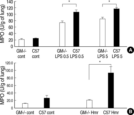
Fig. 1 GM-CSF -/- mice showed decreased infiltration of neutrophils in the lung after LPS injection or hemorrhage. GM-CSF -/- mice and control C57BL mice were treated with low or high doses of LPS (0.5 mg/kg or 5 mg/kg, respectively) injected into the peritoneum. Four hours after the injection of LPS or hemorrhage, mice were sacrificed and lung neutrophil infiltration was evaluated by MPO assay. Each group consisted of five mice. GM-/-, GM-CSF knockout mice; C57, C57BL control mice; Hmr, hemorrhage; LPS 0.5, LPS 0.5 mg/kg i.p.; LPS 5, LPS 5 mg/kg i.p. *, p<0.05.

Fig. 2 GM-CSF-/- mice were resistant to lung leakage after LPS injection or hemorrhage. GM-CSF -/- mice or control C57BL mice were treated with low or high doses of LPS (0.5 mg/kg and 5 mg/kg, respectively) injected into the peritoneum. Three hours after the injection of LPS or hemorrhage, Evans blue dye (50 mg/kg) was injected into the tail vein. One hour after the injection of dye, mice were sacrificed and the lungs were removed. EBD was extracted from the removed lungs by formamide extraction. Each group consisted of five mice. GM-/-, GM-CSF knockout mice; C57, C57BL control mice; Hmr, hemorrhage; LPS 0.5, LPS 0.5 mg/kg i.p.; LPS 5, LPS 5 mg/kg i.p. *, p<0.05.
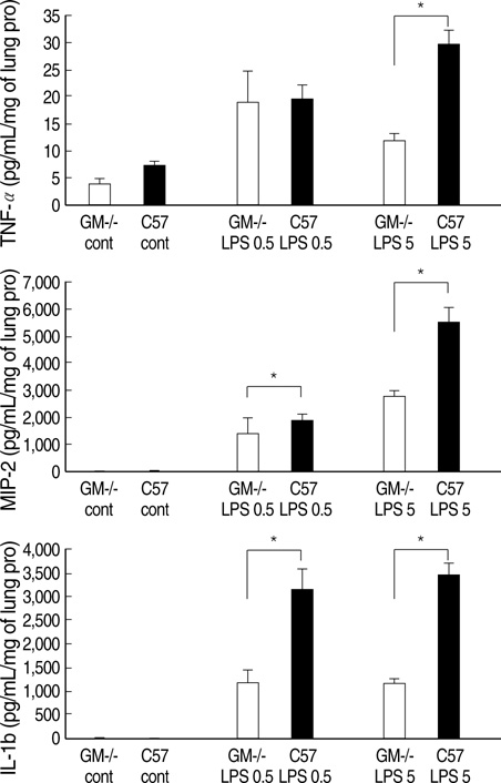
Fig. 3 GM-CSF -/- mice showed decreased levels of TNF-α, MIP-2, and IL-1β in the lung after LPS injection. GM-CSF -/- mice and control C57BL mice were treated with low or high doses of LPS (0.5 mg/kg or 5 mg/kg, respectively) injected into the peritoneum. Four hours after the injection of LPS, mice were sacrificed, and expression of TNF-α, MIP-2 and IL-11β in the lung samples were evaluated by ELISA. Each group consisted of five mice.*, p<0.05.
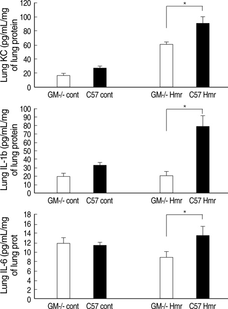
Fig. 4 GM-CSF -/- mice showed decreased levels of KC, IL-6, and IL-1β in the lung after hemorrhage. Four hours after hemorrhage, mice were sacrificed and the expression of KC, IL-6, and IL-1β in the lung samples was evaluated by ELISA. Each group consisted of five mice. *, p<0.05.

Fig. 5 GM-CSF augments TNF-α, MIP-2, and IL-1β expression in mouse bone marrow neutrophils. Bone marrow neutrophils of control mice (5×10⁵/well) were treated with GM-CSF (0, 1, 10, and 100 ng/mL) in combination with LPS (0, 1, 10, and 100 ng/mL). Four hours after stimulation with GM-CSF and/or LPS, the supernatant was collected and the expression of TNF-α, MIP-2, and IL-1β was evaluated by ELISA. This experiment was done three times independently and the displayed data are mean values. *, p<0.05
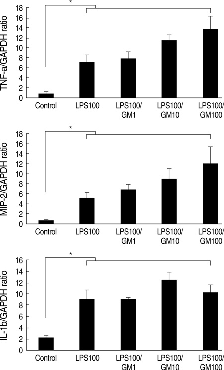
Fig. 6 GM-CSF augments LPS-induced mRNA expression of TNF-α, MIP-2, and IL-1β in mouse bone marrow neutrophils. LPS (100 ng/mL) and incremental concentration of GM-CSF (0, 1, 10, and 100 ng/mL) was added to bone marrow neutrophils of control mice (1×10⁶/microtube). One hour later, the microtube was centrifuged for 12,000 g at 4℃ and total RNA was collected. The mRNA expression of TNF-α, MIP-2, and IL-1β was measured by RT-PCR. This experiment was done three times independently and the displayed data are mean values. LPS 100, LPS 100 ng/mL; LPS 100/GM1, LPS 100 ng/mL+GM-CSF 1 ng/mL; LPS 100/GM10, LPS 100 ng/mL+GM-CSF 10 ng/mL; LPS 100/GM100, LPS 100 ng/mL+GM-CSF 100 ng/mL. *, p<0.05.

Fig. 7 GM-CSF augments LPS-induced nuclear translocation of NF-κB in mouse bone marrow neutrophils. Isolated neutrophils were allocated into 1.5 mL microtubes (107 cells/tube). LPS (100 ng/mL) and incremental concentrations of GM-CSF (0, 1, 10, and 100 ng/mL) were added to bone marrow neutrophils of control mice. One hour later, the microtube was centrifuged at 12,000 g and 4℃ and the nuclear protein was extracted for EMSA. The upper figure and lower graph are representatives of three independent experiments. GM, GM-CSF.
Cited by 1 articles
-
The Role of Keratinocyte-derived Chemokine in Hemorrhage-induced Acute Lung Injury in Mice
Byoung Hoon Lee, Tae Jin Lee, Jae Woo Jung, Dong Jin Oh, Jae Chol Choi, Jong Wook Shin, In Won Park, Byoung Whui Choi, Jae Yeol Kim
J Korean Med Sci. 2009;24(5):775-781. doi: 10.3346/jkms.2009.24.5.775.
Reference
-
1. Fibbe WC, Daha MR, Hiemstra PS, Duinkerken N, Lurvink E, Ralph P, Altrock BW, Kaushansky K, Willemze R, Falkenburg JH. Interleukin1 and poly(rl)poly(rC) induce production of granulocyt CSF, macrophage CSF and granulocyte-macrophage CSF by human endothelial cells. Exp Hematol. 1989. 17:229–234.2. Strieter RM, Kunkel SL, Showell HJ, Remick DG, Phan SH, Ward PA, Marks RM. Endothelial cell gene expression of a neutrophil chemotactic factor by TNF-alpha, LPS, and IL-1 beta. Science. 1989. 243:1467–1469.
Article3. Gasson JC, Weisbart RH, Kaufman SE, Clark SC, Hewick RM, Wong GG, Golde DW. Purified human granulocyte-macrophage colony-stimulating factor: direct effects on neutrophils. Science. 1984. 226:1339–1342.4. Weisbart RH, Golde DW, Clark SC, Wong GG, Gasson JC. Human granulocyte-macrophage colony-stimulating factor is a neutrophil activator. Nature. 1985. 314:361–363.
Article5. Arnberg H, Letocha H, Nou F, Westlin JF, Nilsson S. GM-CSF in chemotherapy-induced febrile neutropenia--a double-blind randomized study. Anticancer Res. 1998. 18:1255–1260.6. Lopez AF, Williamson DJ, Gamble JR, Begley CG, Harlan JM, Klebanoff SJ, Waltersdorph A, Wong G, Clark SC, Vadas MA. Recombinant human granulocyte-macrophage colony-stimulating factor stimulates in vitro mature human neutrophil and eosinophil function, surface receptor expression, and survival. J Clin Invest. 1986. 78:1220–1228.
Article7. Verhoef G, Boogaerts M. Treatment with granulocyte-macrophage colony-stimulating factor and the adult respiratory distress syndrome. Am J Hematol. 1991. 36:285–287.8. Tiegs G, Barsig J, Matiba B, Uhlig S, Wendel A. Potentiation by granulocyte macrophage colony-stimulating factor of lipopolysaccharide toxicity in mice. J Clin Invest. 1994. 93:2616–2622.
Article9. Sauaia A, Moore FA, Moore EE, Lezotte DC. Early risk factors for postinjury multiple organ failure. World J Surg. 1996. 20:392–400.
Article10. Dranoff G, Crawford AD, Sadelain M, Ream B, Rashid A, Bronson RT, Dickersin GR, Bachurski CJ, Mark EL, Whitsett JA. Involvement of granulocyte-macrophage colony-stimulating factor in pulmonary homeostasis. Science. 1994. 264:713–716.
Article11. Kim JY, Park JS, Strassheim S, Douglas I, del Valle FD, Asehnoune K, Mitra S, Kwak SH, Yamada S, Maruyama I, Ishizaka A, Abraham E. HMGB1 contributes to the development of acute lung injury after hemorrhage. Am J Physiol Lung Cell Mol Physiol. 2005. 288:L958–L965.
Article12. Pelaez A, Bechara RI, Joshi PC, Brown LA, Guidot DM. Granulocyte/macrophage colony-stimulating factor treatment improves alveolar epithelial barrier function in alcoholic rat lung. Am J Physiol Lung Cell Mol Physiol. 2004. 286:L106–L111.
Article13. Orozco H, Arch J, Medina-Franco H, Pantoja JP, Gonzalez QH, Vilatoba M, Hinojosa C, Vargas-Vorackova F, Sifuentes-Osornio J. Molgramostim (GM-CSF) associated with antibiotic treatment in nontraumatic abdominal sepsis: a randomized, double-blind, placebo-controlled clinical trial. Arch Surg. 2006. 141:150–153.14. Basu S, Dunn AR, Marino MW, Savoia H, Hodgson G, Lieschke GJ, Cebon J. Increased tolerance to endotoxin by granulocyte-macrophage colony-stimulating factor-deficient mice. J Immunol. 1997. 159:1412–1417.15. Kamiya K, Kono T, Iwamoto J, Yoneda M, Kotani H, Kasai S. The cytoprotective role of lipopolysaccharide-induced nitric oxide against liver damage during early phase of endotoxemia in rats. Shock. 2000. 14:229–233.
Article16. LeVine AM, Reed JA, Kurak KE, Cianciolo E, Whitsett JA. GM-CSF-deficient mice are susceptible to pulmonary group B streptococcal infection. J Clin Invest. 1999. 103:563–569.
Article17. Abraham E, Nick JA, Azam T, Kim SH, Mira JP, Svetkauskaite D, He Q, Zamora M, Murphy J, Park JS, Overdier K, Dinarello CA. Peripheral blood neutrophil activation patterns are associated with pulmonary inflammatory responses to lipopolysaccharide in humans. J Immunol. 2006. 176:7753–7760.
Article
- Full Text Links
-
- Actions
-
Cited
- CITED
-
- Close
- Share
-
- Similar articles
-
- Plasma G-CSF and GM-CSF Concentrations and Expression of their Receptors on the Granulocyte in Children with Leukocytosis
- Plasma G-CSF and GM-CSF Concentration and Amount of Their Receptors on the Granulocyte in Kawasaki Disease
- A Case of Pulmonary Alveolar Proteinosis that Improved with GM-CSF Inhalation Therapy
- Effect of GM-CSF to the M-VAC Chemotherapy Induced Leukopenia in Patients with Urothelial Cancer
- A study of the effects of GM-CSF incubation on long-term human bone marrow cultures
