Evidence for the Presence of Long-Lived Plasma Cells in Nasal Polyps
- Affiliations
-
- 1Department of Otolaryngology-Head and Neck Surgery, Guangzhou Women and Children's Medical Center, Guangzhou, China.
- 2Department of Otolaryngology-Head and Neck Surgery, Tongji Hospital, Tongji Medical College, Huazhong University of Science and Technology, Wuhan, China. zhengliuent@hotmail.com
- 3Department of Otolaryngology-Head and Neck Surgery, Shenzhen Hospital, Southern Medical University, Shenzhen, China.
- KMID: 2468462
- DOI: http://doi.org/10.4168/aair.2020.12.2.274
Abstract
- PURPOSE
Plasma cells and immunoglobulins (Igs) play a pivotal role in the induction and maintenance of chronic inflammation in nasal polyps. During secondary immune responses, plasma cell survival and Ig production are regulated by the local environment. The purpose of the present study was to investigate the presence of long-lived plasma cells (LLPCs) and specific survival niches for LLPCs in human nasal polyps.
METHODS
Nasal mucosal samples were cultured with an air-liquid interface system and the Ig levels in culture supernatants were analyzed by enzyme-linked immunosorbent assay. The characteristics of LLPCs in nasal polyps were determined by immunohistochemistry and immunofluorescence. The expression of neurotrophins as well as their receptors was detected by quantitative real-time polymerase chain reaction, immunohistochemistry, immunofluorescence, and Western blotting.
RESULTS
The numbers of CD138⺠total plasma cells and BCL2⺠plasma cells were increased in both eosinophilic and non-eosinophilic nasal polyps compared with those in normal tissues. The production of IgG, IgA, and IgE was detected in culture supernatants even after a 32-day culture of nasal polyps. Although the total numbers of plasma cells were decreased in nasal polyps after culture, the numbers of BCL2⺠plasma cells remained stable. The expression of nerve growth factor (NGF) as well as tropomyosin receptor kinase (Trk) A, a high-affinity receptor for NGF, was upregulated in both eosinophilic and non-eosinophilic nasal polyps. In addition, BCL2⺠plasma cell numbers were positively correlated with NGF and TrkA mRNA expression in nasal mucosal tissues. Polyp plasma cells had the expression of TrkA.
CONCLUSIONS
Human nasal polyps harbor a population of LLPCs and NGF may be involved in their prolonged survival. LLPCs may be a novel therapeutic target for suppressing the local Ig production in nasal polyps.
MeSH Terms
-
Blotting, Western
Enzyme-Linked Immunosorbent Assay
Eosinophils
Fluorescent Antibody Technique
Humans
Immunoglobulin A
Immunoglobulin E
Immunoglobulin G
Immunoglobulins
Immunohistochemistry
Inflammation
Mucous Membrane
Nasal Polyps*
Nerve Growth Factor
Nerve Growth Factors
Phosphotransferases
Plasma Cells*
Plasma*
Polyps
Real-Time Polymerase Chain Reaction
RNA, Messenger
Tropomyosin
Immunoglobulin A
Immunoglobulin E
Immunoglobulin G
Immunoglobulins
Nerve Growth Factor
Nerve Growth Factors
Phosphotransferases
RNA, Messenger
Tropomyosin
Figure
-
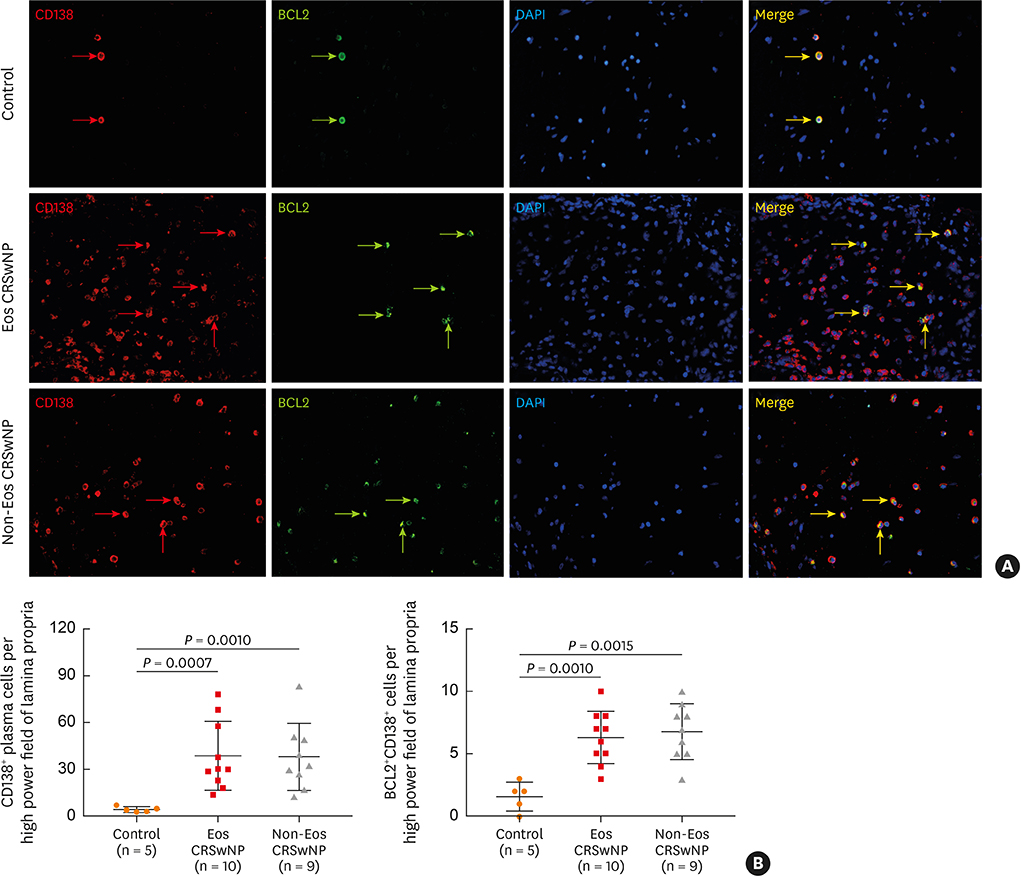
Fig. 1 Increased accumulation of BCL2+CD138+ plasma cells in nasal polyps. (A) Representative photomicrographs showing BCL2+CD138+ plasma cells in control tissues, and in eosinophilic and non-eosinophilic nasal polyp tissues. Red arrows indicate representative CD138+ plasma cells; green arrows indicate representative BCL2+ cells; yellow arrows indicate representative BCL2+CD138+ plasma cells (original magnification ×400). (B) Quantification of CD138+ total plasma cells and BCL2+CD138+ plasma cells in lamina propria. BCL2, B cell lymphoma 2; Eos CRSwNP, eosinophilic chronic rhinosinusitis with nasal polyps; Non-Eos CRSwNP, non-eosinophilic chronic rhinosinusitis with nasal polyps.
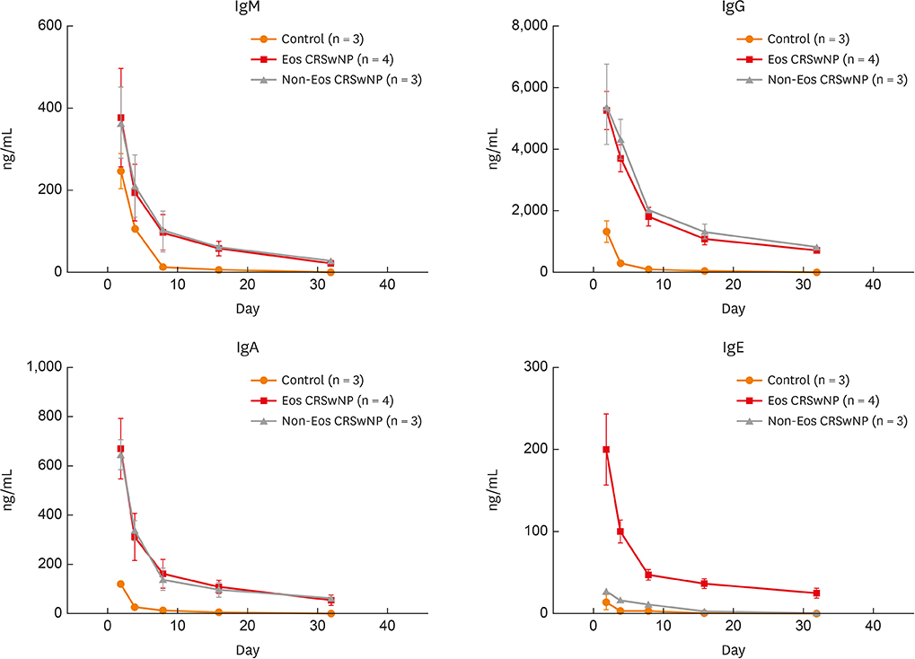
Fig. 2 Ig production from ex vivo cultured sinonasal mucosal samples. Eosinophilic and non-eosinophilic nasal polyps and inferior turbinate mucosal samples were cultured ex vivo for 32 days. Culture supernatants were collected at days 2, 4, 8, 16, and 32, and Ig levels in the culture supernatants were measured by enzyme-linked immunosorbent assay. Ig, immunoglobulin; Eos CRSwNP, eosinophilic chronic rhinosinusitis with nasal polyps; Non-Eos CRSwNP, non-eosinophilic chronic rhinosinusitis with nasal polyps.
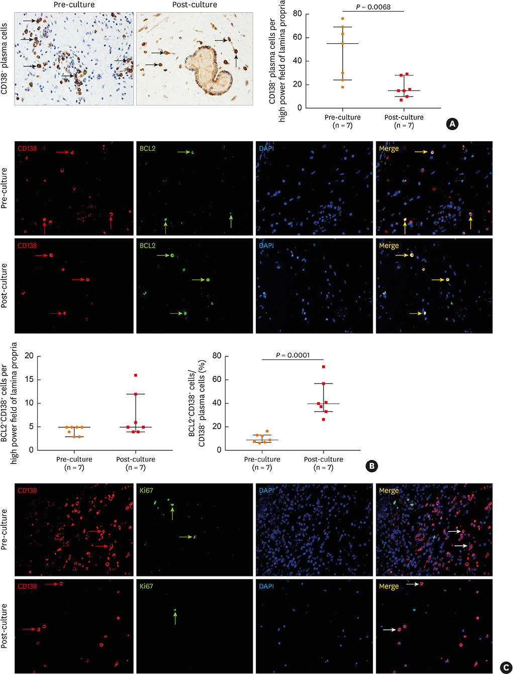
Fig. 3 BCL2+CD138+ plasma cells are retained in nasal polyps after a 32-day ex vivo culture. (A) Representative photomicrographs showing CD138+ plasma cells in nasal polyp tissues before and after culture and the quantification of CD138+ plasma cells. Arrows indicate representative CD138+ plasma cells. (B) Representative photomicrographs and quantitative analysis of BCL2+CD138+ plasma cells in nasal polyp tissues before and after culture. Red arrows indicate representative CD138+ plasma cells; green arrows indicate representative BCL2+ cells; yellow arrows indicate representative BCL2+CD138+ plasma cells. (C) Representative photomicrographs show that CD138+ plasma cells in nasal polyp tissue do not have Ki-67 expression before and after culture. Red arrows indicate representative CD138+ plasma cells; green arrows indicate representative Ki67+ cells; white arrows indicate representative Ki67−CD138+ plasma cells (original magnification ×400). BCL2, B cell lymphoma 2.

Fig. 4 The mRNA expression of NTs (A) as well as their receptors (B), and correlations between BCL2+CD138+ plasma cell count and the expression level of NTs and their receptors (C) in sinonasal mucosa samples as detected by quantitative real-time polymerase chain reaction. NT, neurotrophin; Eos CRSwNP, eosinophilic chronic rhinosinusitis with nasal polyps; Non-Eos CRSwNP, non-eosinophilic chronic rhinosinusitis with nasal polyps; NGF, nerve growth factor; BDNF, brain derived neurotrophic factor; Trk, tropomyosin receptor kinase; BCL2, B cell lymphoma 2; P75, p75 neurotrophin receptor.

Fig. 5 Increased protein expression of NGF in epithelial cells in eosinophilic and non-eosinophilic nasal polyps. Representative photomicrographs showing NGF expression in nasal epithelial cells and infiltrating cells in control tissues and eosinophilic and non-eosinophilic nasal polyp tissues as detected by immunohistochemistry. Isotype control staining is also shown. Arrows indicate representative NGF+ cells. The expression intensity of NGF in epithelial cells was quantified (original magnification ×400). NGF, nerve growth factor; Eos CRSwNP, eosinophilic chronic rhinosinusitis with nasal polyps; Non-Eos CRSwNP, non-eosinophilic chronic rhinosinusitis with nasal polyps; IOD, integrated optical density.
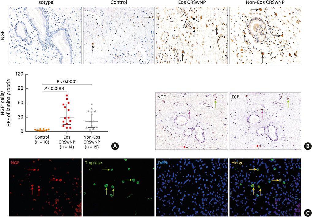
Fig. 6 Increased protein expression of NGF in lamina propria in eosinophilic and non-eosinophilic nasal polyps. (A) Representative photomicrographs showing NGF+ cells in the lamina propria of control tissues, and eosinophilic and non-eosinophilic nasal polyp tissues as detected by immunohistochemistry. Arrows indicate representative NGF+ cells. The numbers of NGF+ cells in the lamina propria were quantified. (B) Representative immunostaining of consecutive tissue sections from a patient with eosinophilic chronic rhinosinusitis with nasal polyps (Eos CRSwNP) showing the expression of NGF by ECP+ eosinophils. Arrows with the same direction indicate the same cells in consecutive serial sections. (C) Representative double immunofluorescence staining of a tissue section from a patient with Eos CRSwNP showing the expression of NGF by tryptase-positive mast cells. Red arrows indicate representative NGF+ cells; green arrows indicate representative tryptase+ cells; yellow arrows indicate representative NGF+tryptase+ cells (original magnification ×400). NGF, nerve growth factor; Eos CRSwNP, eosinophilic chronic rhinosinusitis with nasal polyps; ECP, eosinophilic cationic protein; Non-Eos CRSwNP, non-eosinophilic chronic rhinosinusitis with nasal polyps. HPF, high power field.
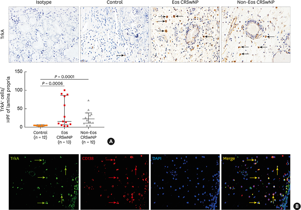
Fig. 7 Increased protein expression of TrkA in eosinophilic and non-eosinophilic nasal polyps. (A) Representative photomicrographs showing TrkA expression in infiltrating cells in the lamina propria of control tissues, and eosinophilic and non-eosinophilic nasal polyp tissues as detected by immunohistochemistry. Arrows indicate representative TrkA+ cells. The numbers of TrkA+ cells in the lamina propria were quantified. (B) Representative double immunofluorescence staining of a tissue section from a patient with eosinophilic chronic rhinosinusitis with nasal polyps (Eos CRSwNP) showing the expression of TrkA by CD138+ plasma cells. Green arrows indicate representative TrkA+ cells; red arrows indicate representative CD138+ cells; yellow arrows indicate representative TrkA+CD138+ plasma cells (original magnification ×400). Trk, tropomyosin receptor kinase; Non-Eos CRSwNP, non-eosinophilic chronic rhinosinusitis with nasal polyps. HPF, high power field.
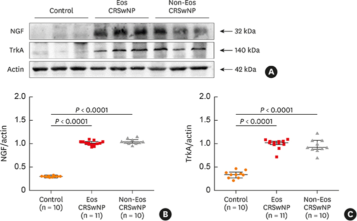
Fig. 8 Increased protein expression of NGF and TrkA in eosinophilic and non-eosinophilic nasal polyps detected by western blot analysis. (A) Representative blots of NGF and TrkA in whole—cell lysates of nasal tissues are shown. (B) NGF relative protein expression was quantified and normalized to β-actin expression. (C) TrkA relative protein expression was quantified and normalized to β-actin. NGF, nerve growth factor; Trk, tropomyosin receptor kinase; Eos CRSwNP, eosinophilic chronic rhinosinusitis with nasal polyps; Non-Eos CRSwNP, non-eosinophilic chronic rhinosinusitis with nasal polyps.
Cited by 1 articles
-
Impact of the Long-Lived Plasma Cells in Patients With Chronic Rhinosinusitis With Nasal Polyps
Roza Khalmuratova, Hyun-Woo Shin
Allergy Asthma Immunol Res. 2020;12(2):173-175. doi: 10.4168/aair.2020.12.2.173.
Reference
-
1. Luger EO, Fokuhl V, Wegmann M, Abram M, Tillack K, Achatz G, et al. Induction of long-lived allergen-specific plasma cells by mucosal allergen challenge. J Allergy Clin Immunol. 2009; 124:819–826.e4.
Article2. van Laar JM, Melchers M, Teng YK, van der Zouwen B, Mohammadi R, Fischer R, et al. Sustained secretion of immunoglobulin by long-lived human tonsil plasma cells. Am J Pathol. 2007; 171:917–927.
Article3. Mesin L, Di Niro R, Thompson KM, Lundin KE, Sollid LM. Long-lived plasma cells from human small intestine biopsies secrete immunoglobulins for many weeks in vitro. J Immunol. 2011; 187:2867–2874.4. Halliley JL, Tipton CM, Liesveld J, Rosenberg AF, Darce J, Gregoretti IV, et al. Long-lived plasma cells are contained within the CD19−CD38hiCD138+ subset in human bone marrow. Immunity. 2015; 43:132–145.5. Cocco M, Stephenson S, Care MA, Newton D, Barnes NA, Davison A, et al. In vitro generation of long-lived human plasma cells. J Immunol. 2012; 189:5773–5785.6. Nguyen DC, Garimalla S, Xiao H, Kyu S, Albizua I, Galipeau J, et al. Factors of the bone marrow microniche that support human plasma cell survival and immunoglobulin secretion. Nat Commun. 2018; 9:3698.
Article7. Tellier J, Kallies A. Finding a home for plasma cells--a niche to survive. Eur J Immunol. 2014; 44:2243–2246.8. Minges Wols HA, Ippolito JA, Yu Z, Palmer JL, White FA, Le PT, et al. The effects of microenvironment and internal programming on plasma cell survival. Int Immunol. 2007; 19:837–846.
Article9. Slifka MK, Antia R, Whitmire JK, Ahmed R. Humoral immunity due to long-lived plasma cells. Immunity. 1998; 8:363–372.
Article10. Corcoran LM, Nutt SL. Long-lived plasma cells have a sweet tooth. Immunity. 2016; 45:3–5.
Article11. Peperzak V, Vikström I, Walker J, Glaser SP, LePage M, Coquery CM, et al. Mcl-1 is essential for the survival of plasma cells. Nat Immunol. 2013; 14:290–297.
Article12. Radbruch A, Muehlinghaus G, Luger EO, Inamine A, Smith KG, Dörner T, et al. Competence and competition: the challenge of becoming a long-lived plasma cell. Nat Rev Immunol. 2006; 6:741–750.
Article13. Abram M, Wegmann M, Fokuhl V, Sonar S, Luger EO, Kerzel S, et al. Nerve growth factor and neurotrophin-3 mediate survival of pulmonary plasma cells during the allergic airway inflammation. J Immunol. 2009; 182:4705–4712.
Article14. Taddeo A, Khodadadi L, Voigt C, Mumtaz IM, Cheng Q, Moser K, et al. Long-lived plasma cells are early and constantly generated in New Zealand Black/New Zealand White F1 mice and their therapeutic depletion requires a combined targeting of autoreactive plasma cells and their precursors. Arthritis Res Ther. 2015; 17:39.
Article15. Schuhmann B, Dietrich A, Sel S, Hahn C, Klingenspor M, Lommatzsch M, et al. A role for brain-derived neurotrophic factor in B cell development. J Neuroimmunol. 2005; 163:15–23.
Article16. Kimata H, Yoshida A, Ishioka C, Kusunoki T, Hosoi S, Mikawa H. Nerve growth factor specifically induces human IgG4 production. Eur J Immunol. 1991; 21:137–141.
Article17. Bayas A, Kruse N, Moriabadi NF, Weber F, Hummel V, Wohleben G, et al. Modulation of cytokine mRNA expression by brain-derived neurotrophic factor and nerve growth factor in human immune cells. Neurosci Lett. 2003; 335:155–158.
Article18. Nassenstein C, Braun A, Erpenbeck VJ, Lommatzsch M, Schmidt S, Krug N, et al. The neurotrophins nerve growth factor, brain-derived neurotrophic factor, neurotrophin-3, and neurotrophin-4 are survival and activation factors for eosinophils in patients with allergic bronchial asthma. J Exp Med. 2003; 198:455–467.
Article19. Hiepe F, Dörner T, Hauser AE, Hoyer BF, Mei H, Radbruch A. Long-lived autoreactive plasma cells drive persistent autoimmune inflammation. Nat Rev Rheumatol. 2011; 7:170–178.
Article20. Fokkens WJ, Lund VJ, Mullol J, Bachert C, Alobid I, Baroody F, et al. European position paper on rhinosinusitis and nasal polyps 2012. Rhinol Suppl. 2012; 23:3 p preceding table of contents1–298.21. Cao PP, Li HB, Wang BF, Wang SB, You XJ, Cui YH, et al. Distinct immunopathologic characteristics of various types of chronic rhinosinusitis in adult Chinese. J Allergy Clin Immunol. 2009; 124:478–484. 484.e1–472.
Article22. Zhang YN, Song J, Wang H, Wang H, Zeng M, Zhai GT, et al. Nasal IL-4+CXCR5+CD4+ T follicular helper cell counts correlate with local IgE production in eosinophilic nasal polyps. J Allergy Clin Immunol. 2016; 137:462–473.23. Tan BK, Li QZ, Suh L, Kato A, Conley DB, Chandra RK, et al. Evidence for intranasal antinuclear autoantibodies in patients with chronic rhinosinusitis with nasal polyps. J Allergy Clin Immunol. 2011; 128:1198–1206.e1.
Article24. Song J, Wang H, Zhang YN, Cao PP, Liao B, Wang ZZ, et al. Ectopic lymphoid tissues support local immunoglobulin production in patients with chronic rhinosinusitis with nasal polyps. J Allergy Clin Immunol. 2018; 141:927–937.
Article25. Zhai GT, Wang H, Li JX, Cao PP, Jiang WX, Song J, et al. IgD-activated mast cells induce IgE synthesis in B cells in nasal polyps. J Allergy Clin Immunol. 2018; 142:1489–1499.e23.
Article26. Shamji MH, Thomsen I, Layhadi JA, Kappen J, Holtappels G, Sahiner U, et al. Broad IgG repertoire in patients with chronic rhinosinusitis with nasal polyps regulates proinflammatory IgE responses. J Allergy Clin Immunol. 2019; 143:2086–2094.e2.
Article27. Hulse KE, Norton JE, Suh L, Zhong Q, Mahdavinia M, Simon P, et al. Chronic rhinosinusitis with nasal polyps is characterized by B-cell inflammation and EBV-induced protein 2 expression. J Allergy Clin Immunol. 2013; 131:1075–1083. 1083.e1–1077.
Article28. Van Zele T, Gevaert P, Holtappels G, van Cauwenberge P, Bachert C. Local immunoglobulin production in nasal polyposis is modulated by superantigens. Clin Exp Allergy. 2007; 37:1840–1847.
Article29. Bateman ED, Hurd SS, Barnes PJ, Bousquet J, Drazen JM, FitzGerald JM, et al. Global strategy for asthma management and prevention: GINA executive summary. Eur Respir J. 2008; 31:143–178.
Article30. Bousquet J, Khaltaev N, Cruz AA, Denburg J, Fokkens WJ, Togias A, AllerGen, et al. Allergic Rhinitis and its Impact on Asthma (ARIA) 2008 update (in collaboration with the World Health Organization, GA(2)LEN and AllerGen). Allergy. 2008; 63:Suppl 86. 8–160.31. Zhang XH, Lu X, Long XB, You XJ, Gao QX, Cui YH, et al. Chronic rhinosinusitis with and without nasal polyps is associated with decreased expression of glucocorticoid-induced leucine zipper. Clin Exp Allergy. 2009; 39:647–654.
Article32. Jia XL, Li SY, Dang SS, Cheng YA, Zhang X, Wang WJ, et al. Increased expression of chondroitin sulphate proteoglycans in rat hepatocellular carcinoma tissues. World J Gastroenterol. 2012; 18:3962–3976.
Article33. Pereira T, Naik S, Tamgadge A. Quantitative evaluation of macrophage expression using CD68 in oral submucous fibrosis: an immunohistochemical study. Ann Med Health Sci Res. 2015; 5:435–441.
Article34. Wang BF, Cao PP, Wang ZC, Li ZY, Wang ZZ, Ma J, et al. Interferon-γ-induced insufficient autophagy contributes to p62-dependent apoptosis of epithelial cells in chronic rhinosinusitis with nasal polyps. Allergy. 2017; 72:1384–1397.
Article35. Liu JX, Liao B, Yu QH, Wang H, Liu YB, Guo CL, et al. The IL-37-Mex3B-Toll-like receptor 3 axis in epithelial cells in patients with eosinophilic chronic rhinosinusitis with nasal polyps. J Allergy Clin Immunol. 2019; S0091-6749(19)30945-5.
Article36. Miguel-García A, Orero T, Matutes E, Carbonell F, Miguel-Sosa A, Linares M, et al. Bcl-2 expression in plasma cells from neoplastic gammopathies and reactive plasmacytosis: a comparative study. Haematologica. 1998; 83:298–304.37. Puthier D, Pellat-Deceunynck C, Barillé S, Robillard N, Rapp MJ, Juge-Morineau N, et al. Differential expression of Bcl-2 in human plasma cell disorders according to proliferation status and malignancy. Leukemia. 1999; 13:289–294.
Article38. Hahn C, Islamian AP, Renz H, Nockher WA. Airway epithelial cells produce neurotrophins and promote the survival of eosinophils during allergic airway inflammation. J Allergy Clin Immunol. 2006; 117:787–794.
Article39. Nassenstein C, Braun A, Nockher WA, Renz H. Neurotrophin effects on eosinophils in allergic inflammation. Curr Allergy Asthma Rep. 2005; 5:204–211.
Article40. Wu X, Myers AC, Goldstone AC, Togias A, Sanico AM. Localization of nerve growth factor and its receptors in the human nasal mucosa. J Allergy Clin Immunol. 2006; 118:428–433.
Article
