Interleukin-17 and Interleukin-22 Induced Proinflammatory Cytokine Production in Keratinocytes via Inhibitor of Nuclear Factor kappaB Kinase-alpha Expression
- Affiliations
-
- 1Department of Microbiology, School of Medicine, Ewha Womans University, Seoul, Korea.
- 2Department of Dermatology, College of Medicine, The Catholic University of Korea, Seoul, Korea. beauty4u@catholic.ac.kr
- 3Department of Psychiatry, College of Medicine, The Catholic University of Korea, Seoul, Korea.
- KMID: 2266033
- DOI: http://doi.org/10.5021/ad.2012.24.4.398
Abstract
- BACKGROUND
The pathogenesis of psoriasis may involve the interleukin (IL)-23 and Th17-mediated immune responses. Th17 cells secret IL-17 and IL-22, which mediates dermal inflammation and acanthosis.
OBJECTIVE
As inhibitor of nuclear factor kappaB kinase-alpha (IKKalpha) has been previously identified as a primary regulator of keratinocyte differentiation and proliferation, we proposed that IL-17 and IL-22 might affect keratinocyte differentiation by changing the expression of IKKalpha.
METHODS
We employed HaCaT cells maintained culture medium at a low calcium concentration (0.06 mM) and induced differentiation by switching to the high concentration (2.8 mM) media with IL-17 or IL-22, then compared the IKKalpha expression and the cell cycle. We employed reconstituted human epidermal skin (Neoderm) and mice ears for the in vivo studies.
RESULTS
Elevated calcium concentration induced IKKalpha expression and terminal differentiation with cell cycle arrest in HaCaT cell cultures. Moreover, IL-17 and IL-22 treatment also induced IKKalpha in HaCaT cells and reconstituted human epidermis. IKKalpha induction was also noted, following the injection of IL-17 and IL-22 into mice ears.
CONCLUSION
Although the induction of IKKalpha was accompanied by keratinocyte differentiation, IL-17 and IL-22 did not affect calcium-mediated differentiation or the cell cycle. Rather, IL-17 and IL-22 appear to contribute to the inflammation occurring via the induction of IKKalpha from keratinocytes or skin layers.
Keyword
MeSH Terms
Figure
-

Fig. 1 IKKα expression is increased under a calcium concentration of 2.8 mM, according to the immunoblot results. HaCaT cells in 0.06 mM of calcium medium were induced to differentiation by replacement with high calcium medium (2.8 mM), and samples were collected every 24 hours for protein extraction. Involucrin, cytokeratin 5, and cytokeratin 10 were detected by the immunoblotting technique described in the Materials and Methods section. Beta-actin blotting was conducted as a loading control.

Fig. 2 Cell cycle analysis demonstrates that HaCaT cells cultured by switching into high calcium concentrations (Ca 2.8 mM) evidenced an elevated proportion of G0/G1 phase, regardless of IL-17 and IL-22 treatment. By way of contrast, HaCaT cells maintained at low calcium concentrations (Ca 0.06 mM) evidenced more cells in S- or G2/M phase compared with those transferred to a medium containing 2.8 mM of calcium. The data are expressed as the means±standard error of the mean and the statistical differences were analyzed between low- and high-calcium treated groups in each of the experiments. PI=(S+G2/M)/(G0/G1+S+G2/M). NS: not significant, PI: proliferation index. **p<0.01; *p<0.05.

Fig. 3 The effects of IL-17 and IL-22 treatment on the expression of IKKα and phosphorylated IκB in HaCaT cells. Cells were treated with IL-17 and IL-22 up to a concentration of 100 ng/ml in a calcium concentration of 2.8 mM and cultured for 24 hours. (A) The immunoblot shown is representative of three independent experiments. (B) The pixel densities are divided by β-actin densities to correct the values, and the data are expressed as the means±standard error of the mean at three independent experiments. *p<0.05.
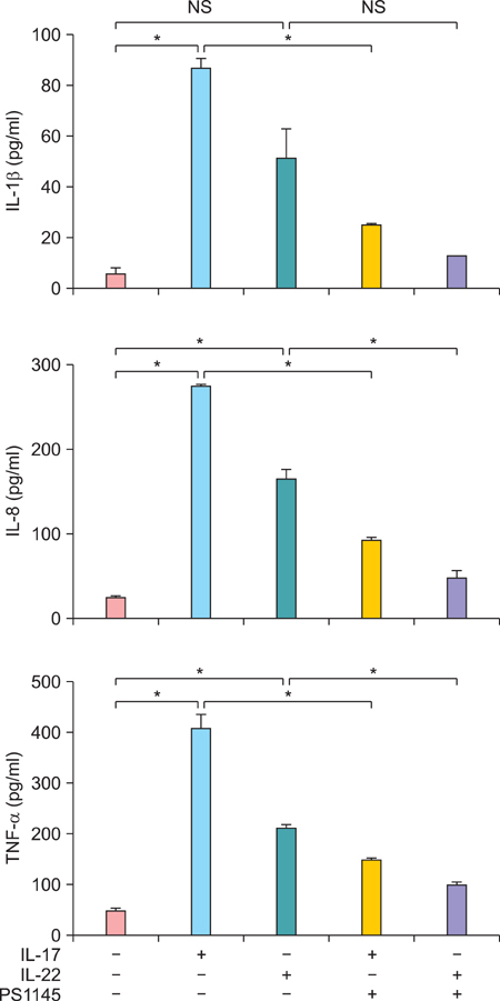
Fig. 4 IL-17 and IL-22 induce pro-inflammatory cytokine production in HaCaT cells. HaCaT cells were cultured under 100 ng/ml of IL-17 or IL-22 for 48 hours, with or without preincubation of PS1145, at a concentration of 10µM for an hour and collected culture supernatant for ELISA. The data are expressed as the means±standard error of the mean at three independent experiments. NS: not significant. *p<0.05.
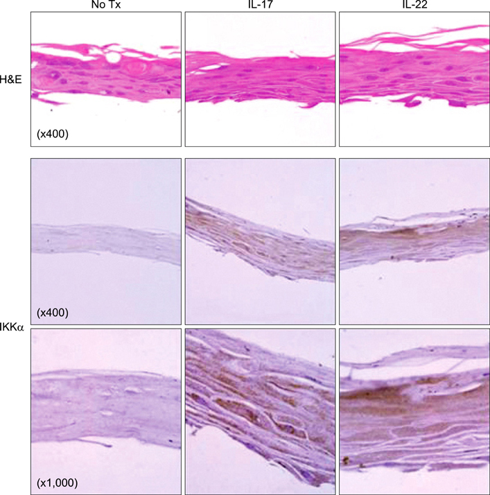
Fig. 5 Both IL-17 and IL-22 induce an increase in IKKα expression in Neoderm. Neoderm tissues were cultured under 100 ng/ml of IL-17 or IL-22 for 48 hours and fixed for H&E and immunohistochemical staining assays. The results shown are representative of the three independent batches of Neoderm experiments.
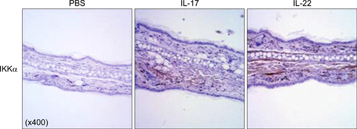
Fig. 6 Elevated IKKα levels in inflammatory mice ears, evoked by IL-17 and IL-22 injection. Skin inflammation, edema, and acanthosis, which were involved in the infiltration of neutrophils and dendritic cells into the ear skin of C57BL/6 WT mice, but less profound inflammatory changes, were noted in the sham controls. Each ear of C57BL/6 WT mice were injected intradermally with 3µg of IL-17 and IL-22 for two consecutive days in a total volume of 30µl. The control group of mice was injected with the same volumes of phosphate-buffered saline (PBS) via an identical route.
Reference
-
1. Tonel G, Conrad C. Interplay between keratinocytes and immune cells--recent insights into psoriasis pathogenesis. Int J Biochem Cell Biol. 2009. 41:963–968.
Article2. Ma HL, Liang S, Li J, Napierata L, Brown T, Benoit S, et al. IL-22 is required for Th17 cell-mediated pathology in a mouse model of psoriasis-like skin inflammation. J Clin Invest. 2008. 118:597–607.
Article3. Nograles KE, Zaba LC, Guttman-Yassky E, Fuentes-Duculan J, Suárez-Fariñas M, Cardinale I, et al. Th17 cytokines interleukin (IL)-17 and IL-22 modulate distinct inflammatory and keratinocyte-response pathways. Br J Dermatol. 2008. 159:1092–1102.
Article4. Zheng Y, Danilenko DM, Valdez P, Kasman I, Eastham-Anderson J, Wu J, et al. Interleukin-22, a T(H)17 cytokine, mediates IL-23-induced dermal inflammation and acanthosis. Nature. 2007. 445:648–651.
Article5. Boniface K, Bernard FX, Garcia M, Gurney AL, Lecron JC, Morel F. IL-22 inhibits epidermal differentiation and induces proinflammatory gene expression and migration of human keratinocytes. J Immunol. 2005. 174:3695–3702.
Article6. Mauro T, Bench G, Sidderas-Haddad E, Feingold K, Elias P, Cullander C. Acute barrier perturbation abolishes the Ca2+ and K+ gradients in murine epidermis: quantitative measurement using PIXE. J Invest Dermatol. 1998. 111:1198–1201.
Article7. Elias PM, Ahn SK, Denda M, Brown BE, Crumrine D, Kimutai LK, et al. Modulations in epidermal calcium regulate the expression of differentiation-specific markers. J Invest Dermatol. 2002. 119:1128–1136.
Article8. Hu Y, Baud V, Oga T, Kim KI, Yoshida K, Karin M. IKKalpha controls formation of the epidermis independently of NF-kappaB. Nature. 2001. 410:710–714.
Article9. Sil AK, Maeda S, Sano Y, Roop DR, Karin M. IkappaB kinase-alpha acts in the epidermis to control skeletal and craniofacial morphogenesis. Nature. 2004. 428:660–664.
Article10. Deyrieux AF, Wilson VG. In vitro culture conditions to study keratinocyte differentiation using the HaCaT cell line. Cytotechnology. 2007. 54:77–83.
Article11. Fuchs E, Byrne C. The epidermis: rising to the surface. Curr Opin Genet Dev. 1994. 4:725–736.
Article12. Hideshima T, Chauhan D, Richardson P, Mitsiades C, Mitsiades N, Hayashi T, et al. NF-kappa B as a therapeutic target in multiple myeloma. J Biol Chem. 2002. 277:16639–16647.13. Liu B, Zhu F, Xia X, Park E, Hu Y. A tale of terminal differentiation: IKKalpha, the master keratinocyte regulator. Cell Cycle. 2009. 8:527–531.
Article14. Swaidani S, Bulek K, Kang Z, Liu C, Lu Y, Yin W, et al. The critical role of epithelial-derived Act1 in IL-17- and IL-25-mediated pulmonary inflammation. J Immunol. 2009. 182:1631–1640.
Article15. Leis H, Sanchis A, Pérez P. Deletion of the N-terminus of IKKgamma induces apoptosis in keratinocytes and impairs the AKT/PTEN signaling pathway. Exp Cell Res. 2007. 313:742–752.
Article16. Wolk K, Kunz S, Witte E, Friedrich M, Asadullah K, Sabat R. IL-22 increases the innate immunity of tissues. Immunity. 2004. 21:241–254.
Article
- Full Text Links
-
- Actions
-
Cited
- CITED
-
- Close
- Share
-
- Similar articles
-
- Regulation of Interleukin-17 Production in Patients with Rheumatoid Arthritis by Phosphoinositide 3-kinase (PI3K)/ Akt and Nuclear Factor KappaB (NF-kappaB) Dependent Signal Transduction Pathway
- Blockade of p38 Mitogen-activated Protein Kinase Pathway Inhibits Interleukin-6 Release and Expression in Primary Neonatal Cardiomyocytes
- Induction of Interleukin-22 (IL-22) production in CD4+ T Cells by IL-17A Secreted from CpG-Stimulated Keratinocytes
- Jak1/Stat3 Is an Upstream Signaling of NF-kappaB Activation in Helicobacter pylori-Induced IL-8 Production in Gastric Epithelial AGS Cells
- Pyrrolidine Dithiocarbamate Inhibits Nuclear Factor kappaB and Toll-Like Receptor 4 Expression in Rats with Acute Necrotizing Pancreatitis
