Lab Anim Res.
2011 Mar;27(1):59-62. 10.5625/lar.2011.27.1.62.
Cronobacter sakazakii Infection Induced Fatal Clinical Sequels Including Meningitis in Neonatal ICR Mice
- Affiliations
-
- 1Center for Animal Resources Development, Wonkwang University, Iksan, Republic of Korea.
- 2Division of Human Environmental Sciences, College of Life Science, Wonkwang University, Iksan, Republic of Korea. hoikyung@wku.ac.kr
- 3Institute of Biotechnology, Wonkwang University, Iksan, Republic of Korea.
- KMID: 2391860
- DOI: http://doi.org/10.5625/lar.2011.27.1.62
Abstract
- Cronobacter sakazakii (C. sakazakii), formerly Enterobacter sakazakii, is an emerging pathogen associated with the ingestion of contaminated reconstituted formula that causes serious illnesses such as bacteremia, septicemia, necrotizing enterocolitis, meningitis and death in low-birth-weight preterm neonatal infants. The objective of this study was to develop an animal model for human neonatal C. sakazakii infections. We acquired timed-pregnant ICR mice and allowed them to give birth naturally. On postnatal day 3.5, each pup was administered orally a total dose of approximately 107 CFU C. sakazakii strain 3439. Mice were observed twice daily for morbidity and mortality. At postnatal day 10.5, the remaining pups were euthanized, and brain, liver, and cecum were excised and analyzed for the presence of C. sakazakii. C. sakazakii was isolated from cecum and other tissues in inoculated mice. In the tissues of C. sakazakii infected mice, meningitis and gliosis were detected in brain. In this study, we confirmed the neonatal ICR mice may be used a very effective animal model for human neonatal C. sakazakii infections.
MeSH Terms
Figure
-
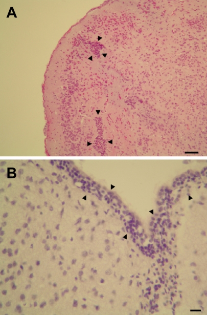
Figure 1 Histopathological findings of the cerebrum of Cronobacter sakazakii-infected mice. A: gliosis (black arrows), bar=100 µm. B: meningitis (black arrows), bar=50 µm. Hematoxylin-eosin stain.
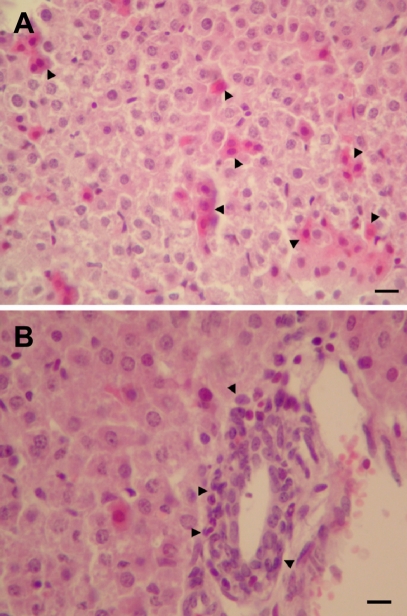
Figure 2 Histopathological findings of the liver of Cronobacter sakazakii-infected mice. A: hepatocytic degeneration (black arrows), bar=50 µm. B: inflammation (black arrows), bar=50 µm. Hematoxylin-eosin stain.
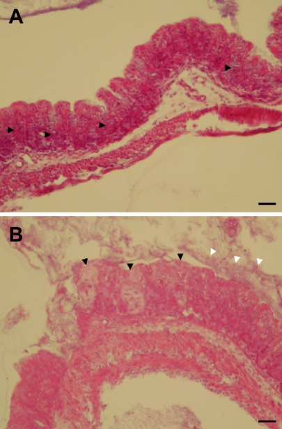
Figure 3 Histopathological findings of the cecum of Cronobacter sakazakii-infected mice. A: inflammation (black arrows), bar=100 µm. B: degeneration (black arrows) and bacterial colonies (white arrows), bar=100 µm. Hematoxylin-eosin stain.
Reference
-
1. Bar-Oz B, Preminger A, Peleg O, Block C, Arad I. Enterobacter sakazakii infection in the newborn. Acta Paediatr. 2001; 90(3):356–358. PMID: 11332182.2. Beuchat LR, Kim H, Gurtler JB, Lin LC, Ryu JH, Richards GM. Cronobacter sakazakii in foods and factors affecting its survival, growth, and inactivation. Int J Food Microbiol. 2009; 136(2):204–213. PMID: 19346021.3. Drudy D, Mullane NR, Quinn T, Wall PG, Fanning S. Enterobacter sakazakii: an emerging pathogen in powdered infant formula. Clin Infect Dis. 2006; 42(7):996–1002. PMID: 16511766.4. Iversen C, Forsythe SJ. Risk profile of Enterobacter sakazakii, an emergent pathogen associated with infant milk formula. Trends Food Sci Technol. 2003; 14(11):443–454.5. Iversen C, Mullane N, McCardel B, Tal BD, Lehner A, Fannin S, Stephan R, Joosten H. Cronobacter gen. nov., a new genus to accommodate the biogroups of Enterobacter sakazakii, and proposal of Cronobacter sakazakii gen. nov., comb. nov., Cronobacter malonaticus sp. nov., Cronobacter turicensis sp. nov., Cronobacter muytjensii sp. nov., Cronobacter dublinensis sp. nov., Cronobacter genomospecies 1, and of three subspecies, Cronobacter dublinensis subsp. dublinensis subsp. nov., Cronobacter dublinensis subsp. lausannensis subsp. nov. and Cronobacter dublinensis subsp. lactaridi subsp. nov. Int J Syst Evol Microbiol. 2008; 58(6):1442–1447. PMID: 18523192.6. Iversen C, Waddington M, On SL, Forsythe S. Identification and phylogeny of Enterobacter sakazakii relative to Enterobacter and Citrobacter species. J Clin Microbiol. 2004; 42(11):5368–5370. PMID: 15528745.7. Lai KK. Enterobacter sakazakii infections among neonates, infants, children, and adults: case reports and a review of the literature. Medicine (Baltimore). 2001; 80(2):113–122. PMID: 11307587.8. Nazarowec-White M, Farber JM. Enterobacter sakazakii: a review. Int J Food Microbiol. 1997; 34(2):103–113. PMID: 9039558.9. Pagotto FJ, Nazarowec-White M, Bidawid S, Farber JM. Enterobacter sakazakii: infectivity and enterotoxin production in vitro and on vivo. J Food Prot. 2003; 66(3):370–377. PMID: 12636287.10. Richardson AN, Lambert S, Smith MA. Neonatal mice as models for Cronobacter sakazakii infection in infants. J Food Prot. 2009; 72(11):2363–2367. PMID: 19903401.11. Urmenyi AMC, Franklin AW. Neonatal death from pigmented coliform infection. Lancet. 1961; 1(7172):313–315. PMID: 13779326.
Article12. Willis-Owen SA, Flint J. The genetic basis of emotional behaviour in mice. Eur J Hum Genet. 2006; 14(6):721–728. PMID: 16721408.
Article13. Zak O. Zak O, editor. Introductory background to animal models of infection. Handbook of Animal Models and Infection: Experimental Models in Antimicrobial Chemotherapy. 1999. 1st ed. San Diego: Academic Press;p. 1–117.
- Full Text Links
-
- Actions
-
Cited
- CITED
-
- Close
- Share
-
- Similar articles
-
- A Case of Enterobacter sakazakii Epidural Abscess in Neonate
- Evaluation of commercial probiotic lactic cultures against biofilm formation by Cronobacter sakazakii
- A Case of Neonatal Sepsis with Meningitis due to Gardnerella vaginalis
- Human Herpesvirus 6 Meningitis in a Neonatal Case
- Group B Streptococcal Meningitis in Neonate: 2001-2011
