Suppressive Effects of Mesenchymal Stem Cells in Adipose Tissue on Allergic Contact Dermatitis
- Affiliations
-
- 1Department of Dermatology, The Jikei University School of Medicine, Tokyo, Japan. kikutyini@jikei.ac.jp
- 2Department of Regenerative Medicine, Research Institute, National Center for Global Health and Medicine, Tokyo, Japan.
- 3Department of Dermatology, Nippon Medical School, Tokyo, Japan.
- 4Department of Dermatology, Teikyo University School of Medicine, Tokyo, Japan.
- KMID: 2388935
- DOI: http://doi.org/10.5021/ad.2017.29.4.391
Abstract
- BACKGROUND
Allergic contact dermatitis (ACD), which is accelerated by interferon (IFN)-γ and suppressed by interleukin (IL)-10 as regulators, is generally self-limited after removal of the contact allergen. Adipose tissue-derived multipotent mesenchymal stem cells (ASCs) potentially exert immunomodulatory effects. Considering that subcutaneous adipose tissue is located close to the site of ACD and includes mesenchymal stem cells (MSCs), the MSCs in adipose tissue could contribute to the self-limiting course of ACD.
OBJECTIVE
The aims of the present study were to elucidate the effects of MSCs in adipose tissue on ACD and to examine any cytokine-mediated mechanisms involved.
METHODS
Ear thickness in a C57BL/6 mouse model of ACD using contact hypersensitivity (CHS) elicited by 2,4,6-trinitro-1-chlorobenzene was evaluated as a marker of inflammation level. Five and nine mice were injected with ASCs and phosphate-buffered saline (PBS), respectively. After ASC or PBS injection, real-time reverse transcription-polymerase chain reaction and enzyme-linked immunosorbent assay were performed.
RESULTS
Histology showed that CHS was self-limited and ear thickness was suppressed by ASCs in a dose-dependent manner. IFN-γ expression in the elicited skin site and regional lymph nodes was significantly lower in ASC-treated mice than in control mice. IL-10 expression did not differ between treated and control mice. The suppressive effects of ASCs on CHS response did not differ between IL-10 knock-out C57BL/6 mice and wild-type mice.
CONCLUSION
The present findings suggest that MSCs in adipose tissue may contribute to the self-limiting course of ACD through decreased expression of IFN-γ, but not through increased expression of IL-10.
Keyword
MeSH Terms
Figure
-
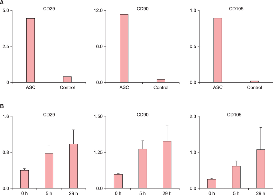
Fig. 1 Increased levels of mesenchymal stem cell (MSC) markers in the adipose tissue-derived multipotent MSCs (ASCs) and contact hypersensitivity. (A) Expressions of CD29, CD90, and CD105 in the ASCs were examined with real-time reverse transcription-polymerase chain reaction (RT-PCR). Vertical axis indicates expression levels standardized with OAZ1 expression. Mouse ear tissues were used as controls. (B) Expression of CD29, CD90, and CD105 in the ASCs were examined with RT-PCR. Values are presented as means±standard error of the mean. Vertical and horizontal axes indicate expression levels standardized with OAZ1 expression and 0, 5, and 29 h after elicitation, respectively.
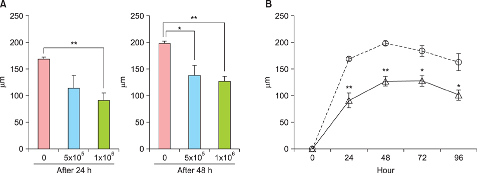
Fig. 2 Suppressive effects of adipose tissue-derived multipotent mesenchymal stem cells (ASCs) on contact hypersensitivity (CHS) response. Values are presented as mean change and standard error of the mean. Each treatment group included ≥5 C57BL/6 mice sensitized with 2,4,6-trinitro-1-chlorobenzene (TNCB). The change in ear thickness was determined as the pre-treatment thickness subtracted from that at the examined time point. Significant differences between samples are indicated: *p<0.05, **p<0.01. (A) Dose-dependent effects of ASCs on CHS response. Mean changes in ear thicknesses of mice injected with 0, 5×10⁵, or 1×10⁶ ASCs at 24 and 48 h after elicitation. Vertical and horizontal axes indicate changes in ear thickness (µm) and ASC dose, respectively. (B) Continuous effects of ASCs on CHS response. Mean changes in ear thickness of mice injected with 1×10⁶ ASCs or phosphate-buffered saline (PBS) at 0, 24, 48, 72, and 96 h after elicitation. Vertical and horizontal axes indicate changes in ear thickness (µm) and examined time points, respectively. Circle symbol and dotted line indicate values for PBS-treated mice. Triangle symbol and solid line indicate values for ASC-treated mice.
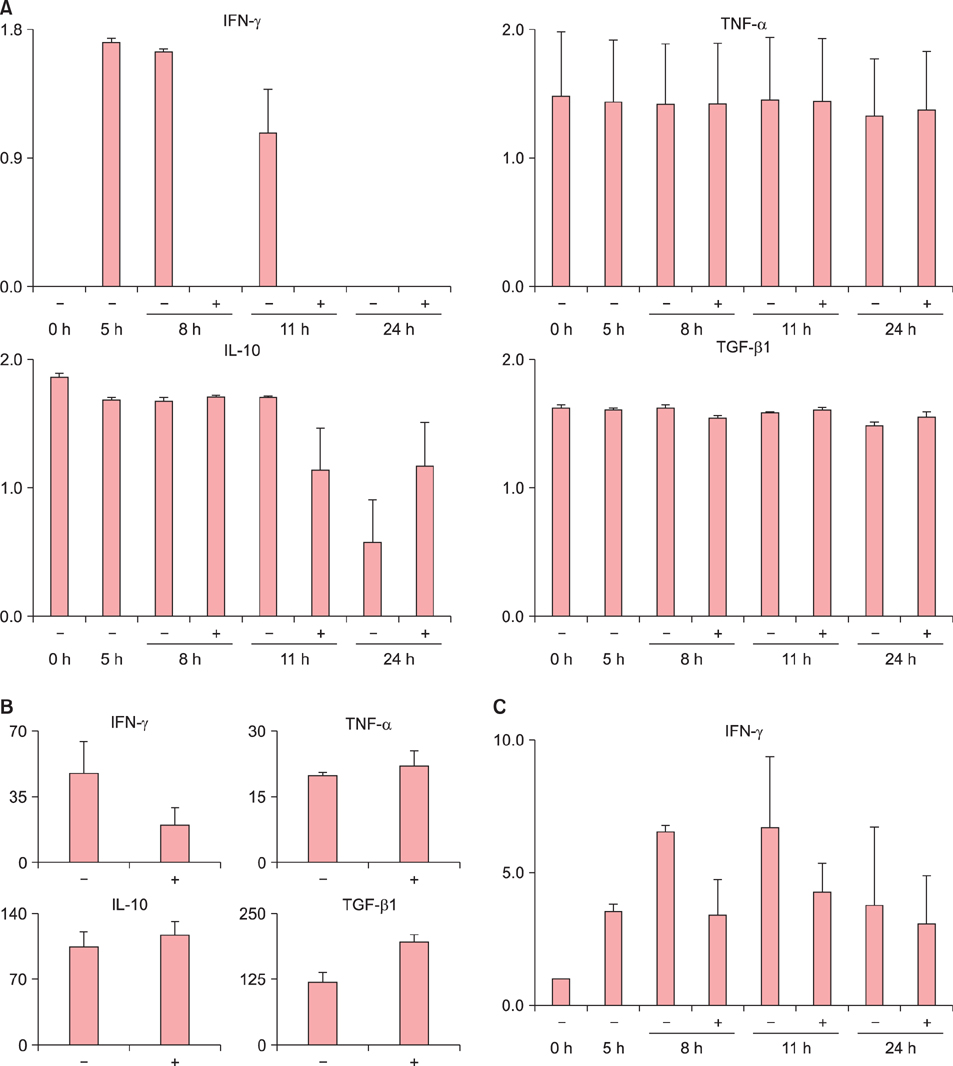
Fig. 3 Change of cytokine expression with adipose tissue-derived multipotent mesenchymal stem cell (ASC) treatment. (A) Expressions of interferon (IFN)-γ, tumor necrosis factor (TNF)-α, interleukin (IL)-10 and transforming growth factor (TGF)-β1 mRNA in the elicited skin sites were examined with real-time reverse transcription-polymerase chain reaction (RT-PCR). Vertical and horizontal axes indicate expression levels standardized with OAZ1 expression and time points (hours) with treatment of ASC(+) or phosphate-buffered saline (PBS)(−), respectively. Values are presented as means±standard error of the mean (SEM). Each treatment group included ≥3 C57BL/6 mice sensitized with 2,4,6-trinitro-1-chlorobenzene (TNCB). (B) Serum levels of IFN-γ, TNF-α, IL-10, and TGF-β1 were examined with enzyme-linked immunosorbent assay. Vertical and horizontal axes indicate concentration level and treatment as ASC(+) or PBS(−), respectively. Each treatment group included ≥3 C57BL/6 mice sensitized with TNCB. (C) mRNA expression of IFN-γ in regional lymph nodes was examined with RT-PCR. Vertical and horizontal axes indicate expression levels standardized with OAZ1 expression and time points with the treatment as ASC(+) or PBS(−), respectively. Values are presented as means±SEM. Each treatment group included ≥3 C57BL/6 mice sensitized with TNCB.

Fig. 4 Comparison between wild-type and interleukin-10−/− mice in adipose tissue-derived multipotent mesenchymal stem cell (ASC) effects on contact hypersensitivity. Change rates in ear thicknesses in wild-type (WT) and interleukin-10-knockout (KO) mice treated by ASCs are indicated. The change rate was defined as the difference between average ear thickness of ASC-treated mice and that of phosphate-buffered saline (PBS)-treated mice divided by average ear thickness of the PBS-treated mice. Equivalence between WT and KO at each examined time point was tested using the chi-square statistics. Each treatment group included ≥10 C57BL/6 mice sensitized with 2,4,6-trinitro-1-chlorobenzene (TNCB). Standard error of the mean was not given, because change rate was calculated using average values in every treatment group.
Reference
-
1. Saint-Mezard P, Rosieres A, Krasteva M, Berard F, Dubois B, Kaiserlian D, et al. Allergic contact dermatitis. Eur J Dermatol. 2004; 14:284–295.
Article2. Gaspari AA, Katz SI. Contact hypersensitivity. Curr Protoc Immunol. 2001; Chapter 4. Unit 4.2.
Article3. Tončić RJ, Lipozenčić J, Martinac I, Gregurić S. Immunology of allergic contact dermatitis. Acta Dermatovenerol Croat. 2011; 19:51–68.4. Sebastiani S, Albanesi C, De PO, Puddu P, Cavani A, Girolomoni G. The role of chemokines in allergic contact dermatitis. Arch Dermatol Res. 2002; 293:552–559.
Article5. Cavani A, Albanesi C, Traidl C, Sebastiani S, Girolomoni G. Effector and regulatory T cells in allergic contact dermatitis. Trends Immunol. 2001; 22:118–120.
Article6. Cavani A, Nasorri F, Prezzi C, Sebastiani S, Albanesi C, Girolomoni G. Human CD4+ T lymphocytes with remarkable regulatory functions on dendritic cells and nickel-specific Th1 immune responses. J Invest Dermatol. 2000; 114:295–302.
Article7. Williams AR, Hare JM. Mesenchymal stem cells: biology, pathophysiology, translational findings, and therapeutic implications for cardiac disease. Circ Res. 2011; 109:923–940.8. Dominici M, Le Blanc K, Mueller I, Slaper-Cortenbach I, Marini F, Krause D, et al. Minimal criteria for defining multipotent mesenchymal stromal cells. The international society for cellular therapy position statement. Cytotherapy. 2006; 8:315–317.
Article9. Konno M, Hamazaki TS, Fukuda S, Tokuhara M, Uchiyama H, Okazawa H, et al. Efficiently differentiating vascular endothelial cells from adipose tissue-derived mesenchymal stem cells in serum-free culture. Biochem Biophys Res Commun. 2010; 400:461–465.
Article10. Zhou Y, Yuan J, Zhou B, Lee AJ, Lee AJ, Ghawji M Jr, et al. The therapeutic efficacy of human adipose tissue-derived mesenchymal stem cells on experimental autoimmune hearing loss in mice. Immunology. 2011; 133:133–140.
Article11. Lin CS, Lin G, Lue TF. Allogeneic and xenogeneic transplantation of adipose-derived stem cells in immunocompetent recipients without immunosuppressants. Stem Cells Dev. 2012; 21:2770–2778.
Article12. Fang B, Song Y, Liao L, Zhang Y, Zhao RC. Favorable response to human adipose tissue-derived mesenchymal stem cells in steroid-refractory acute graft-versus-host disease. Transplant Proc. 2007; 39:3358–3362.
Article13. Berg DJ, Leach MW, Kühn R, Rajewsky K, Müller W, Davidson NJ, et al. Interleukin 10 but not interleukin 4 is a natural suppressant of cutaneous inflammatory responses. J Exp Med. 1995; 182:99–108.
Article14. Bobr A, Igyarto BZ, Haley KM, Li MO, Flavell RA, Kaplan DH. Autocrine/paracrine TGF-β1 inhibits Langerhans cell migration. Proc Natl Acad Sci U S A. 2012; 109:10492–10497.
Article15. Wernersson S, Pejler G. Mast cell secretory granules: armed for battle. Nat Rev Immunol. 2014; 14:478–494.
Article16. Watanabe H, Numata K, Ito T, Takagi K, Matsukawa A. Innate immune response in Th1- and Th2-dominant mouse strains. Shock. 2004; 22:460–466.
Article17. Mikami N, Matsushita H, Kato T, Kawasaki R, Sawazaki T, Kishimoto T, et al. Calcitonin gene-related peptide is an important regulator of cutaneous immunity: effect on dendritic cell and T cell functions. J Immunol. 2011; 186:6886–6893.
Article
