Dexamethasone Inhibits TGF-β1-Induced Cell Migration by Regulating the ERK and AKT Pathways in Human Colon Cancer Cells Via CYR61
- Affiliations
-
- 1Department of Internal Medicine, Jeju National University School of Medicine, Jeju, Korea.
- 2Department of Biochemistry, Jeju National University School of Medicine, Jeju, Korea. moonjcho@jejunu.ac.kr
- 3Department of Internal Medicine, Gachon University Gil Medical Center, Incheon, Korea. dbs@gilhospital.com
- KMID: 2344088
- DOI: http://doi.org/10.4143/crt.2015.209
Abstract
- PURPOSE
One of the features in cancer development is the migration of cancer cells to form metastatic lesions. CYR61 protein promotes migration and the epithelial-mesenchymal transition in several cancer cell types. Evidence suggests that CYR61 and dexamethasone are relevant to colorectal cancer. However, relationships between them and colorectal cancer are still unclear. Understanding the molecular mechanism of colorectal cancer progression related with CYR61 and dexamethasone, which is widely used for combination chemotherapy, is necessary for improved therapy.
MATERIALS AND METHODS
We used colorectal cancer cells, HCT116, co-treated with transforming growth factor β1 (TGF-β1) and dexamethasone to examine the inhibitory migration effect of dexamethasone by migratory assay. Alternatively, both migratory pathways, expression of AKT and ERK, and the target factor CYR61 was also tested by co-treatment with TGF-β1 and dexamethasone.
RESULTS
We report that dexamethasone significantly inhibited TGF-β1-induced cell migration, without affecting cell proliferation. Importantly, we observed that TGF-β1 promoted the epithelial-mesenchymal transition process and that dexamethasone co-treatment abolished this effect. ERK and AKT signaling pathways were found to mediate TGF-β1-induced migration, which was inhibited by dexamethasone. In addition, TGF-β1 treatment induced CYR61 expression whereas dexamethasone reduced it. These observations were compatible with the modulation of migration observed following treatment of HCT116 cells with human recombinant CYR61 and anti-CYR61 antibody. Our results also indicated that TGF-β1 enhanced collagen I and reduced matrix metalloproteinase 1 expression, which was reversed by dexamethasone treatment.
CONCLUSION
These findings suggested that dexamethasone inhibits AKT and ERK phosphorylation, leading to decreased CYR61 expression, which in turn blocks TGF-β1-induced migration.
Keyword
MeSH Terms
-
Cell Movement*
Cell Proliferation
Collagen
Colon*
Colonic Neoplasms*
Colorectal Neoplasms
Cysteine-Rich Protein 61
Dexamethasone*
Drug Therapy, Combination
Epithelial-Mesenchymal Transition
HCT116 Cells
Humans*
Matrix Metalloproteinase 1
Phosphorylation
Transforming Growth Factors
Collagen
Cysteine-Rich Protein 61
Dexamethasone
Matrix Metalloproteinase 1
Transforming Growth Factors
Figure
-
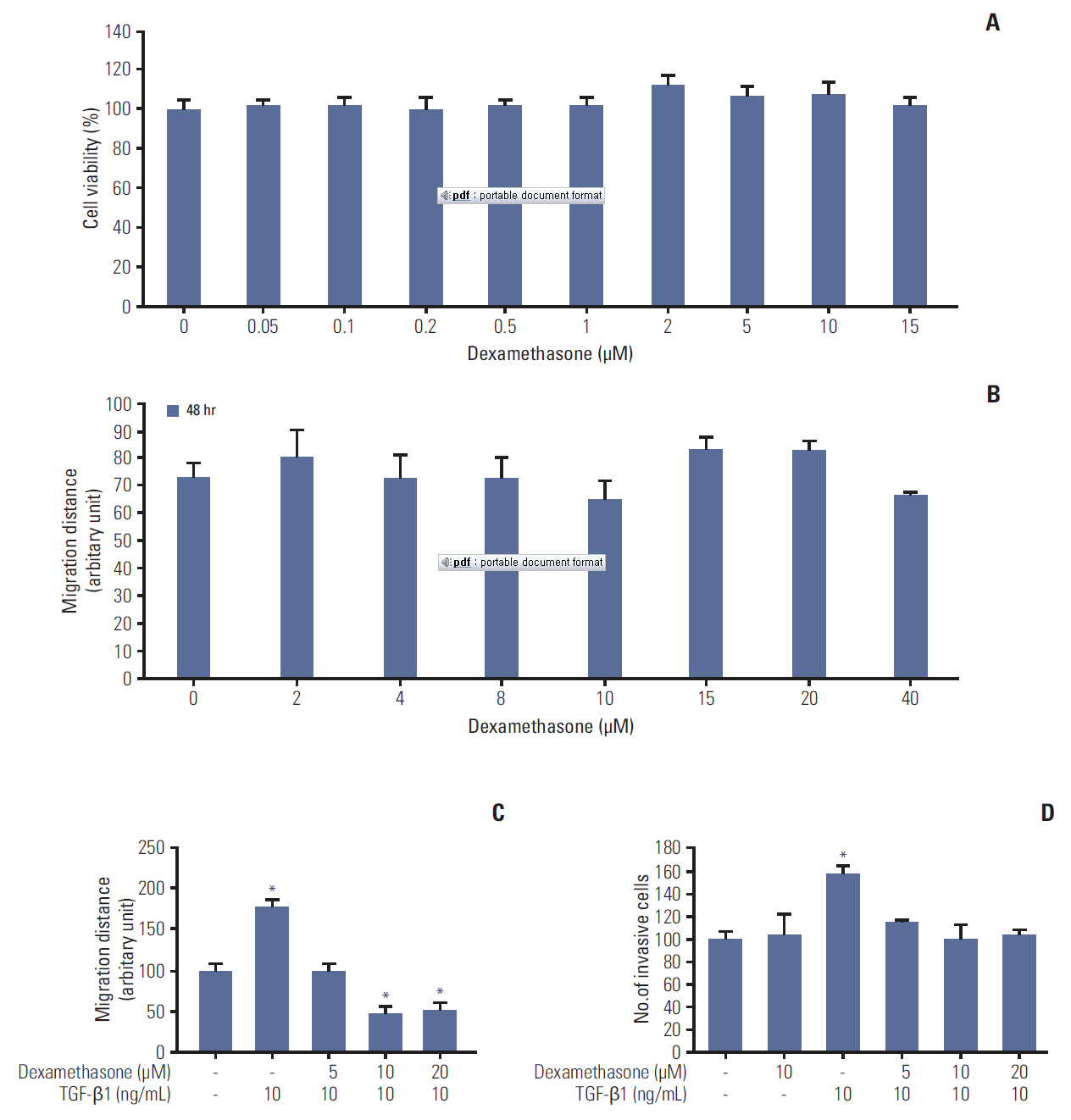
Fig. 1. Effects of dexamethasone on migration and proliferation of HCT116 cells. (A) HCT116 cells were seeded at a density of 3×10⁴ cells/mL and grown for 24 hours, followed by incubation of the cells with dexamethasone for 48 hours at the indicated concentrations. Cells were then incubated with 3-(4,5-dimethylthiazol-2-yl)-2,5-diphenyltetrazolium bromide solution for 4 hours at 37ºC, and the absorbance was measured at 570 mm. (B) HCT116 cells were seeded in 48-well plates at 7×10⁴ cells/mL overnight and then scratched and treated with the indicated concentrations of dexamethasone. The length of the scratches was measured after 24 and 48 hours. (C) The scratch wound healing assay was performed as described previously. HCT116 cells were treated with dexamethasone (1, 2, and 5 μM) alone or co-treated with transforming growth factor β1 (TGF-β1, 10 ng/mL). (D) Transwell invasion assay. HCT116 cells were seeded overnight as described in the "Materials and Methods" section. The cells were then treated with TGF-β1 (10 ng/mL) or co-treated with dexamethasone for 48 hours at a final concentration of 5, 10, or 20 μM. Invasive cells were collected by trypsinization and counted. The data shown are the mean±SD from at least three independent experiments. *p < 0.05, compared to control cells.
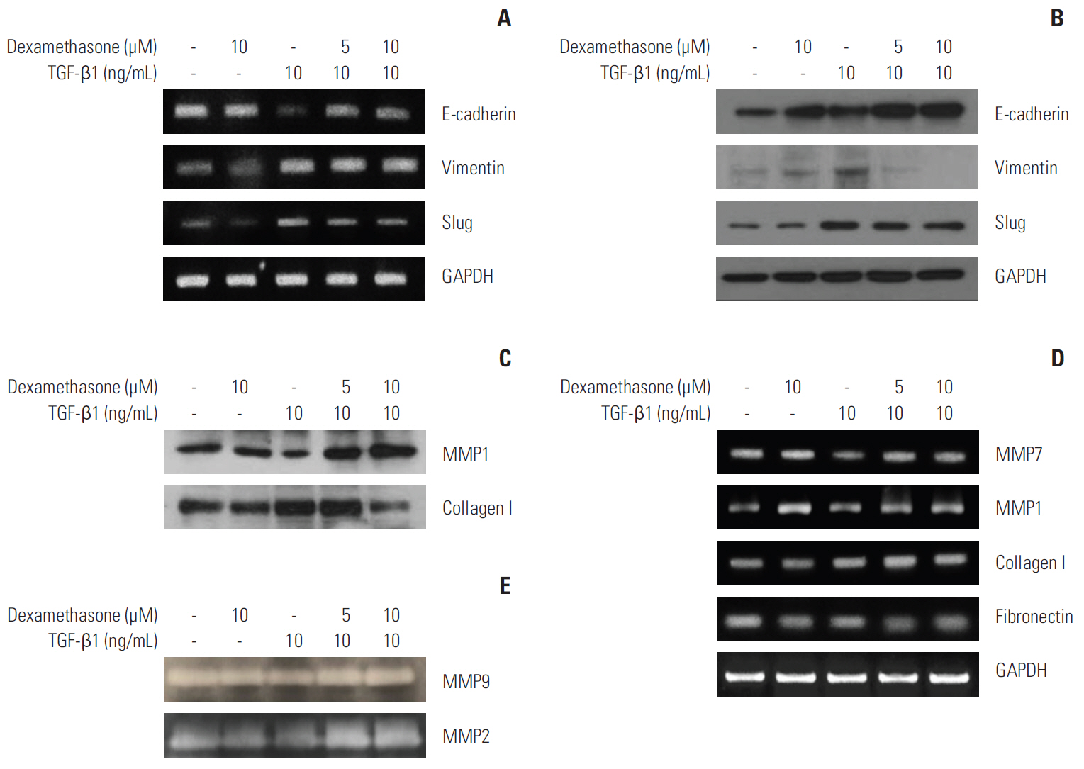
Fig. 2. Downregulation of transforming growth factor β1 (TGF-β1)–induced epithelial-mesenchymal transition markers by dexamethasone treatment. HCT116 cells were seeded overnight and treated for 48 hours with 10 ng/mL TGF-β1, or co-treated with 5 or 10 μM dexamethasone. (A, D) mRNA expression was examined by reverse-transcriptase polymerase chain reaction using primers designed to amplify the indicated targets. (B) Protein expression was examined by western blot analysis with the indicated antibodies. (C) HCT116 cells were treated in serum-free media with the indicated concentrations of TGF-β1 and dexamethasone. Conditioned media were removed and concentrated by Amicon-filter centrifugation. Proteins in the media were analyzed by western blot for the expression of collagen I and matrix metalloproteinase (MMP) 1, using the indicated antibodies. (E) Conditioned media were analyzed by zymography in gelatin gels, as described in the "Materials and Methods" section. GAPDH, glyceraldehyde 3-phosphate dehydrogenase.
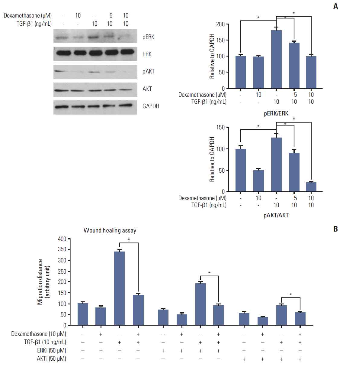
Fig. 3. Transforming growth factor β1 (TGF-β1) induced ERK and AKT phosphorylation and cell migration, which was reversed by dexamethasone treatment. (A) HCT116 cells were treated for 48 hours with 10 ng/mL TGF-β1 or co-treated with 5 or 10 μM dexamethasone. ERK and AKT phosphorylation were detected by western blot analysis with the indicated antibodies. (B) Scratch wound healing assays were performed as described previously. HCT116 cells were treated with dexamethasone alone (1, 2, or 5 μM), or co-treated with TGF-β1 (10 ng/mL) and/or the ERK inhibitor PD98059 (50 μM) or the AKT inhibitor LY49002 (50 μM). Scratch migration distances were measured after 24 and 48 hours. The histogram shows the results obtained after ImageJ data analysis. The data shown represent the mean percent migration distance±standard deviation from at least three replicates, *p < 0.05 in all experiments. The statistical analysis was performed between the TGF-β1 treatment alone with the control (no treat) or TGF-β1 treatment alone with co-treatment with dexamethasone, respectively.
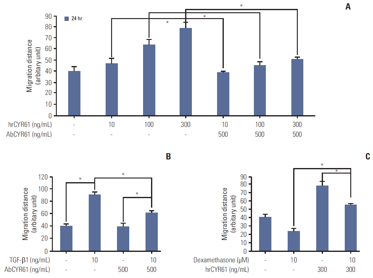
Fig. 4. The mediated role of CYR61 in dexamethasone-inhibited cell migration. (A) HCT116 cells were seeded overnight in 48-well plates at a density of 7×10⁴ cells/mL and then scratched and treated with the indicated concentrations of hrCYR61, with or without AbCYR61 (500 ng/mL). Scratch migration distances were measured after 24 hours and 48 hours. (B, C) Scratch wound healing assays were performed as described previously. HCT116 cells were treated with dexamethasone (10 μM) or transforming growth factor β1 (TGF-β1, 10 ng/mL) alone, or co-treated with hrCYR61 (330 ng/mL) or AbCYR61 (500 ng/mL). The values shown represent the mean±standard deviation of three independent experiments, *p < 0.05, compared with control cells in all experiments. The statistical analysis was performed between the TGF-β1 treatment alone with the control (no treat) or TGF-β1 treatment alone with co-treatment with AbCYR61 or hrCYR61, respectively.
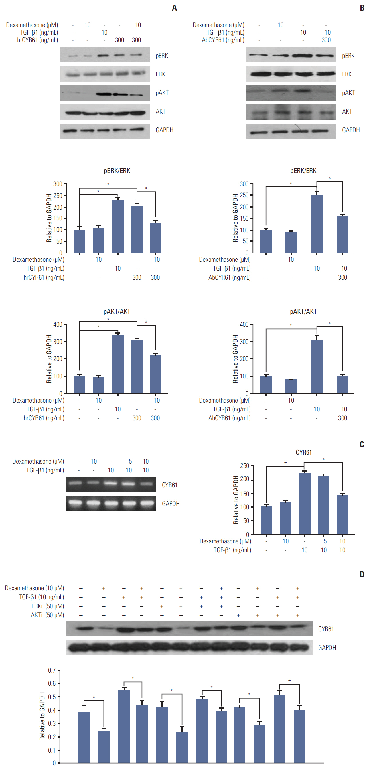
Fig. 5. CYR61 regulation of ERK and AKT phosphorylation and dexamethasone-inhibited cell migration. HCT116 cells were treated for 48 hours with transforming growth factor β1 (TGF-β1, 10 ng/mL), or with dexamethasone (10 μM) combined with hrCYR61 (300 ng/mL) (A) or AbCYR61 (500 ng/mL) (B). ERK and AKT phosphorylation were detected by western blot analysis with the indicated antibodies. (C) CYR61 mRNA expression levels were examined by reverse-transcriptase polymerase chain reaction using primers designed against the indicated targets. (D) HCT116 cells were treated with dexamethasone (1, 2, or 5 μM) alone, or co-treated with TGF-β1 (10 ng/mL) and/or the ERK inhibitor PD98059 (50 μM) or the AKT inhibitor LY49002 (50 μM). CYR61 protein expression was examined by western blot analysis with the indicated antibodies. Data are represented as the mean percent migration distance±standard deviation of at least three replicates. *p < 0.05 in all experiments. GAPDH, glyceraldehyde 3-phosphate dehydrogenase.
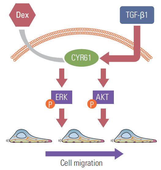
Fig. 6. Proposed model for modulation of CYR61 expression by transforming growth factor β1 (TGF-β1) and dexamethasone to activate or inhibit ERK and AKT phosphorylation, which promote cell migration. Dex, dexamethasone.
Reference
-
References
1. Cunningham D, Atkin W, Lenz HJ, Lynch HT, Minsky B, Nordlinger B, et al. Colorectal cancer. Lancet. 2010; 375:1030–47.
Article2. Kalluri R, Weinberg RA. The basics of epithelial-mesenchymal transition. J Clin Invest. 2009; 119:1420–8.
Article3. Wells A, Yates C, Shepard CR. E-cadherin as an indicator of mesenchymal to epithelial reverting transitions during the metastatic seeding of disseminated carcinomas. Clin Exp Metastasis. 2008; 25:621–8.
Article4. Iwatsuki M, Mimori K, Yokobori T, Ishi H, Beppu T, Nakamori S, et al. Epithelial-mesenchymal transition in cancer development and its clinical significance. Cancer Sci. 2010; 101:293–9.
Article5. Steelman LS, Chappell WH, Abrams SL, Kempf RC, Long J, Laidler P, et al. Roles of the Raf/MEK/ERK and PI3K/PTEN/Akt/mTOR pathways in controlling growth and sensitivity to therapy-implications for cancer and aging. Aging (Albany NY). 2011; 3:192–222.
Article6. Holbourn KP, Acharya KR, Perbal B. The CCN family of proteins: structure-function relationships. Trends Biochem Sci. 2008; 33:461–73.
Article7. Sun ZJ, Wang Y, Cai Z, Chen PP, Tong XJ, Xie D. Involvement of Cyr61 in growth, migration, and metastasis of prostate cancer cells. Br J Cancer. 2008; 99:1656–67.
Article8. Terada N, Kulkarni P, Getzenberg RH. Cyr61 is a potential prognostic marker for prostate cancer. Asian J Androl. 2012; 14:405–8.
Article9. Jeong D, Heo S, Ahn TS, Lee S, Park S, Kim H, et al. Cyr61 expression is associated with prognosis in patients with colorectal cancer. BMC Cancer. 2014; 14:164.
Article10. Auphan N, DiDonato JA, Rosette C, Helmberg A, Karin M. Immunosuppression by glucocorticoids: inhibition of NF-kappa B activity through induction of I kappa B synthesis. Science. 1995; 270:286–90.11. Rinehart J, Keville L, Measel J, Spiekerman AM, Burke K. Corticosteroid alteration of carboplatin-induced hematopoietic toxicity in a murine model. Blood. 1995; 86:4493–9.
Article12. Gundisch S, Boeckeler E, Behrends U, Amtmann E, Ehrhardt H, Jeremias I. Glucocorticoids augment survival and proliferation of tumor cells. Anticancer Res. 2012; 32:4251–61.13. Rinehart J, Arnold S, Kloecker G, Lim A, Zaydan MA, Baeker T, et al. Phase II randomized trial of carboplatin and gemcitabine with or without dexamethasone pre-treatment in patients with Stage IV non-small cell lung cancer. Cancer Chemother Pharmacol. 2013; 71:1375–83.
Article14. Singh PP, Lemanu DP, Taylor MH, Hill AG. Association between preoperative glucocorticoids and long-term survival and cancer recurrence after colectomy: follow-up analysis of a previous randomized controlled trial. Br J Anaesth. 2014; 113 Suppl 1:i68–73.
Article15. Seo GY, Park S, Huh JS, Cho M. The protective effect of glycitin on UV-induced skin photoaging in human primary dermal fibroblast. J Korean Soc Appl Biol Chem. 2014; 57:463–8.
Article16. Nicolas FJ, Lehmann K, Warne PH, Hill CS, Downward J. Epithelial to mesenchymal transition in Madin-Darby canine kidney cells is accompanied by down-regulation of Smad3 expression, leading to resistance to transforming growth factor-beta-induced growth arrest. J Biol Chem. 2003; 278:3251–6.17. Suman S, Kurisetty V, Das TP, Vadodkar A, Ramos G, Lakshmanaswamy R, et al. Activation of AKT signaling promotes epithelial-mesenchymal transition and tumor growth in colorectal cancer cells. Mol Carcinog. 2014; 53 Suppl 1:E151–60.
Article18. Lau LF. CCN1/CYR61: the very model of a modern matricellular protein. Cell Mol Life Sci. 2011; 68:3149–63.
Article19. Haque I, Mehta S, Majumder M, Dhar K, De A, McGregor D, et al. Cyr61/CCN1 signaling is critical for epithelial-mesenchymal transition and stemness and promotes pancreatic carcinogenesis. Mol Cancer. 2011; 10:8.
Article20. Chen J, Song Y, Yang J, Gong L, Zhao P, Zhang Y, et al. The up-regulation of cysteine-rich protein 61 induced by transforming growth factor beta enhances osteosarcoma cell migration. Mol Cell Biochem. 2013; 384:269–77.
Article21. Lee YJ, Lee DM, Lee SH. Production of Cyr61 protein is modulated by extracellular acidification and PI3K/Akt signaling in prostate carcinoma PC-3 cells. Food Chem Toxicol. 2013; 58:169–76.
Article22. Shim SH, Hah JH, Hwang SY, Heo DS, Sung MW. Dexamethasone treatment inhibits VEGF production via suppression of STAT3 in a head and neck cancer cell line. Oncol Rep. 2010; 23:1139–43.
Article23. Bernardi RJ, Trump DL, Yu WD, McGuire TF, Hershberger PA, Johnson CS. Combination of 1alpha,25-dihydroxyvitamin D(3) with dexamethasone enhances cell cycle arrest and apoptosis: role of nuclear receptor cross-talk and Erk/Akt signaling. Clin Cancer Res. 2001; 7:4164–73.24. Jang YH, Shin HS, Choi HS, Ryu ES, Kim MJ, Min SK, et al. Effects of dexamethasone on the TGF-β1-induced epithelialto-mesenchymal transition in human peritoneal mesothelial cells. Lab Invest. 2013; 93:194–206.
Article25. Sabile AA, Arlt MJ, Muff R, Husmann K, Hess D, Bertz J, et al. Caprin-1, a novel Cyr61-interacting protein, promotes osteosarcoma tumor growth and lung metastasis in mice. Biochim Biophys Acta. 2013; 1832:1173–82.
Article
- Full Text Links
-
- Actions
-
Cited
- CITED
-
- Close
- Share
-
- Similar articles
-
- 4-O-Methylhonokiol Protects HaCaT Cells from TGF-β1-Induced Cell Cycle Arrest by Regulating Canonical and Non-Canonical Pathways of TGF-β Signaling
- Parthenolide inhibits transforming growth factor β1-induced epithelial-mesenchymal transition in colorectal cancer cells
- Synergistic effect of ERK inhibition on tetrandrine-induced apoptosis in A549 human lung carcinoma cells
- Effects of Curcumin on Apoptosis in SW480 Human Colon Cancer Cell Line
- Aspirin-Triggered Resolvin D1 Inhibits TGF-β1-Induced EndMT through Increasing the Expression of Smad7 and Is Closely Related to Oxidative Stress
