J Korean Neurosurg Soc.
2014 Sep;56(3):261-264. 10.3340/jkns.2014.56.3.261.
Value of Perfusion Weighted Magnetic Resonance Imaging in the Diagnosis of Supratentorial Anaplastic Astrocytoma
- Affiliations
-
- 1Department of Radiology, Seoul National University Bundang Hospital, Seongnam, Korea.
- 2Department of Radiology, Kyung Hee University Hospital, School of Medicine, Kyung Hee University, Seoul, Korea. euijkim@hanmail.net
- 3Department of Radiology, Kyung Hee University Hospital at Gangdong, School of Medicine, Kyung Hee University, Seoul, Korea.
- 4Department of Neurosurgery, Kyung Hee University Hospital at Gangdong, School of Medicine, Kyung Hee University, Seoul, Korea.
- KMID: 2339968
- DOI: http://doi.org/10.3340/jkns.2014.56.3.261
Abstract
- We report perfusion weighted imaging (PWI) findings of nonenhanced anaplastic astrocytoma in a 30-year-old woman. Brain magnetic resonance imaging showed a nonenhanced brain tumor with mild peritumoral edema on the right medial frontal lobe and right genu of corpus callosum, suggesting a low-grade glioma. However, PWI showed increased relative cerebral blood volume, relative cerebral blood flow, and permeability of nonenhanced brain tumor compared with contralateral normal brain parenchyma, suggesting a high-grade glioma. After surgery, final histopathological analysis revealed World Health Organization grade III anaplastic astrocytoma. This case demonstrates the importance of PWI for preoperative evaluation of nonenhanced brain tumors.
Keyword
MeSH Terms
Figure
-
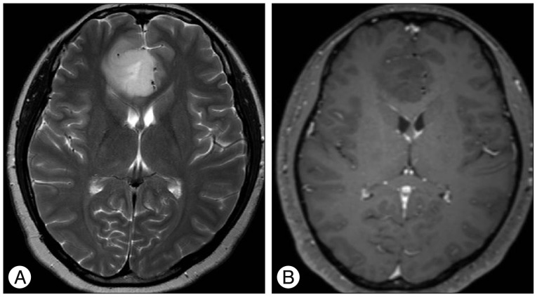
Fig. 1 T2-weighted image (A) shows homogenously hyperintense mass with mild peritumoral edema and contrast-enhanced T1-weighted image (B) shows homogenously hypointense mass without enhancement on the right medial frontal lobe and right genu of corpus callosum.
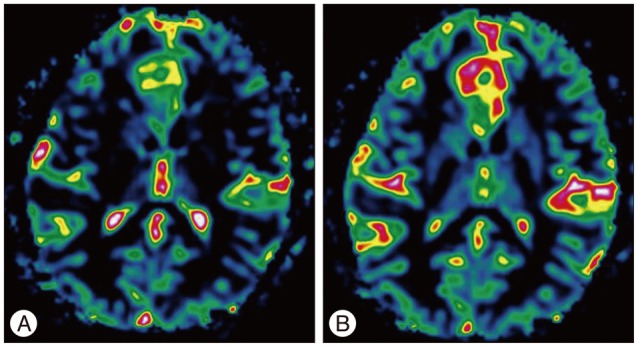
Fig. 2 Perfusion color maps derived from dynamic susceptibility contrast-enhanced perfusion weighted imaging show that the visual increase in the relative cerebral blood volume of 3.6 (A) and relative cerebral blood flow of 2.0 (B).
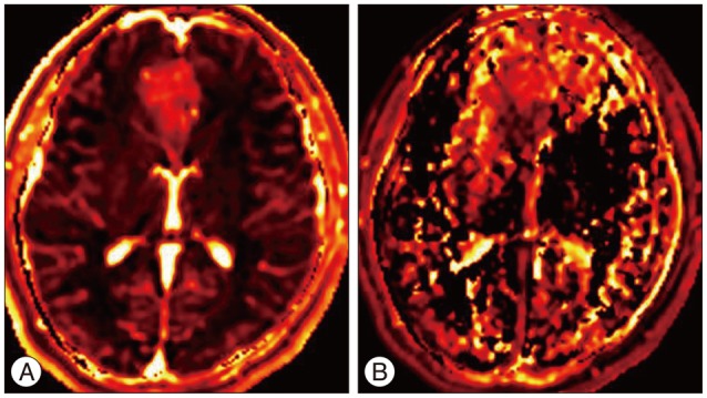
Fig. 3 Perfusion color maps derived from dynamic contrast-enhanced perfusion weighted imaging show that the visual increase in the area under curve (A) and permeability (ktrans) (B).
Reference
-
1. Awasthi R, Rathore RK, Soni P, Sahoo P, Awasthi A, Husain N, et al. Discriminant analysis to classify glioma grading using dynamic contrast-enhanced MRI and immunohistochemical markers. Neuroradiology. 2012; 54:205–213. PMID: 21541688.
Article2. Bruner JM. Neuropathology of malignant gliomas. Semin Oncol. 1994; 21:126–138. PMID: 8153659.3. Fan GG, Deng QL, Wu ZH, Guo QY. Usefulness of diffusion/perfusion-weighted MRI in patients with non-enhancing supratentorial brain gliomas : a valuable tool to predict tumour grading? Br J Radiol. 2006; 79:652–658. PMID: 16641420.
Article4. Kassner A, Thornhill R. Measuring the integrity of the human blood-brain barrier using magnetic resonance imaging. Methods Mol Biol. 2011; 686:229–245. PMID: 21082374.
Article5. Law M, Yang S, Babb JS, Knopp EA, Golfinos JG, Zagzag D, et al. Comparison of cerebral blood volume and vascular permeability from dynamic susceptibility contrast-enhanced perfusion MR imaging with glioma grade. AJNR Am J Neuroradiol. 2004; 25:746–755. PMID: 15140713.6. Lupo JM, Cha S, Chang SM, Nelson SJ. Dynamic susceptibility-weighted perfusion imaging of high-grade gliomas : characterization of spatial heterogeneity. AJNR Am J Neuroradiol. 2005; 26:1446–1454. PMID: 15956514.7. Maia AC Jr, Malheiros SM, da Rocha AJ, da Silva CJ, Gabbai AA, Ferraz FA, et al. MR cerebral blood volume maps correlated with vascular endothelial growth factor expression and tumor grade in nonenhancing gliomas. AJNR Am J Neuroradiol. 2005; 26:777–783. PMID: 15814920.8. Mihara F, Numaguchi Y, Rothman M, Kristt D, Fiandaca M, Swallow L. Non-enhancing supratentorial malignant astrocytomas : MR features and possible mechanisms. Radiat Med. 1995; 13:11–17. PMID: 7597198.9. Petrella JR, Provenzale JM. MR perfusion imaging of the brain : techniques and applications. AJR Am J Roentgenol. 2000; 175:207–219. PMID: 10882275.10. Rees J. Advances in magnetic resonance imaging of brain tumours. Curr Opin Neurol. 2003; 16:643–650. PMID: 14624071.
Article
- Full Text Links
-
- Actions
-
Cited
- CITED
-
- Close
- Share
-
- Similar articles
-
- Supratentorial Extraventricular Anaplastic Ependymoma Presenting with Repeated Intratumoral Hemorrhage
- Perfusion MR Imaging of Cerebral Gliomas: Comparison with Histologic Tumor Grade
- A Case of Anaplastic Astrocytoma in Term Pregnancy
- Arterial Spin Labelling Perfusion, Proton MR Spectroscopy and Susceptibility-Weighted MR Findings of Acute Necrotizing Encephalopathy: a Case Report
- Advanced Magnetic Resonance Imaging for Pediatric Brain Tumors: Current Imaging Techniques and Interpretation Algorithms
