Arterial Spin Labelling Perfusion, Proton MR Spectroscopy and Susceptibility-Weighted MR Findings of Acute Necrotizing Encephalopathy: a Case Report
- Affiliations
-
- 1Department of Radiology, Gyeongsang National University Hospital and Gyeongsang National University School of Medicine, Jinju, Korea. choids@gnu.ac.kr
- KMID: 2452530
- DOI: http://doi.org/10.13104/imri.2019.23.2.157
Abstract
- In this study, we report arterial spin labelling perfusion, proton MR spectroscopy and susceptibility-weighted MR findings of acute necrotizing encephalopathy in a child with rotavirus infection.
Keyword
MeSH Terms
Figure
-

Fig. 1 An 18-month-old girl with a sudden generalized tonic-clonic (GTC) seizure. (a, b) Axial T2 and (c, d) diffusion-weighted images show symmetric hyper intense lesions in the bilateral cerebellum, thalami and basal ganglia. (e) On ADC map image, the lesions revealed restrict diffusion except for the peripheral portion. (f) There are multiple hemorrhagic foci in the bilateral thalami on the susceptibility-weighted image.
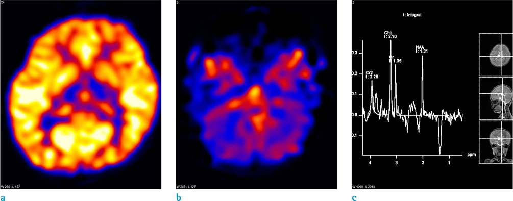
Fig. 2 (a, b) ASL-perfusion images show decreased perfusion in both thalami and cerebellum. (c) Proton MR spectroscopy obtained from the right thalamus reveals an inverted lactate doublet at 1.33 ppm.
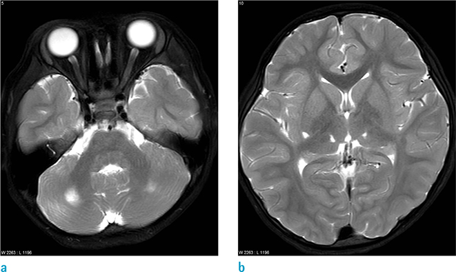
Fig. 3 The follow-up MR obtained 3 years later. (a, b) Axial T2-weighted images show focal high signal intensity lesions indicating tissue losses in both cerebellum, right basal ganglia and both thalami.
Reference
-
1. Mizuguchi M, Abe J, Mikkaichi K, et al. Acute necrotising encephalopathy of childhood: a new syndrome presenting with multifocal, symmetric brain lesions. J Neurol Neurosurg Psychiatry. 1995; 58:555–561.
Article2. Mizuguchi M. Acute necrotizing encephalopathy of childhood: a novel form of acute encephalopathy prevalent in Japan and Taiwan. Brain Dev. 1997; 19:81–92.
Article3. Hoshino A, Saitoh M, Oka A, et al. Epidemiology of acute encephalopathy in Japan, with emphasis on the association of viruses and syndromes. Brain Dev. 2012; 34:337–343.
Article4. Albayram S, Bilgi Z, Selcuk H, et al. Diffusion-weighted MR imaging findings of acute necrotizing encephalopathy. AJNR Am J Neuroradiol. 2004; 25:792–797.5. Kim JH, Kim IO, Lim MK, et al. Acute necrotizing encephalopathy in Korean infants and children: imaging findings and diverse clinical outcome. Korean J Radiol. 2004; 5:171–177.
Article6. Wong AM, Simon EM, Zimmerman RA, Wang HS, Toh CH, Ng SH. Acute necrotizing encephalopathy of childhood: correlation of MR findings and clinical outcome. AJNR Am J Neuroradiol. 2006; 27:1919–1923.7. Goo HW, Choi CG, Yoon CH, Ko TS. Acute necrotizing encephalopathy: diffusion MR imaging and localized proton MR spectroscopic findings in two infants. Korean J Radiol. 2003; 4:61–65.
Article8. Oki J, Yoshida H, Tokumitsu A, et al. Serial neuroimages of acute necrotizing encephalopathy associated with human herpesvirus 6 infection. Brain Dev. 1995; 17:356–359.
Article9. Hayakawa J, Fujino O, Murakami M, Fukunaga Y. Unusual findings in single-photon emission computed tomography in a 1-year-old boy with acute necrotizing encephalopathy. Pediatr Int. 2007; 49:94–96.
Article10. Okumura A, Mizuguchi M, Kidokoro H, et al. Outcome of acute necrotizing encephalopathy in relation to treatment with corticosteroids and gammaglobulin. Brain Dev. 2009; 31:221–227.
Article11. Manara R, Franzoi M, Cogo P, Battistella PA. Acute necrotizing encephalopathy: combined therapy and favorable outcome in a new case. Childs Nerv Syst. 2006; 22:1231–1236.
Article
- Full Text Links
-
- Actions
-
Cited
- CITED
-
- Close
- Share
-
- Similar articles
-
- Acute Necrotizing Encephalopathy: Diffusion MR Imaging and Localized Proton MR Spectroscopic Findings in Two Infants
- MR Imaging of Acute Necrotizing Encephalopathy:A Report of Two Cases
- Diagnosis of Meniscal Tear of the Knee Using Proton-weighted Fast Spin-Echo MR Imaging: Can be an Alternative to Conventional Spin-Echo Imaging?
- MR Spectroscopy and Diffusion Weighted Imaging Findings of Primary Non-Hodgkin Lymphoma of the Breast: Two Case Reports
- Diffusion-Weighted MR Imaging in Acute Wernicke's Encephalopathy Associated with Pseudomembranous Colitis: A Case Report and Review of the Literature
