Supratentorial Extraventricular Anaplastic Ependymoma Presenting with Repeated Intratumoral Hemorrhage
- Affiliations
-
- 1Department of Neurosurgery, School of Medicine, Kyungpook National University, Daegu, Korea. jhwang@knu.ac.kr
- KMID: 2134276
- DOI: http://doi.org/10.14791/btrt.2014.2.2.81
Abstract
- Supratentorial extraventricular anaplastic ependymomas are extremely rare. We report the case of a 23-year-old male with a supratentorial extraventricular anaplastic ependymoma that presented with repeated intratumoral hemorrhage. The patient was diagnosed with an intracerebral hematoma in the left occipital lobe and underwent operation. The hematoma did not reveal tumor cells, but a new tumor grew in the same location 5 years later. Magnetic resonance imaging showed a tumor with intratumoral hemorrhage. The patient underwent the tumor resection. Intraoperative findings showed that the tumor had no continuity with the ventricle. Histopathological examinations confirmed an anaplastic ependymoma. The spinal evaluation was unremarkable, and radiotherapy was administered to the left occipital lobe. Four years later, the tumor recurred at the cervicomedullary junction and T8-T9 levels. This case demonstrates that anaplastic ependymomas should be included in the differential diagnoses of supratentorial extraventricular tumors presenting with repeated intratumoral hemorrhage.
Keyword
MeSH Terms
Figure
-
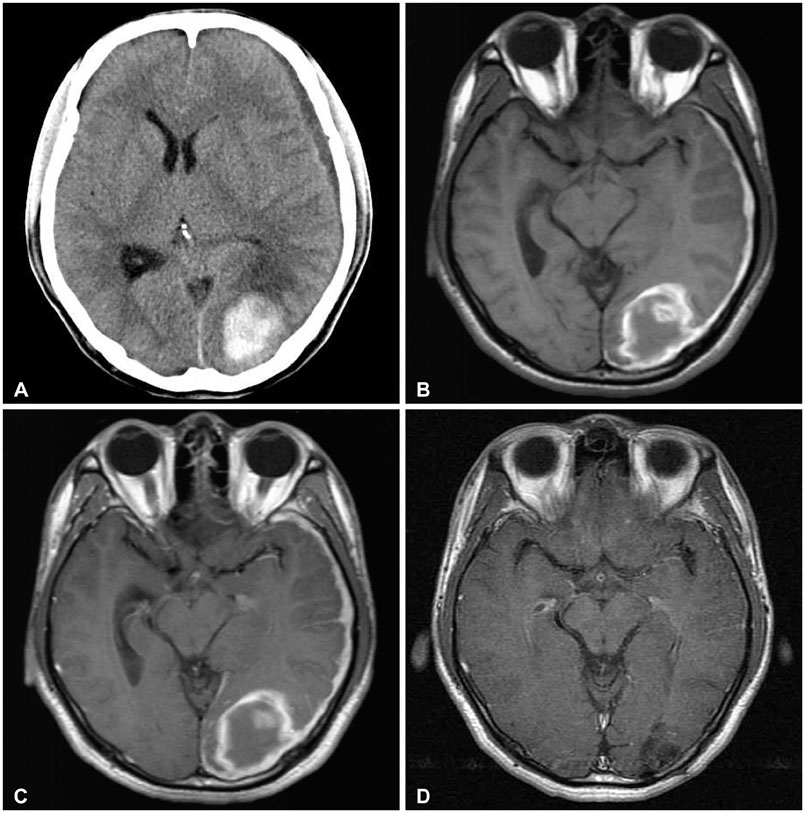
Fig. 1 Preoperative imaging studies and a follow-up MRI after surgery. A: CT scan showing an intracerebral hematoma in the left occipital lobe and a chronic subdural hemorrhage along the left cerebral convexity. B and C: T1-weighted and contrast-enhanced T1-weighted MRI revealing no enhanced lesions. D: A follow-up MRI obtained 4 years after surgery demonstrating no new lesion.
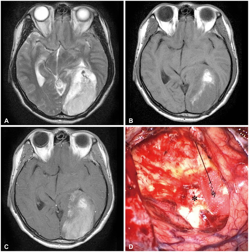
Fig. 2 Magnetic resonance images demonstrating a new mass with intratumoral hemorrhage, and an intraoperative photograph. A and B: T2-weighted and T1-weighted MRI showing a mass with mixed intensity. C: Contrast-enhanced T1-weighted MRI revealing a strong enhancement in the same location as the previous hematoma. D: Intraoperative photograph demonstrating that the tumor showed no continuity with the ventricular system (asterisk: intact wall of the lateral ventricle).
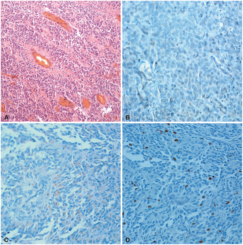
Fig. 3 Histopathological findings. A: Hematoxylin and eosin-stained section showing increased cellularity with perivascualr pseudorosettes (×100). B, C, and D: Immunohistochemical stainings for glial fibrillary acidic protein, epithelial membrane antigen (×200) and Ki-67 labeling index revealing approximately 10% (×200).
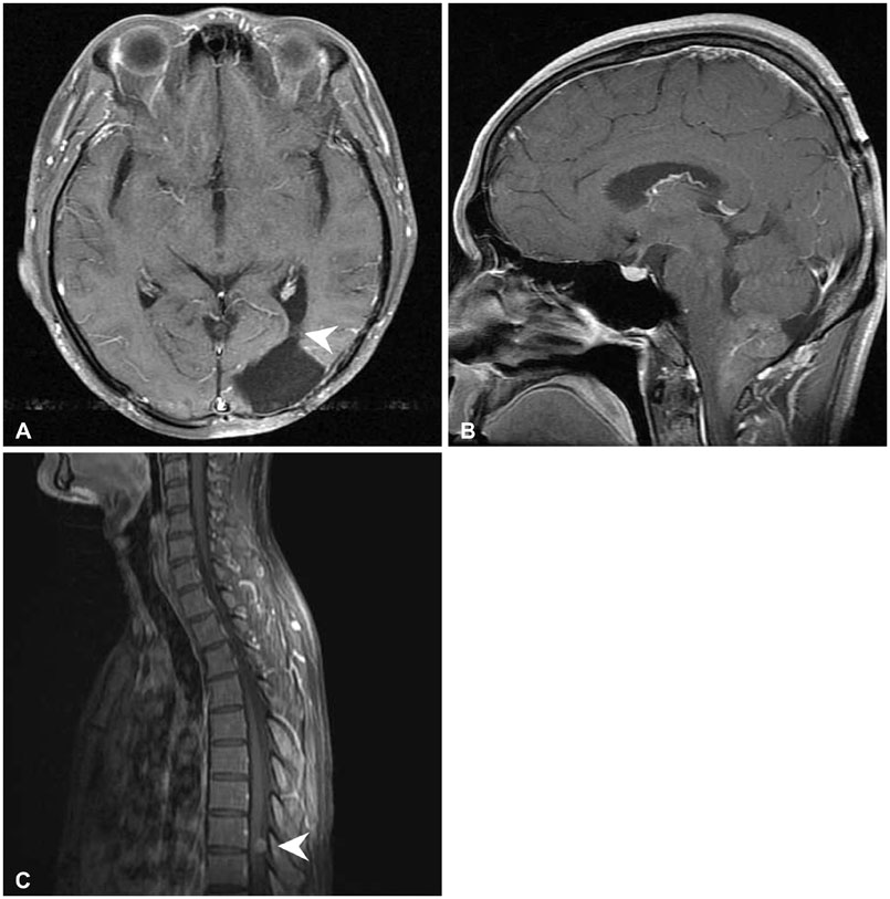
Fig. 4 Magnetic resonance images showing a recurred tumor. A: Axial contrast-enhanced T1-weighted image showing no recurrence at the primary site (arrowhead: intact wall of the lateral ventricle). B and C: Sagittal contrast-enhanced T1-weighted images of brain and thoracic spine demonstrating the tumor recurrence and dissemination (arrowhead: dissemination at the intradural space of the eighth to ninth thoracic levels).
Reference
-
1. Nazar GB, Hoffman HJ, Becker LE, Jenkin D, Humphreys RP, Hendrick EB. Infratentorial ependymomas in childhood: prognostic factors and treatment. J Neurosurg. 1990; 72:408–417.
Article2. Oppenheim JS, Strauss RC, Mormino J, Sachdev VP, Rothman AS. Ependymomas of the third ventricle. Neurosurgery. 1994; 34:350–352. discussion 352-3.
Article3. Van Gompel JJ, Koeller KK, Meyer FB, et al. Cortical ependymoma: an unusual epileptogenic lesion. J Neurosurg. 2011; 114:1187–1194.
Article4. Iwamoto N, Murai Y, Yamamoto Y, Adachi K, Teramoto A. Supratentorial extraventricular anaplastic ependymoma in an adult with repeated intratumoral hemorrhage. Brain Tumor Pathol. 2014; 31:138–143.
Article5. Moritani S, Kushima R, Bamba M, et al. Highly anaplastic extraventricular ependymoma arising in an adult, mimicking metastatic adenocarcinoma with heavy stromal inflammation and emperiporesis. Pathol Int. 2003; 53:539–546.
Article6. Vernet O, Farmer JP, Meagher-Villemure K, Montes JL. Supratentorial ectopic ependymoma. Can J Neurol Sci. 1995; 22:316–319.
Article7. Kojima A, Yamaguchi N, Okui S, Kamiya M, Hirato J, Nakazato Y. Parenchymal anaplastic ependymoma with intratumoral hemorrhage: a case report. Brain Tumor Pathol. 2003; 20:85–88.
Article8. Ernestus RI, Schröder R, Klug N. Spontaneous intracerebral hemorrhage from an unsuspected ependymoma in early infancy. Childs Nerv Syst. 1992; 8:357–360.
Article9. Saito R, Kumabe T, Kanamori M, Sonoda Y, Tominaga T. Dissemination limits the survival of patients with anaplastic ependymoma after extensive surgical resection, meticulous follow up, and intensive treatment for recurrence. Neurosurg Rev. 2010; 33:185–191. discussion 191-2.
Article10. Kano H, Niranjan A, Kondziolka D, Flickinger JC, Lunsford LD. Outcome predictors for intracranial ependymoma radiosurgery. Neurosurgery. 2009; 64:279–287. discussion 287-8.
Article
- Full Text Links
-
- Actions
-
Cited
- CITED
-
- Close
- Share
-
- Similar articles
-
- A Case of Recurrent Supratentorial Extraventricular Anaplastic Ependymoma in Adult
- Supratentorial Anaplastic Ependymoma Mimicking an Extra-Axial Tumor: A Case Report
- A Case of Supratentorial Intra-axial Ependymoma Showing Exophytic Growth
- Supratentorial Cortical Ependymoma in a 21-Month-Old Boy
- Rapidly Enlarging Pediatric Cortical Ependymoma
