Imaging Appearance of Human Immunodeficiency Virus Encephalitis on the Diffusion Weighted Images: A Case Report
- Affiliations
-
- 1Department of Radiology, Daejeon Eulji University Hospital, Daejeon, Korea. midosyu@eulji.ac.kr
- 2Department of Neurology, Daejeon Eulji University Hospital, Daejeon, Korea.
Abstract
- Imaging finding of human immunodeficiency virus (HIV) encephalitis contain bilateral, symmetric, patchy, or diffuse increased T2WI signal intensities in the basal ganglia, cerebellum, brainstem, and centrum semiovale. In particular, the centrum semiovale is most commonly involved. Most of the HIV encephalitis cases are accompanied by brain atrophy. No previous study has reported symmetric increased signal intensity at the bilateral centrum semiovale without brain atrophy on diffusion weighted images in HIV encephalitis patients. Here, we report a case of this. We suggest that radiologists should consider the possibility of HIV encephalitis if there are symmetric increases in signal intensity at the bilateral centrum semiovale on diffusion weighted images of patients with a history of HIV infection.
MeSH Terms
Figure
-
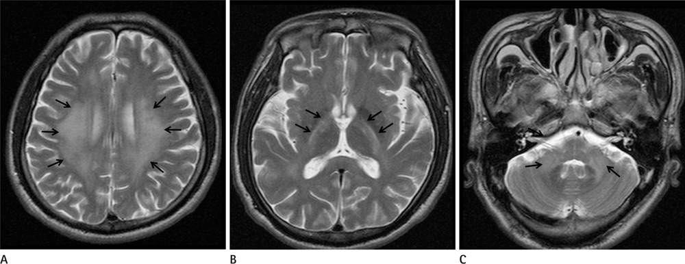
Fig. 1 T2 weighted images. The T2 weighted images of the patient showed diffuse "hazy" increased T2 signal intensity at the bilateral frontoparietal centrum semiovale (arrows) (A), both basal ganglia, mainly in the posterior internal capsules (arrows) (B), and the middle (arrows) and superior cerebellar peduncles (C). There was no significant cerebral white matter volume reduction, ventricular widening, or cortical atrophy. The imaging findings were consistent with HIV encephalitis, except for cerebral white matter atrophy and a previous history of HIV infection. Additionally, there was no evidence of other opportunistic cerebral infections in the patient affecting the cerebral white matter. Note.-HIV = human immunodeficiency virus

Fig. 2 Diffusion weighted images. There was increased signal intensity on the diffusion weighted images at the bilateral frontoparietal centrum semiovale (A), and both basal ganglia, mainly in both posterior internal capsules (B); this was consistent with the increased area of T2 signal. Diffusion weighted images suggested active and acute brain parenchymal HIV activity. Note.-HIV = human immunodeficiency virus
Reference
-
1. Offiah CE, Turnbull IW. The imaging appearances of intracranial CNS infections in adult HIV and AIDS patients. Clin Radiol. 2006; 61:393–401.2. Provenzale JM, Jinkins JR. Brain and spine imaging findings in AIDS patients. Radiol Clin North Am. 1997; 35:1127–1166.3. Ramsey RG, Geremia GK. CNS complications of AIDS: CT and MR findings. AJR Am J Roentgenol. 1988; 151:449–454.4. Chrysikopoulos HS, Press GA, Grafe MR, Hesselink JR, Wiley CA. Encephalitis caused by human immunodeficiency virus: CT and MR imaging manifestations with clinical and pathologic correlation. Radiology. 1990; 175:185–191.5. Olsen WL, Longo FM, Mills CM, Norman D. White matter disease in AIDS: findings at MR imaging. Radiology. 1988; 169:445–448.6. Pomara N, Crandall DT, Choi SJ, Johnson G, Lim KO. White matter abnormalities in HIV-1 infection: a diffusion tensor imaging study. Psychiatry Res. 2001; 106:15–24.7. Bergui M, Bradac GB, Oguz KK, Boghi A, Geda C, Gatti G, et al. Progressive multifocal leukoencephalopathy: diffusion-weighted imaging and pathological correlations. Neuroradiology. 2004; 46:22–25.
- Full Text Links
-
- Actions
-
Cited
- CITED
-
- Close
- Share
-
- Similar articles
-
- Toxoplasma Encephalitis in a Patient with Human Immunodeficiency Virus Infection
- Recurrent Clinically Mild Encephalitis/Encephalopathy with a Reversible Splenial Lesion (MERS) on Diffusion Weighted Imaging: A Case Report
- MR Imaging Features of Acute Enterovirus 71 Encephalitis in a Patient with Hand-Foot-Mouth Disease: A Case Report
- A Case of Encephalitis with a Reversible Splenial Lesion on a Diffusion Weighted MRI Image
- Relapsing Herpes Simplex Encephalitis
