J Korean Soc Radiol.
2011 Mar;64(3):213-216. 10.3348/jksr.2011.64.3.213.
Recurrent Clinically Mild Encephalitis/Encephalopathy with a Reversible Splenial Lesion (MERS) on Diffusion Weighted Imaging: A Case Report
- Affiliations
-
- 1Department of Pediatrics, Jeju National University College of Medicine, Jeju, Korea.
- 2Department of Radiology, Jeju National University College of Medicine, Jeju, Korea. jkcontrast@naver.com
- KMID: 2097939
- DOI: http://doi.org/10.3348/jksr.2011.64.3.213
Abstract
- We report serial MR imaging of an 11-year-old boy who had a recurrent episode of clinically mild encephalitis/encephalopathy with a reversible splenial lesion. During the first episode, brain lesions were limited to the corpus callosum. However, for the second episode, the lesions were distributed in the corpus callosum and bilateral deep white matter. No abnormality remained in the follow-up MR images obtained after full recovery.
MeSH Terms
Figure
-

Fig. 1 MR imaging of the first episode. Diffusion-weighted imaging (DWI) and ADC map (A, B) indicated that the lesions in the genu and splenium of the CC (arrows) show a pattern of water diffusion restriction (high signal intensity on the DWI, low signal intensity on the ADC map).

Fig. 2 MR imaging of the second episode. DWI (A-C) and ADC map (D-F) demonstrate the lesion in the entire CC and deep periventricular white matter, which show a pattern of restricted water diffusion.
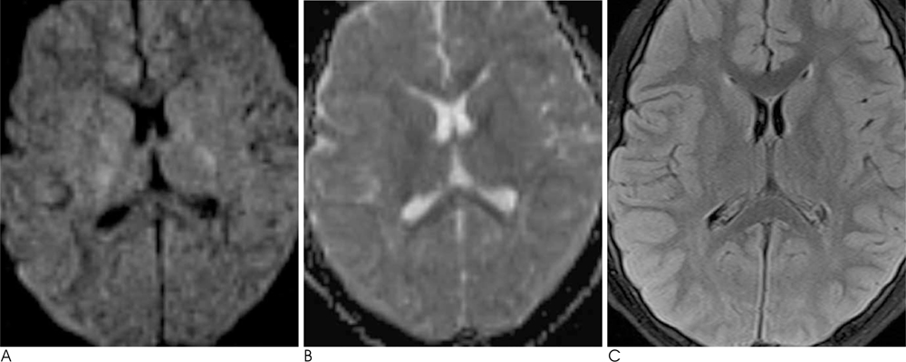
Fig. 3 Follow-up MR imaging after 4 months. There is no remarkable residual lesion in the corpus callosum on axial DWI, ADC map (A, B), and FLAIR image (C).
Reference
-
1. Tada H, Takanashi J, Barkovich AJ, Oba H, Maeda M, Tsukahara H, et al. Clinically mild encephalitis/encephalopathy with a reversible splenial lesion. Neurology. 2004; 63:1854–1858.2. Gurtler S, Ebner A, Tuxhorn I, Ollech I, Pohlmann-Eden B, Woermann FG. Transient lesion in the splenium of the corpus callosum and antiepileptic drug withdrawal. Neurology. 2005; 65:1032–1036.3. Wada A, Yoshida R, Oda K, Fukuba E, Uchida N, Kitagaki H. Acute encephalopathy associated with intravenous immunoglobulin therapy. AJNR Am J Neuroradiol. 2005; 26:2311–2315.4. Garcia-Monco JC, Cortina IE, Ferreira E, Martinez A, Ruiz L, Cabrera A, et al. Reversible Splenial Lesion Syndrome (RESLES): What's in a Name? J Neuroimaging. 2011; 21:e1–e14.5. Takanashi J, Barkovich AJ, Shiihara T, Tada H, Kawatani M, Tsukahara H, et al. Widening spectrum of a reversible splenial lesion with transiently reduced diffusion. AJNR Am J Neuroradiol. 2006; 27:836–838.6. Bulakbasi N, Kocaoglu M, Tayfun C, Ucoz T. Transient splenial lesion of the corpus callosum in clinically mild influenza-associated encephalitis/encephalopathy. AJNR Am J Neuroradiol. 2006; 27:1983–1986.7. Takanashi J. Two newly proposed infectious encephalitis/encephalopathy syndromes. Brain Dev. 2009; 31:521–528.8. Takanashi J, Imamura A, Hayakawa F, Terada H. Differences in the time course of splenial and white matter lesions in clinically mild encephalitis/encephalopathy with a reversible splenial lesion (MERS). J Neurol Sci. 2010; 292:24–27.9. Narita M, Tanaka H, Togashi T, Abe S. Cytokines involved in CNS manifestations caused by Mycoplasma pneumoniae. Pediatr Neurol. 2005; 33:105–109.10. Aiba H, Mochizuki M, Kimura M, Hojo H. Predictive value of serum interleukin-6 level in influenza virus-associated encephalopathy. Neurology. 2001; 57:295–299.
- Full Text Links
-
- Actions
-
Cited
- CITED
-
- Close
- Share
-
- Similar articles
-
- The Changes in Axial and Radial Diffusivity in a Patient with Clinically Mild Encephalitis/Encephalopathy with a Reversible Splenial Lesion
- A Case of Encephalitis with a Reversible Splenial Lesion on a Diffusion Weighted MRI Image
- Lithium-Induced Downbeat Nystagmus with Reversible Splenial Lesion
- A Case of Scrub Typhus Related Encephalopathy Presenting as Rapidly Progressive Dementia
- Mild Encephalopathy with Reversible Lesion in the Splenium of the Corpus Callosum and Bilateral Frontal White Matter
