Korean J Urol.
2011 Apr;52(4):295-297.
Extensive Systemic Sarcoidosis with Testicular Involvement Mimicking Metastatic Testicular Cancer
- Affiliations
-
- 1Department of Urology, Seoul Veterans Hospital, Seoul, Korea.
- 2Department of Urology, Yonsei University College of Medicine, Seoul, Korea.
- 3Department of Urology, Ajou University School of Medicine, Suwon, Korea. sikimuro@ajou.ac.kr
- 4Department of Pathology, Ajou University School of Medicine, Suwon, Korea.
Abstract
- Sarcoidosis is an idiopathic, multisystem disease that rarely involves the genitourinary tract. Here we present an unusual case of testicular sarcoidosis with extensive lymphadenopathy that mimicked a metastatic testicular tumor. A 27-year-old male presented with a palpable right testicular mass accompanied by multiple palpable inguinal lymph nodes. The scrotal ultrasound showed a hypoechoic lesion at the inferior portion of the right testis. Extensive enlarged lymph nodes were noted in multiple areas on the abdominal computed tomography. Preoperative testicular tumor markers were within the normal range. Exploration of the right testis with a frozen section analysis of the right testicular mass and of a palpable right inguinal lymph node showed granulomatous inflammation. The testis was salvaged and the final pathological diagnosis was sarcoidosis. Treatment with high-dose corticosteroids resulted in complete resolution of the intratesticular mass and a significant decrease in the extent of the lymphadenopathy.
Keyword
MeSH Terms
Figure
-
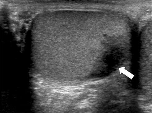
FIG. 1 Scrotal ultrasound showing ill-defined, irregular, hypoechoic lesion at the inferior portion of the right testis (arrow).
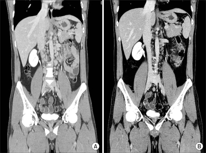
FIG. 2 A 3D coronal image computed tomography scan of the abdomen after administration of intravenous contrast medium. (A) Extensive enlarged lymph nodes were seen in the retroperitoneal space. (B) Four months after the start of high-dose corticosteroid therapy, multiple, extensive retroperitoneal lymph nodes had considerably decreased in size.

FIG. 3 Histopathological examination of the intratesticular lesion showed non-caseating epithelioid cell granulomas with giant cells (H&E, ×200). (A) Testis. (B) Right superficial inguinal node.
Reference
-
1. Kodama K, Hasegawa T, Egawa M, Tomosugi N, Mukai A, Namiki M. Bilateral epididymal sarcoidosis presenting without radiographic evidence of intrathoracic lesion: review of sarcoidosis involving the male reproductive tract. Int J Urol. 2004. 11:345–348.2. Reineks EZ, MacLennan GT. Sarcoidosis of the testis and epididymis. J Urol. 2008. 179:1147.3. Geller RA, Kuremskey DA, Copeland JS, Stept R. Sarcoidosis and testicular neoplasm: an unusual association. J Urol. 1977. 118:487–488.4. Blacher EJ, Maynard JF. Seminoma and sarcoidosis: an unusual association. Urology. 1985. 26:288–289.5. Rayson D, Burch PA, Richardson RL. Sarcoidosis and testicular carcinoma. Cancer. 1998. 83:337–343.6. Massarweh NN, Bhalani VK, Shaw KK, Crawford B, Lang E, Davis R. Testicular presentation of sarcoidosis and organ preservation: case report and review of management strategies. Urology. 2006. 67:200.7. Miyazaki E, Tsuda T, Mochizuki A, Sugisaki K, Ando M, Matsumoto T, et al. Sarcoidosis presenting as bilateral hydronephrosis. Intern Med. 1996. 35:579–582.8. Datta SN, Freeman A, Amerasinghe CN, Rosenbaum TP. A case of scrotal sarcoidosis that mimicked tuberculosis. Nat Clin Pract Urol. 2007. 4:227–230.
- Full Text Links
-
- Actions
-
Cited
- CITED
-
- Close
- Share
-
- Similar articles
-
- An Unusual Case of Testicular Seminoma Mimicking Segmental Testicular Infarction
- Unusual Presentation of a Testicular Lymphoma Mimicking a Missed Testicular Torsion: A Case Report
- Neonatal Testicular Torsion Mimicking a Testicular Tumor
- Solitary Testicular Metastasis of Prostate Cancer Mimicking Primary Testicular Cancer
- A Case of Antenatal Testicular Torsion Mimicking Inflammation and Tumor in the Neonate
