Neonatal Testicular Torsion Mimicking a Testicular Tumor
- Affiliations
-
- 1Department of Urology, Kosin University College of Medicine, Korea.
- 2Department of Urology, College of Medicine, Pusan National University, Busan, Korea. lsd@pusan.ac.kr
- KMID: 1204328
- DOI: http://doi.org/10.4111/kju.2008.49.10.957
Abstract
- The large, hard, non-tender features of neonatal testicular torsion may sometimes lead to the misdiagnosis of a testicular tumor. The authors present the case of a 1-week old male neonate in whom the differential diagnosis between testicular torsion and a tumor proved difficult. Based on serial physical examinations and serial color Doppler ultrasonography findings, the initial diagnosis was a testicular tumor with/without torsion and a concomitant communicating hydrocele. However, the intraoperative findings did not support the existence of testicular torsion. A left radical orchiectomy and contralateral orchiopexy with herniorrhaphy were undertaken, but somewhat surprisingly, the final pathologic findings demonstrated a left testicular torsion.
Keyword
Figure
-

Fig. 1. Serial color Doppler ultrasonographic findings at first visit (A) and 1 week later (B). The left testis showed an ovoid intratesticular lesion with heterogeneous echogenecity, and was five times larger than the right testis. The right testis was normal with a hydrocele. There were no interval changes in ultrasonographic findings between the first visit and follow-up 1 week later.
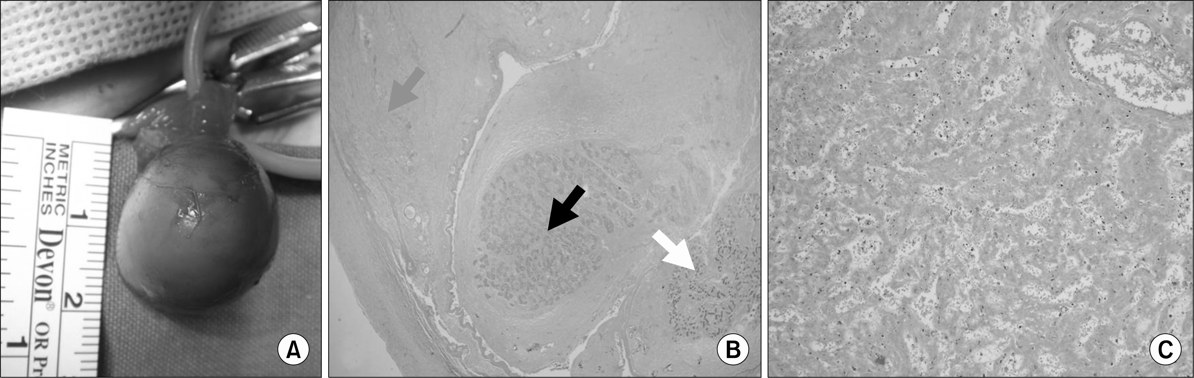
Fig. 2. Intraoperative gross and microscopic findings of the left testis. (A) A large mass wrapped around the testis, spermatic cord, and epididymis. (B) On the right side of the epididymis, the nucleus showed the normal structure (white arrow), however on the lower center of the epididymis, the nucleus disappeared due to the infarction (black arrow). The left testis demonstrated the necrotic findings that the normal seminiferous tubules are not visible due to the infarction (gray arrow). (C) Seminiferous tubule structures were lost with congestion on the intermediate center (H&E; B: x20; C: x200).
Reference
-
References
1. Führer S, May M, Koch A, Marusch F, Gunia S, Erler T, et al. Intrauterine torsion of a testicular teratoma: a case report. J Perinatol. 2005; 25:220–2.
Article2. Ricci P, Cantisani V, Drudi FM, Carbone I, Coniglio M, Bosco S, et al. Prenatal testicular torsion: sonographic appearance in the newborn infant. Eur Radiol. 2001; 11:2589–92.
Article3. Cartwright PC, Snow BW, Reid BS, Shultz PK. Color Doppler ultrasound in newborn testis torsion. Urology. 1995; 45:667–70.
Article4. Wu JT, Book L, Sudar K. Serum alpha fetoprotein (AFP) levels in normal infants. Pediatr Res. 1981; 15:50–2.
Article
- Full Text Links
-
- Actions
-
Cited
- CITED
-
- Close
- Share
-
- Similar articles
-
- A Case of Antenatal Testicular Torsion Mimicking Inflammation and Tumor in the Neonate
- Unusual Presentation of a Testicular Lymphoma Mimicking a Missed Testicular Torsion: A Case Report
- An Unusual Case of Testicular Seminoma Mimicking Segmental Testicular Infarction
- The Pathological Anatomy of Intermittent Testicular Torsion
- A Case or Synchronous Bilateral Testicular Torsion in the Neonate
