Dement Neurocogn Disord.
2013 Mar;12(1):29-32. 10.12779/dnd.2013.12.1.29.
Serial Changes in Diffusion-Weighted Magnetic Resonance Images with Hypoperfusion on Brain SPECT in a Case of Hashimoto's Encephalopathy: Understanding Pathophysiology of Hashimoto's Encephalopathy
- Affiliations
-
- 1Department of Neurology, Dong-A University College of Medicine, Busan, Korea. neuropark@dau.ac.kr
- 2Department of Endocrinology, Dong-A University College of Medicine, Busan, Korea.
- 3Department of Radiology, Dong-A University College of Medicine, Busan, Korea.
- KMID: 2222358
- DOI: http://doi.org/10.12779/dnd.2013.12.1.29
Abstract
- Diffuse or focal white matter hyperintensity lesions on MRI have been reported in only a few patients with Hashimoto's encephalopathy (HE), and anti-TPO antibody level is high in most cases. We report a 59-year-old woman who presented with acute onset of disorientation with confusion. Anti-thyroglobulin antibody was detected in high titer, although anti-TPO antibody titer was not high. Thyroid sonography and biopsy revealed Hashimoto's thyroiditis. Initial fluid-attenuated inversion recovery (FLAIR) image and diffusion-weighted imaging (DWI) revealed ill-defined, diffuse, high-signal intensity lesions on the deep white matters and globus pallidus. Brain SPECT showed significant hypoperfusion in both basal ganglia (especially globus pallidus), frontal and temporal lobes. With the impression of HE, the patient was treated on a high-dose steroid. Over the next 15 weeks, her cognition improved to a nearly normal state and the MRI findings on DWI and FLAIR showed resolution paralleling her clinical improvement. Our case illustrates the peculiar changes in the MR findings, especially in DWI, with hypoperfusion on brain SPECT in patients with HE and allows for a greater understanding of the pathophysiology of HE.
MeSH Terms
-
Autoantibodies
Basal Ganglia
Biopsy
Brain
Brain Diseases
Cognition
Female
Globus Pallidus
Hashimoto Disease
Humans
Magnetic Resonance Imaging
Magnetic Resonance Spectroscopy
Magnetics
Magnets
Temporal Lobe
Thyroid Gland
Thyroiditis
Tomography, Emission-Computed, Single-Photon
Autoantibodies
Brain Diseases
Hashimoto Disease
Figure
-

Fig. 1 Cone biopsy of thyroid. Infiltration of lymphoid foillicle (A) and Hurthle cell change of surrounding follicular epithelial cells (B).

Fig. 2 Initial and followup brain MRI findings. Initial diffusionweighted, FLAIR, T2weighted, T1weighted and contrast images (A and B) showing illdefined diffuse high signal intensity lesions on the deep white matter and globus pallidus. After 3 months (C and D), DWI, FLAIR images show the extent of the lesions, which are markedly decreased compared to the previous MRI and cystic changes occur in bilateral globus pallidus.
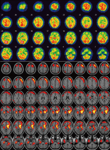
Fig. 3 Tc99m HMPAO SPECT shows significant hypoperfusion in both basal ganglia (especially globus pallidus), frontal and temporal lobes at admission.
Reference
-
1. Schiess N, Pardo CA. Hashimoto's Encephalopathy. Ann NY Acad Sci. 2008. 1142:254–265.
Article2. Ferracci F, Moretto G, Candeago RM, Cimini N, Conte F, Gentile M, et al. Anti-thyroid antibodies in the CSF: their role in the pathogenesis of Hashimoto's encephalopathy. Neurology. 2003. 60:712–714.
Article3. Grommes C, Griffin C, Downes KA, Lerner AJ. Steroid-responsive encephalopathy associated with autoimmune thyroiditis presenting with diffusion MR imaging changes. AJNR Am J Neuroradiol. 2008. 29:1550–1551.
Article4. Chen C, Qin W, Wei C, Wang X, Li K. Time course of Hashimoto's encephalopathy revealed by MRI: Report of two cases. J Neurol Sci. 2011. 300:169–172.
Article5. Mijajlovic M, Mirkovic M, Dackovic J, Zidverc-Trajkovic J, Sternic N. Clinical manifestations, diagnostic criteria and therapy of Hashimoto's encephalopathy: report of two cases. J Neurol Sci. 2010. 288:194–196.
Article6. Blanchin S, Coffin C, Viader F, Ruf J, Carayon P, Potier F, et al. Anti-thyroperoxidase antibodies from patients with Hashimoto's encephalopathy bind to cerebellar astrocytes. J Neuroimmunol. 2007. 192:13–20.7. Kothbauer-Margreiter I, Sturzenegger M, Komor J, Baumgartner R, Hess CW. Encephalopathy associated with Hashimoto thyroiditis: diagnosis and treatment. J Neurol. 1996. 243:585–593.8. Chang T, Riffsy MT, Gunaratne PS. Hashimoto encephalopathy: clinical and MRI improvement following high-dose corticosteroid therapy. Neurologist. 2010. 16:394–396.
Article9. Forchetti CM, Katsamakis G, Garron DC. Autoimmune thyroiditis and a rapidly progressive dementia: global hypoperfusion on SPECT scanning suggests a possible mechanism. Neurology. 1997. 49:623–626.
Article
- Full Text Links
-
- Actions
-
Cited
- CITED
-
- Close
- Share
-
- Similar articles
-
- Nonconvulsive Status Epilepticus as the First Manifestation of Hashimoto's Encephalopathy
- Hashimoto's Encephalopathy Presenting With Memory Disturbance and Complex Partial Seizure
- Uremic Encephalopathy with Atypical Magnetic Resonance Features on Diffusion-Weighted Images
- Two Cases of Hypertensive Brainstem Encephalopathy
- Hashimoto's Encephalopathy Presented with Nonspecific Vasogenic Edema in Brain Magnetic Resonance Imaging
