The Posterior Choroidal Profiles Measured by Spectral Domain Optical Coherence Tomography in Healthy Korean Children
- Affiliations
-
- 1Cheil Eye Hospital, Daegu, Korea. eyejholee@hotmail.com
- KMID: 2217918
- DOI: http://doi.org/10.3341/jkos.2013.54.11.1708
Abstract
- PURPOSE
We assessed changes of the choroidal thickness in healthy Korean children using enhanced depth imaging (EDI) optical coherence tomography (OCT) and evaluated the association of choroidal thickness and axial length.
METHODS
Seventy-nine eyes (79 children) within +/-1 diopter spherical equivalent underwent horizontal and vertical scan using EDI OCT. Two observers determined independently the choroidal thickness at 1 mm intervals from 3 mm nasal and 4 mm temporal to the fovea and 1 mm superior and inferior to the fovea using the manual caliper provided by the device software. Statistical analysis was performed to evaluate variations of choroidal thickness at each location and to correlate choroidal thickness and axial length. Intraclass correlation coefficient (ICC) was calculated.
RESULTS
The subjects' mean age was 7.67 years. The mean axial length was 22.96 mm and mean subfoveal choroidal thickness was 296.13 microm. The thinnest choroidal thickness was 160.57 microm at 3 mm nasal to the fovea and the thickest was 319.49 microm at 4 mm temporal to the fovea. The choroidal thickness at 1 mm superior (294.70 microm) and inferior (287.11 microm) to the fovea showed no statistical significance compared with the subfoveal choroidal thickness. The mean choroidal thickness was thicker at 3 mm and 2 mm nasal to the fovea in eyes with shorter than the mean axial length (p < 0.05). For the assessment of intra-observer reproducibility, the ICC ranged from 0.995 to 0.998 (p < 0.001).
CONCLUSIONS
The choroidal thickness increased from the nasal to the temporal direction at the posterior pole and eyes with shorter axial lengths tended to present thicker choroids at the nasal area in healthy Korean children.
Keyword
Figure
-
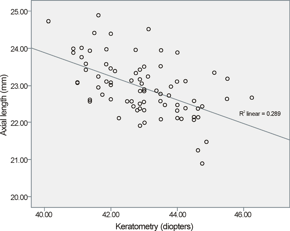
Figure 1. Scatterplot of axial length and keratometry of all subjects shows a significant negative correlation (p < 0.05).
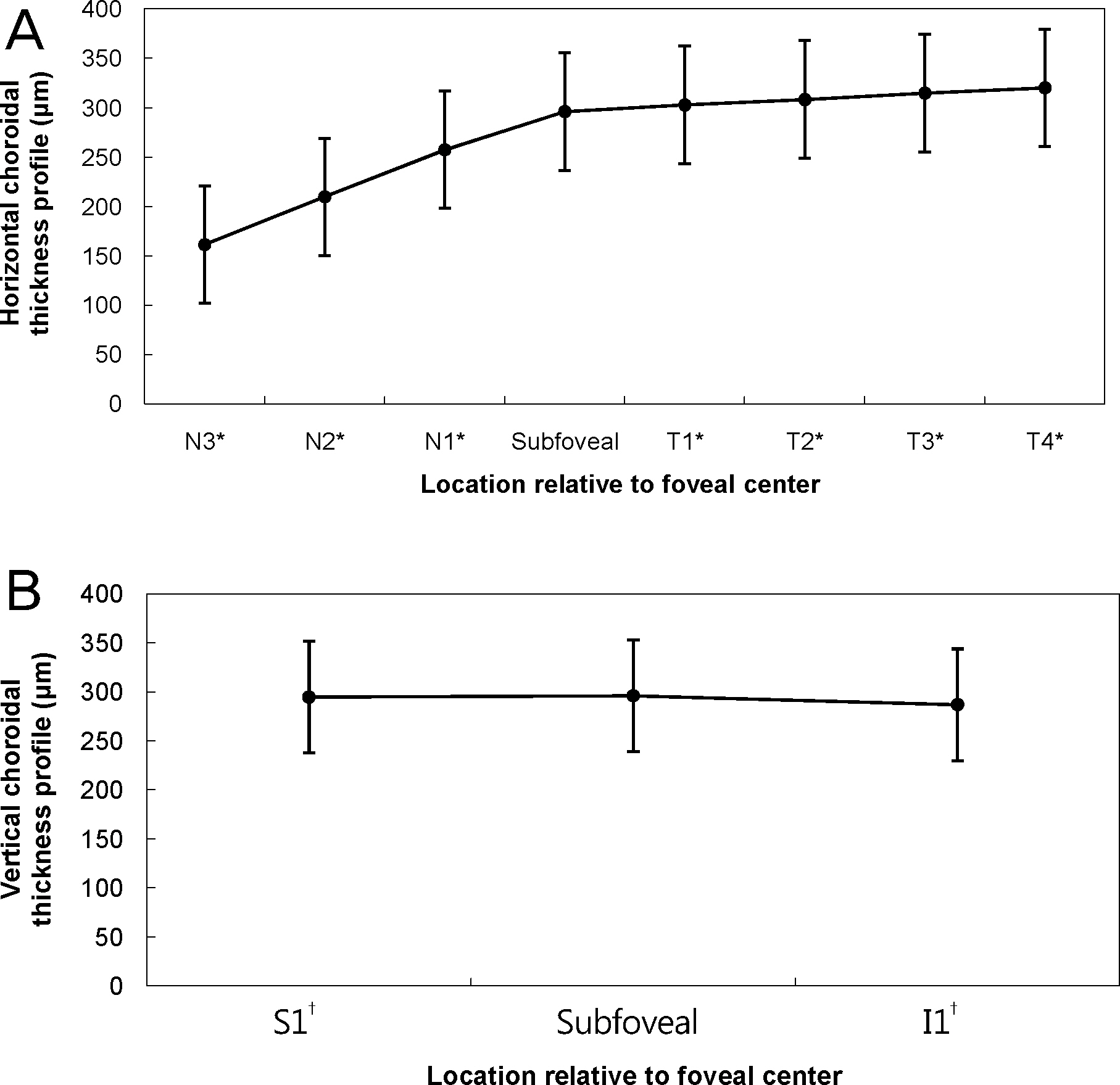
Figure 2. Horizontal (A) and vertical (B) choroidal thickness profile. N3* = nasal 3000 μm; N2* = nasal 2000 μm; N1* = nasal 1000 μm; T1* = temporal 1000 μm; T2* = temporal 2000 μm; T3* = temporal 3000 μm; T4* = temporal 4000 μm; S1 = superior 1000 μm; I1 † = inferior 1000 μm.
Cited by 2 articles
-
Changes in Choroidal Thickness after Panretinal Photocoagulation in Diabetic Retinopathy Patients
Sung Yu, Yong Il Kim, Kyoo Won Lee, Hyun Gu Kang
J Korean Ophthalmol Soc. 2016;57(2):256-263. doi: 10.3341/jkos.2016.57.2.256.A Retrospective Study of Choroidal Thickness in Children with Unilateral High Myopia
Yong Wun Cho, Che Ron Kim, Woong Sun Yoo, Ji Myong Yoo
J Korean Ophthalmol Soc. 2015;56(10):1624-1629. doi: 10.3341/jkos.2015.56.10.1624.
Reference
-
References
1. Kim EJ, Kim JH, Koo SH, et al. Choroidal thickness changes ac- cording to the refractive errors and axial length in Korean myopia patients. J Korean Ophthalmol Soc. 2012; 53:1814–22.2. Spaide RF, Koizumi H, Pozonni MC. Enhanced depth imaging spectral-domain optical coherence tomography. Am J Ophthalmol. 2008; 146:496–500.
Article3. Margolis R, Spaide RF. A pilot study of enhanced depth imaging optical coherence tomography of the choroid in normal eyes. Am J Ophthalmol. 2009; 147:811–5.
Article4. Kim KH, Kim DG. The relationship among refractive power, axial length and choroidal thickness measured by SD-OCT in myopia. J Korean Ophthalmol Soc. 2012; 53:626–31.
Article5. Ikuno Y, Kawaguchi K, Nouchi T, Yasuno Y. Choroidal thickness in healthy Japanese subjects. Invest Ophthalmol Vis Sci. 2010; 51:2173–6.
Article6. Fujiwara T, Imamura Y, Margolis R, et al. Enhanced depth imaging optical coherence tomography of the choroid in highly myopic eyes. Am J Ophthalmol. 2009; 148:445–50.
Article7. Agawa T, Miura M, Ikuno Y, et al. Choroidal thickness measure- ment in healthy Japanese subj ects by three-dimensional high-pene- tration optical coherence tomography. Graefes Arch Clin Exp Ophthalmol. 2011; 249:1485–92.8. Li XQ, Larsen M, Munch IC. Subfoveal choroidal thickness in re- lation to sex and axial length in 93 Danish university students. Invest Ophthalmol Vis Sci. 2011; 52:8438–41.9. Ruiz-Moreno JM, Flores-Moreno I, Lugo F, et al. Macular choroi- dal thickness in normal pediatric population measured by Swept-Source Optical Coherence Tomography. Invest Ophthalmol Vis Sci. 2013; 54:353–9.10. Harris A, Bingaman DP, Ciulla TA, Martin BJ. Retina and choroi- dal blood flow in health and disease. Ryan SJ, Ogden TE, Hinton DR, editors. Retina. 4th ed.Philadelphia: Elsevier;2006. p. 83–102.11. Kim SW, Oh J, Kwon SS, et al. Comparison of choroidal thickness among patients with healthy eyes, early age-related maculopathy, neovascular age-related macular degeneration, central serous cho- rioretinopathy, and polypoidal choroidal vasculopathy. Retina. 2011; 31:1904–11.12. Cho JH, Bae SH, Han JR, et al. Comparison of choroidal thickness in eyes with central serous chorioretinopathy, asymptomatic fellow eyes and normal eyes. J Korean Ophthalmol Soc. 2012; 53:87–93.
Article13. Lee SH, Chung H, Kim HC. Subfoveal choroidal thickness in fel- low eyes of patients with central serous chorioretinopathy. J Korean Ophthalmol Soc. 2012; 53:982–7.14. Chung SE, Kang SW, Lee JH, Kim YT. Choroidal thickness in pol- ypoidal choroidal vasculopathy and exudative age-related macular degeneration. Ophthalmology. 2011; 118:840–5.15. Manjunath V, Goren J, Fujimoto JG, Duker JS. Analysis of choroi- dal thickness in age-related macular degeneration using spec- tral-domain optical coherence tomography. Am J Ophthalmol. 2011; 152:663–8.16. Switzer DW Jr, Mendonca LS, Saito M, et al. Segregation of oph- thalmoscopic characteristics according to choroidal thickness in patients with early age-related macular degeneration. Retina. 2012; 32:1265–71.17. Ueta T, Obata R, Inoue Y, et al. Background comparison of typical age-related macular degeneration and polypoidal choroidal vascul- opathy in Japanese patients. Ophthalmology. 2009; 116:2400–6.18. Ikuno Y, Tano Y. Retinal and choroidal biometry in highly myopic eyes with spectral-domain optical coherence tomography. Invest Ophthalmol Vis Sci. 2009; 50:3876–80.
Article19. Fong AH, Li KK, Wong D. Choroidal evaluation using enhanced depth imaging spectral-domain optical coherence tomography in Vogt-Koyanagi-Harada disease. Retina. 2011; 31:502–9.
Article20. Say EA, Shah SU, Ferenczy S, Shields CL. Optical coherence to- mography of retinal and choroidal tumors. J Ophthalmol. 2011; 2011:385058.21. Shields CL, Perez B, Materin MA, et al. Optical coherence tomog- raphy of choroidal osteoma in 22 cases: evidence for photoreceptor atrophy over the decalcified portion of the tumor. Ophthalmology. 2007; 114:e53–8.22. Maul EA, Friedman DS, Chang DS, et al. Choroidal thickness measured by spectral domain optical coherence tomography: fac- tors affecting thickness in glaucoma patients. Ophthalmology. 2011; 118:1571–9.23. Esmaeelpour M, Povazay B, Hermann B, et al. Mapping choroidal and retinal thickness variation in type 2 diabetes using three-di- mensional 1060-nm optical coherence tomography. Invest Ophthalmol Vis Sci. 2011; 52:5311–6.24. Vance SK, Imamura Y, Freund KB. The effects of sildenafil citrate on choroidal thickness as determined by enhanced depth imaging optical coherence tomography. Retina. 2011; 31:332–5.
Article25. Brown JS, Flitcroft DI, Ying GS, et al. In vivo human choroidal thickness measurements: evidence for diurnal fluctuations. Invest Ophthalmol Vis Sci. 2009; 50:5–12.
Article26. Fujiwara A, Shiragami C, Shirakata Y, et al. Enhanced depth imaging spectral-domain optical coherence tomography of subfoveal choroidal thickness in normal Japanese eyes. Jpn J Ophthalmol. 2012; 56:230–5.
Article27. Chen FK, Yeoh J, Rahman W, et al. Topographic variation and in- terocular symmetry of macular choroidal thickness using enhanced depth imaging optical coherence tomography. Invest Ophthalmol Vis Sci. 2012; 53:975–85.28. Atchison DA, Jones CE, Schmid KL, et al. Eye shape in emme- tropia and myopia. Invest Ophthalmol Vis Sci. 2004; 45:3380–6.
- Full Text Links
-
- Actions
-
Cited
- CITED
-
- Close
- Share
-
- Similar articles
-
- Choroidal Thickness at the Outside of Fovea in Diabetic Retinopathy Using Spectral-Domain Optical Coherence Tomography
- The Relationship among Refractive Power, Axial Length and Choroidal Thickness Measured by SD-OCT in Myopia
- A Case of Ocular Toxoplasmosis Imaged with Spectral Domain Optical Coherence Tomography
- Comparison of Choroidal Thickness in Patients with Diabetes by Spectral-domain Optical Coherence Tomography
- Reproducibility of Choroidal Thickness in Normal Korean Eyes Using Two Spectral Domain Optical Coherence Tomography
