A Case of Ocular Toxoplasmosis Imaged with Spectral Domain Optical Coherence Tomography
- Affiliations
-
- 1Department of Ophthalmology, Kangnam Sacred Heart Hospital, Hallym University College of Medicine, Seoul, Korea. wooho.nam@gmail.com
- KMID: 1120184
- DOI: http://doi.org/10.3341/kjo.2012.26.1.58
Abstract
- A 54-year-old man presented with blurred central vision in the right eye of two weeks' duration. On presentation, visual acuity was 40 / 50 in the right eye and fundus examination showed a whitish-yellow inflammatory lesion near an atrophic, pigmented retinochoroidal scar located in the superotemporal quadrant. Serologic assessment was negative for IgM, but serum IgG to toxoplasma was elevated. Spectral domain optical coherence tomography (SD-OCT) revealed increased reflectivity from the inner retinal layer, retinal thickening, and choroidal shadowing while focal posterior hyaloid thickening and detachment were observed in the new lesion. He was treated with trimethoprim/sulfamethoxazole, clindamycin, and prednisone. SD-OCT is helpful for definitively differentiating ocular toxoplasmosis from other retinal diseases.
MeSH Terms
Figure
-
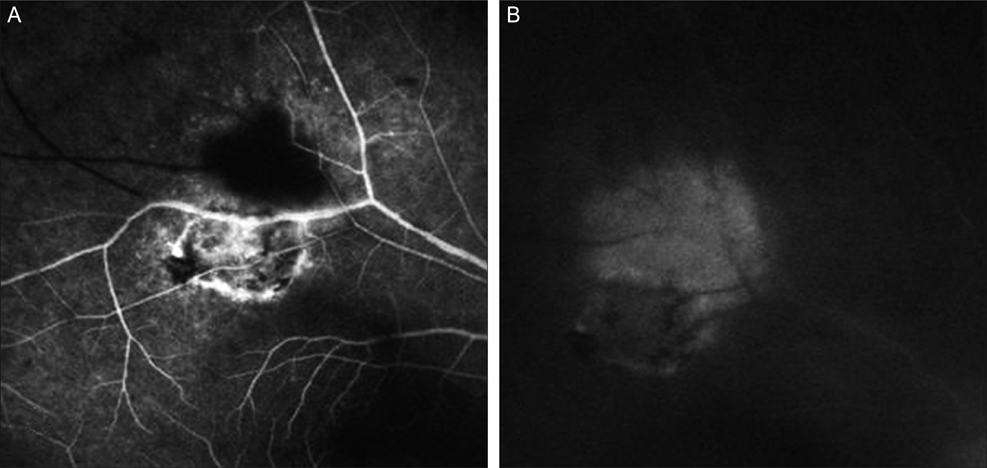
Fig. 1 (A) Hyperfluorescence was observed at the margin of the old lesion and in the new lesion on early phase fluorescein angiography. (B) The new lesion showed hyperfluorescence in the late-phase.

Fig. 2 (A) Fundus photography revealed a whitish-yellow inflammatory lesion near an atrophic, pigmented retinochoroidal scar located in the superotemporal quadrant. (B) Spectral domain optical coherence tomography showed increased reflectivity from the inner retinal layer, retinal thickening and choroidal shadowing. Focal posterior hyaloid thickening and detachment were also observed (arrow). (C) Hyperreflectivity, thinning of the neurosensory retina and a disorganized retinal pigment epithelium layer were observed.
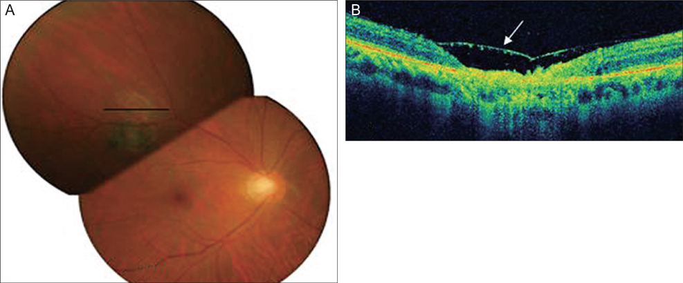
Fig. 3 (A) The lesion resolved and had a scar-like appearance. (B) Spectral domain optical coherence tomography showed thinning of the retina, loss of normal striations, and a disorganized retinal pigment epithelium layer in addition to posterior hyaloid detachment (arrow).
Reference
-
1. McCannel CA, Holland GN, Helm CJ, et al. UCLA Community-Based Uveitis Study Group. Causes of uveitis in the general practice of ophthalmology. Am J Ophthalmol. 1996. 121:35–46.2. Pierce EA, D'amico DJ. Ocular toxoplasmosis: pathogenesis, diagnosis, and management. Semin Ophthalmol. 1993. 8:40–52.3. Smith JR, Cunningham ET Jr. Atypical presentations of ocular toxoplasmosis. Curr Opin Ophthalmol. 2002. 13:387–392.4. Singh M, Chee CK. Spectral domain optical coherence tomography imaging of retinal diseases in Singapore. Ophthalmic Surg Lasers Imaging. 2009. 40:336–341.5. Orefice JL, Costa RA, Orefice F, et al. Vitreoretinal morphology in active ocular toxoplasmosis: a prospective study by optical coherence tomography. Br J Ophthalmol. 2007. 91:773–780.
- Full Text Links
-
- Actions
-
Cited
- CITED
-
- Close
- Share
-
- Similar articles
-
- Characterization of Peripapillary Atrophy Using Spectral Domain Optical Coherence Tomography
- Fundus Autofluorescence, Fluorescein Angiography and Spectral Domain Optical Coherence Tomography Findings of Retinal Astrocytic Hamartomas in Tuberous Sclerosis
- Short-Term Clinical Observation of Acute Retinal Pigment Epitheliitis Using Spectral-Domain Optical Coherence Tomography
- Comparison of the Efficacy between Time and Spectral Domain Optical Coherence Tomography for the Identification of Vitreomacular Interface
- Spectral Domain Optical Coherence Tomography Findings of Butterfly Shaped Pigment Dystrophy
