Human coagulation factor VIII domain-specific recombinant polypeptide expression
- Affiliations
-
- 1Department of Biological Science, College of Natural Sciences, Ajou University, Suwon, Korea. hsunkim@ajou.ac.kr
- 2National Institute of Arthritis and Musculoskeletal and Skin Diseases, National Institutes of Health, Bethesda, MD, USA.
- KMID: 2172774
- DOI: http://doi.org/10.5045/br.2015.50.2.103
Abstract
- BACKGROUND
Hemophilia A is caused by heterogeneous mutations in F8. Coagulation factor VIII (FVIII), the product of F8, is composed of multiple domains designated A1-A2-B-A3-C1-C2. FVIII is known to interact with diverse proteins, and this characteristic may be important for hemostasis. However, little is known about domain-specific functions or their specific binding partners.
METHODS
To determine F8 domain-specific functions during blood coagulation, the FVIII domains A1, A2, A3, and C were cloned from Hep3B hepatocytes. Domain-specific recombinant polypeptides were glutathione S-transferase (GST)- or polyhistidine (His)-tagged, over-expressed in bacteria, and purified by specific affinity chromatography.
RESULTS
Recombinant polypeptides of predicted sizes were obtained. The GST-tagged A2 polypeptide interacted with coagulation factor IX, which is known to bind the A2 domain of activated FVIII.
CONCLUSION
Recombinant, domain-specific polypeptides are useful tools to study the domain-specific functions of FVIII during the coagulation process, and they may be used for production of domain-specific antibodies.
Keyword
MeSH Terms
Figure
-
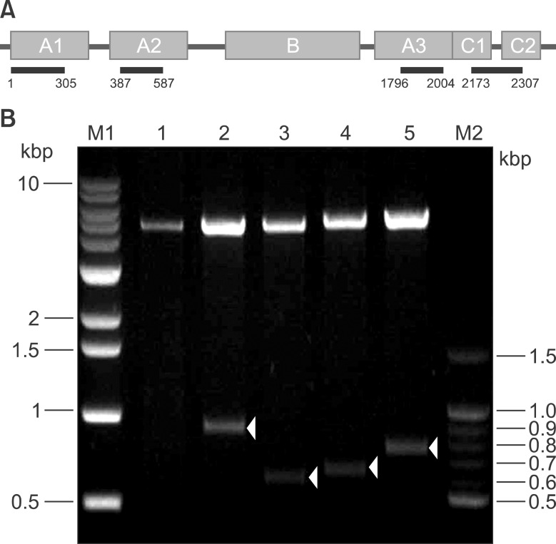
Fig. 1 Cloning of each F8 domain. (A) A1, A2, B, A3, C1, and C2 represent the domains of human FVIII, and the bars indicate the cloning region of each domain. The numbers under each bar indicate the corresponding sequence of amino acids in each domain. (B) pET-28a(+) vectors containing each recombinant F8 domain were digested with BamHI and XhoI. Each domain was subcloned into pGEX4T-2, a GST expression vector. The plasmids were purified, and the products were subjected to BamHI and XhoI digestion. Lane 1, pGEX only; lane 2, pGEX-A1; lane 3, pGEX-A2; lane 4, pGEX-A3; and lane 5, pGEX-C. Arrowheads indicate the products of each domain. M1: 1 kb DNA marker, M2: 100 bp DNA marker.
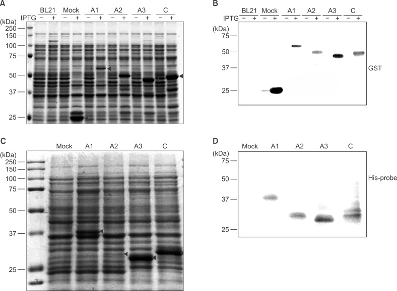
Fig. 2 Expression of recombinant FVIII polypeptides tagged with GST (A, B) or His (C, D). (A) Expression of recombinant FVIII polypeptides was induced by incubation with 0.5 mM IPTG for 4 hr at 25℃. To assess expression, whole-cell lysates were separated in 10% polyacrylamide gels. Gels were stained with Coomassie Brilliant Blue. (B) Proteins in the gel were transferred to a PVDF membrane and immunoblotted with an anti-GST antibody. BL21: host bacterial strain, Mock: pGEX4T-2 vector introduced into BL21. (C) To express His-tagged polypeptides corresponding to FVIII domains, the DNA fragment of each domain was subcloned into pET28a, a His-tagging expression vector. Expression of the His-tagged, domain-specific polypeptides was induced with 1 mM IPTG. (D) His-tagged polypeptides corresponding to FVIII domains were identified by western blotting using an anti-His-tag antibody. Arrowheads in (A) and (C) indicate the corresponding domain polypeptides.
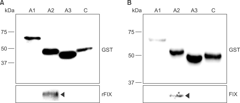
Fig. 3 Interaction of recombinant FVIII domain polypeptides with FIX. To characterize recombinant FVIII domain polypeptides, a pull-down assay was performed with FIX. GST-tagged polypeptides were purified and incubated with either recombinant FIX (A) or standard human plasma (B). The interacting proteins were precipitated by incubation with a 50% slurry of glutathione-agarose beads. The proteins bound to the GST-FVIII complexes were eluted and analyzed by western blotting with anti-GST (upper box) or anti-FIX (lower box) antibodies. Specific lanes: GST pull-down with GST-FVIII-A1 (lane 1), GST-FVIII-A2 (lane 2), GST-FVIII-A3 (lane 3), and GST-FVIII-C (lane 4).
Reference
-
1. Björkman S, Carlsson M, Berntorp E, Stenberg P. Pharmacokinetics of factor VIII in humans. Obtaining clinically relevant data from comparative studies. Clin Pharmacokinet. 1992; 22:385–395. PMID: 1505144.2. Bowen DJ. Haemophilia A and haemophilia B: molecular insights. Mol Pathol. 2002; 55:127–144. PMID: 11950963.
Article3. Pipe SW, Kaufman RJ. Factor VIII C2 domain missense mutations exhibit defective trafficking of biologically functional proteins. J Biol Chem. 1996; 271:25671–25676. PMID: 8810344.
Article4. Fang H, Wang L, Wang H. The protein structure and effect of factor VIII. Thromb Res. 2007; 119:1–13. PMID: 16487577.
Article5. Lenting PJ, van Mourik JA, Mertens K. The life cycle of coagulation factor VIII in view of its structure and function. Blood. 1998; 92:3983–3996. PMID: 9834200.
Article6. De Brasi CD, Slavutsky IR, Larripa IB. Molecular genetics of hemophilia A. Medicina (B Aires). 1996; 56:509–517. PMID: 9239887.7. Miners AH, Sabin CA, Tolley KH, Lee CA. The changing patterns of factor VIII (FVIII) and factor IX (FIX) clotting factor usage in a comprehensive care centre between 1980 and 1994. Haemophilia. 1998; 4:4–9. PMID: 9873858.
Article8. van't Veer C, Hackeng TM, Delahaye C, Sixma JJ, Bouma BN. Activated factor X and thrombin formation triggered by tissue factor on endothelial cell matrix in a flow model: effect of the tissue factor pathway inhibitor. Blood. 1994; 84:1132–1142. PMID: 8049429.9. Pipe SW. Functional roles of the factor VIII B domain. Haemophilia. 2009; 15:1187–1196. PMID: 19473417.
Article10. Pipe SW, Morris JA, Shah J, Kaufman RJ. Differential interaction of coagulation factor VIII and factor V with protein chaperones calnexin and calreticulin. J Biol Chem. 1998; 273:8537–8544. PMID: 9525969.
Article11. Hwang SH, Kim MJ, Lim JA, Kim HC, Kim HS. Profiling of factor VIII mutations in Korean haemophilia A. Haemophilia. 2009; 15:1311–1317. PMID: 19719548.12. Seo JY, Jang MA, Kim HJ, Lee KO, Kim SH, Kim HJ. Sequence variation data of F8 and F9 genes in functionally validated control individuals: implications on the molecular diagnosis of hemophilia. Blood Res. 2013; 48:206–210. PMID: 24086941.13. Hwang SH, Lim JA, Kim MJ, et al. Profiling of differentially expressed genes in haemophilia A with inhibitor. Haemophilia. 2012; 18:e247–e253. PMID: 22176207.
Article14. Lollar P, Parker CG. Subunit structure of thrombin-activated porcine factor VIII. Biochemistry. 1989; 28:666–674. PMID: 2496750.
Article15. Davidson CJ, Hirt RP, Lal K, et al. Molecular evolution of the vertebrate blood coagulation network. Thromb Haemost. 2003; 89:420–428. PMID: 12624623.
Article16. Hoffman M, Monroe DM 3rd, Roberts HR. Activated factor VII activates factors IX and X on the surface of activated platelets: thoughts on the mechanism of action of high-dose activated factor VII. Blood Coagul Fibrinolysis. 1998; 9(Suppl 1):S61–S65. PMID: 9819030.17. Kappelmayer J, Bernabei A, Edmunds LH Jr, Edgington TS, Colman RW. Tissue factor is expressed on monocytes during simulated extracorporeal circulation. Circ Res. 1993; 72:1075–1081. PMID: 8097439.
Article
- Full Text Links
-
- Actions
-
Cited
- CITED
-
- Close
- Share
-
- Similar articles
-
- A Case of Hemophilia A Diagnosed in a Premature Infant
- Synthesis of recombinant blood coagulation factor VIII (FVIII) heavy and light chains and reconstitution of active form of FVIII
- Recurrent Cerebral Venous Thrombosis Associated with Elevated Factor VIII
- Spontaneous Retroperitoneal Hemorrhage Caused by Idiopathic Acquired Hemophilia A Misdiagnosed as a Delayed Traumatic Hematoma: A Case Report
- Post-operative Bleeding due to Acquired Hemophilia Successfully Treated with Recombinant Factor VIIa: Case Report
