Korean J Ophthalmol.
2014 Feb;28(1):83-85. 10.3341/kjo.2014.28.1.83.
A Korean Patient with Lattice Corneal Dystrophy Type IV with Leu527Arg Mutation in the TGFBI Gene
- Affiliations
-
- 1Institute of Vision Research, Department of Ophthalmology, Yonsei University College of Medicine, Seoul, Korea. shadik@yuhs.ac
- 2Department of Clinical Diagnosis, Yonsei University College of Medicine, Seoul, Korea.
- 3Corneal Dystrophy Research Institute, Yonsei University, Seoul, Korea.
- KMID: 1792097
- DOI: http://doi.org/10.3341/kjo.2014.28.1.83
Abstract
- An 87-year-old woman visited our clinic for a scheduled cataract surgery. At the time of preoperative evaluation, slit lamp examination showed lattice-shaped and granular deposits with asymmetrical patterns in the stroma of both corneas. Genomic DNA samples of the patient, amplified by polymerase chain reaction, showed a single nucleotide substitution, c. 1580T>G (p.L527R), in the transforming growth factor-beta-induced TGFBI gene. We also found two additional SNP mutations, c.1620T>C (p.F540F) and c.1678+23G>A, along with the well-known L527R mutation. This is the first report of lattice corneal dystrophy type IV with an L527R mutation outside of Japan, and could challenge the idea that L527R is caused by a mutation from a single Japanese ancestor.
MeSH Terms
-
Aged, 80 and over
Corneal Dystrophies, Hereditary/diagnosis/*genetics/metabolism
DNA/*genetics
DNA Mutational Analysis
Extracellular Matrix Proteins/*genetics/metabolism
Female
Humans
*Mutation
Pedigree
Polymerase Chain Reaction
Transforming Growth Factor beta/*genetics/metabolism
DNA
Extracellular Matrix Proteins
Transforming Growth Factor beta
Figure
-

Fig. 1 Corneal photographs of lattice corneal dystrophy type IV patients. (A) The right eye was the more severely affected eye, displaying nodulolinear amyloid deposits (arrow). (B) The deposits are mainly located in anterior stroma (arrowhead). (C) The left eye showed less linear and macular opacity than the right.
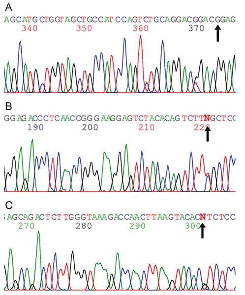
Fig. 2 Open-frame sequencing of the TGFBI gene from the patient. Direct sequencing of polymerase chain reaction products corresponding to exon 12 of TGFBI. (A) Heterozygous mutation, c.1580T>G (arrow) was detected in the TGFBI gene. (B) Heterozygous c.1620T>C p.Phe540Phe (rs4669) was detected (arrow). (C) Heterozygous c.1678+23G>A (rs2072239) was detected (arrow).
Reference
-
1. Munier FL, Korvatska E, Djemai A, et al. Kerato-epithelin mutations in four 5q31-linked corneal dystrophies. Nat Genet. 1997; 15:247–251.2. Munier FL, Frueh BE, Othenin-Girard P, et al. BIGH3 mutation spectrum in corneal dystrophies. Invest Ophthalmol Vis Sci. 2002; 43:949–954.3. Chakravarthi SV, Kannabiran C, Sridhar MS, Vemuganti GK. TGFBI gene mutations causing lattice and granular corneal dystrophies in Indian patients. Invest Ophthalmol Vis Sci. 2005; 46:121–125.4. Fujiki K, Hotta Y, Nakayasu K, et al. A new L527R mutation of the betaIGH3 gene in patients with lattice corneal dystrophy with deep stromal opacities. Hum Genet. 1998; 103:286–289.5. Fukuoka H, Kawasaki S, Yamasaki K, et al. Lattice corneal dystrophy type IV (p.Leu527Arg) is caused by a founder mutation of the TGFBI gene in a single Japanese ancestor. Invest Ophthalmol Vis Sci. 2010; 51:4523–4530.
- Full Text Links
-
- Actions
-
Cited
- CITED
-
- Close
- Share
-
- Similar articles
-
- A Case of Lattice Corneal Dystrophy Type 1 Initially Showing Phenotypic Characteristics of Granular Corneal Dystrophy Type 2 in One Eye and Dot and Map Lesions in the Contralateral Eye
- Mutation Analysis of the TGFBI Gene in Consecutive Korean Patients With Corneal Dystrophies
- A Case of Corneal Transplantation for Type III Lattice Corneal Dystrophy with Aphakia
- Lattice Corneal Dystrophy, Gelsolin Type: The First Case Report in Korea
- Phenotypes of Granular Corneal Dystrophy Type 2 among Koreans in Their Twenties
