The Changes of Expression of Survivin by Butyrate in HCT116 Colon Cancer Cells
- Affiliations
-
- 1Department of Surgery, School of Medicine, Dankook University, Cheonan, Korea. dkpark@dankook.ac.kr
- KMID: 1750697
- DOI: http://doi.org/10.4174/jkss.2009.77.5.297
Abstract
- PURPOSE
The causes of colon cancer can be divided into genetic and environmental components. A high-fiber diet is known to reduce the risk of colon cancer. Dietary fiber is converted to short chain fatty acid, butyrate, in the colon by bacteria. Butyrate is used as an energy source for the colonic epithelial cells, and is known to induce apoptosis in colon cancer cell lines. Survivin, a recently discovered member of the IAP (inhibitor of apoptosis) family, is known to suppress apoptosis. Not only does it suppress cell apoptosis, but it also has a protective effect from disabling G2/M phase of the cell cycle by attaching to the microtubule of the mitotic spindle. The purpose of this study is to evaluate the effect of butyrate on the expression of survivin, in HCT116 colon cancer cell lines.
METHODS
Cytotoxicity of butyrate was measured by MTS method. Cell cycle phase and apoptosis was analyzed by flowcytometry. Protein expression of survivin was evaluated by Western blot analysis, and the mRNA expression by RT-PCR.
RESULTS
Butyrate can induce apoptosis in HCT116 colon cancer cell line at a concentration of 6 mM. Butyrate suppressed the expression of survivin mRNA and also the expression of cytosolic and nuclear survivin. In flowcytometric analysis, the apoptotic portion was increased and the proportions of S and M phase were decreased when cultured with butyrate.
CONCLUSION
We concluded that butyrate could induce cellular apoptosis partially by suppressing the expression of survivin in HCT116 colon cancer cells.
Keyword
MeSH Terms
Figure
-

Fig. 1 Cytotoxicity to HCT116 colon cancer cell by butyrate. HCT116 cells were incubated with different concentrations of butyrate for 72 hours. Cytotoxicity was measured by MTS method.
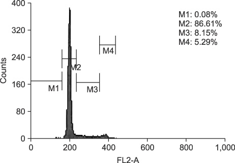
Fig. 2 G1/S phase synchronization was induced by serum withdrawal for 3 days. The cell cycle phases were evaluated by flowcytometry. M1 = subG1 phase; M2 = G0/G1 phase; M3 = S phase fraction; M4 = M phase.

Fig. 3 DNA fragmentation by butyrate. The HCT116 cells were treated with or without butyrate. DNA fragmentation was induced only in 6 mM butyrate treated cells, which indicates cellular apoptosis. M = DNA marker; 1 = without butyrate, 0 hr; 2 = without butyrate, 72 hr; 3 = with 6 mM butyrate, 0 hr; 4 = with 6 mM butyrate, 72 hr incubation.

Fig. 4 Expression of survivin, Bcl-2, and Bax proteins. HCT116 cells were incubated with 6 mM butyrate for 72 hr. The expression of survivin, Bcl-2, and Bax protein was evaluated by Western blotting. Immunoblot analysis revealed that survivin completely lost its expression by butyrate, but on the contrary, Bcl-2 expression was increased by butyrate.
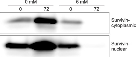
Fig. 5 Expression of cytoplasmic and nuclear survivin protein after 72 hour incubation with or without butyrate (6 mM). Butyrate can inhibit both cytoplasmic and nuclear survivin expression.
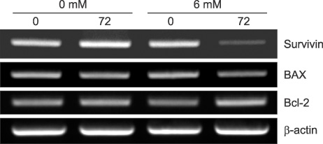
Fig. 6 Expression of apoptosis-related gene mRNA induced by butyrate. There was no difference in expression of Bcl-2, Bax mRNA transcripts by butyrate. But survivin mRNA transcript was reduced by butyrate, indicating that the absent expression of survivin protein by butyrate is due to suppression of mRNA transcription.

Fig. 7 Cell cycle analysis with flowcytometry. HCT116 cells were incubated for 1, 2, and 3 days with 6 mM butyrate. (A) 0 hr, without butyrate, (B) incubated for 1 day without butyrate, (C) incubated for 1 day with 6 mM of butyrate, (D) incubated for 2 day without butyrate, (E) incubated for 2 day with 6 mM of butyrate, (F) incubated for 3 day without butyrate, (G) incubated for 3 day with 6 mM of butyrate.

Fig. 8 Apoptosis analysis with flowcytometry. HCT116 cells were incubated for 1, 2, and 3 days with 6 mM butyrate. (A) incubated for 1 day without butyrate, (B) incubated for 1 day with 6 mM of butyrate, (C) incubated for 2 days without butyrate, (D) incubated for 2 days with 6 mM of butyrate, (E) incubated for 3 days without butyrate, (F) incubated for 3 days with 6 mM of butyrate. The apoptotic proportion of cell cycle was greatly increased by butyrate (from 3.13% to 47.86% on 2nd day of incubation, from 0.70% to 18.8% on 3rd day of incubation with butyrate).
Reference
-
1. Hague A, Manning AM, Hanlon KA, Huschtscha LI, Hart D, Paraskeva C. Sodium butyrate induces apoptosis in human colonic tumour cell lines in a p53-independent pathway: implications for the possible role of dietary fibre in the prevention of large-bowel cancer. Int J Cancer. 1993. 55:498–505.2. Bonnotte B, Favre N, Reveneau S, Micheau O, Droin N, Garrido C, et al. Cancer cell sensitization to fas-mediated apoptosis by sodium butyrate. Cell Death Differ. 1998. 5:480–487.3. Giardina C, Boulares H, Inan MS. NSAIDs and butyrate sensitize a human colorectal cancer cell line to TNF-alpha and Fas ligation: the role of reactive oxygen species. Biochim Biophys Acta. 1999. 1448:425–438.4. McMillan L, Butcher SK, Pongracz J, Lord JM. Opposing effects of butyrate and bile acids on apoptosis of human colon adenoma cells: differential activation of PKC and MAP kinases. Br J Cancer. 2003. 88:748–753.5. Sandler RS, Lyles CM, Peipins LA, McAuliffe CA, Woosley JT, Kupper LL. Diet and risk of colorectal adenomas: macronutrients, cholesterol, and fiber. J Natl Cancer Inst. 1993. 85:884–891.6. Radley S, lmray CHE, Davis A, Hendrickse CW, Donovan IA, Lawson AM, et al. Duodenal bile acid profiles in patients with colorectal cancer or polyps. Eur J Gastroenterol Hepatol. 1993. 5:721–730.7. Bayerdorffer E, Mannes GA, Richter WO, Ochsenkuhn T, Wiebecke B, Kopcke W, et al. Increased serum deoxycholic acid levels in men with colorectal adenomas. Gastroenterology. 1993. 104:145–151.8. Mahmoud NN, Dannenberg AJ, Bilinski RT, Mestre JR, Chadburn A, Churchill M, et al. Administration of an unconjugated bile acid increases duodenal tumors in a murine model of familial adenomatous polyposis. Carcinogenesis. 1999. 20:299–303.9. Narisawa T, Magadia NE, Weisburger JH, Wynder EL. Promoting effect of bile acids on colon carcinogenesis after intrarectal instillation of N-methyl-N-nitro-N'-nitrosoguanidine in rats. J Natl Cancer Inst. 1974. 53:1093–1097.10. Reddy BS, Wynder EL. Large-bowel carcinogenesis: fecal constituents of populations with diverse incidence rates of colon cancer. J Natl Cancer Inst. 1973. 50:1437–1442.11. Sutherland LAM, Bird RP. The effect of chenodeoxycholic acid on the development of aberrant crypt foci in the rat colon. Cancer Lett. 1994. 76:101–107.12. DeRubertis FR, Craven PA, Saito R. Bile salt stimulation of colonic epithelial proliferation. Evidence for involvement of lipoxygenase products. J Clin Invest. 1984. 74:1614–1624.13. Martinez-Diez MC, Serrano MA, Monte MJ, Marin JJ. Comparison of the effects of bile acids on cell viability and DNA synthesis by rat hepatocytes in primary culture. Biochim Biophys Acta. 2000. 1500:153–160.14. Cummings JH. Short chain fatty acids in the human colon. Gut. 1981. 22:763–779.15. Kruh J. Effects of sodium butyrate, a new pharmacological agent, on cells in culture. Mol Cell Biochem. 1982. 42:65–82.16. Tsao D, Shi ZR, Wong A, Kim YS. Effect of sodium butyrate on carcinoembryonic antigen production by human colonic adenocarcinoma cells in culture. Cancer Res. 1983. 43:1217–1222.17. Boffa LC, Lupton JR, Mariani MR, Ceppi M, Newmark HL, Scalmati A, et al. Modulation of colonic epithelial cell proliferation, histone acetylation, and luminal short chain fatty acids by variation of dietary fiber (wheat bran) in rats. Cancer Res. 1992. 52:5906–5912.18. Barnard JA, Warwick G. Butyrate rapidly induces growth inhibition and differentiation in HT-29 cells. Cell Growth Differ. 1993. 4:495–501.19. Berry RD, Paraskeva C. Expression of carcinoembryonic antigen by adenoma and carcinoma derived epithelial cell lines: possible marker of tumour progression and modulation of expression by sodium butyrate. Carcinogenesis. 1988. 9:447–450.20. Rothe M, Pan MG, Henzel WJ, Ayres TM, Goeddel DV. The TNFR2-TRAF signaling complex contains two novel proteins related to baculoviral inhibitor of apoptosis proteins. Cell. 1995. 83:1243–1252.21. Ambrosini G, Adida C, Altieri DC. A novel anti-apoptosis gene, survivin, expressed in cancer and lymphoma. Nat Med. 1997. 3:917–921.22. Gianani R, Jarboe E, Orlicky D, Frost M, Bobak J, Lehner R, et al. Expression of survivin in normal, hyperplastic, and neoplastic colonic mucosa. Hum Pathol. 2001. 32:119–125.23. Chantalat L, Skoufias DA, Kleman JP, Jung B, Dideberg O, Margolis RL. Crystal structure of human survivin reveals a bow tie-shaped dimer with two unusual alpha-helical extensions. Mol Cell. 2000. 6:183–189.24. Li F, Altieri DC. Transcriptional analysis of human survivin gene expression. Biochem J. 1999. 344:305–311.25. Swana HS, Grossman D, Anthony JN, Weiss RM, Altieri DC. Tumor content of the antiapoptosis molecule survivin and recurrence of bladder cancer. N Engl J Med. 1999. 341:452–453.26. Lu CD, Altieri DC, Tanigawa N. Expression of a novel antiapoptosis gene, survivin, correlated with tumor cell apoptosis and p53 accumulation in gastric carcinomas. Cancer Res. 1998. 58:1808–1812.27. Okada E, Murai Y, Matsui K, Isizawa S, Cheng C, Masuda M, et al. Survivin expression in tumor cell nuclei is predictive of a favorable prognosis in gastric cancer patients. Cancer Lett. 2001. 163:109–116.28. Yamamoto T, Manome Y, Nakamura M, Tanigawa N. Downregulation of survivin expression by induction of the effector cell protease receptor-1 reduces tumor growth potential and results in an increased sensitivity to anticancer agents in human colon cancer. Eur J Cancer. 2002. 38:2316–2324.29. Miller AA, Kurschel E, Osieka R, Schmidt CG. Clinical pharmacology of sodium butyrate in patients with acute leukemia. Eur J Cancer Clin Oncol. 1987. 23:1283–1287.
- Full Text Links
-
- Actions
-
Cited
- CITED
-
- Close
- Share
-
- Similar articles
-
- Change in Expression of Survivin Caused by Using Oxaliplatin in HCT116 Colon Cancer Cells
- YM155, specific survivin inhibitor, can enhance artesunate-induced cytotoxicity in HCT116 colon cancer cells
- Antichemosensitizing effect of resveratrol in cotreatment with oxaliplatin in HCT116 colon cancer cell
- Effect of Deoxycholic Acid on the Proliferation and Apoptosis of HCT116 Colon Cancer Cells
- Cucurbitacin E’s Anti-Cancer Effects on HCT116 Human Colon Cancer Cells by Controlling Expression and Phosphorylation Levels of Caspase-9, eIF-2α, and ATF-4
