MR Diagnosis of a Pulmonary Embolism: Comparison of P792 and Gd-DOTA for First-Pass Perfusion MRI and Contrast-Enhanced 3D MRA in a Rabbit Model
- Affiliations
-
- 1Department of Biomedical Engineering, Emory University and Georgia Institute of Technology, Atlanta, GA, USA.
- 2Department of Radiology, University of Virginia Health System, Charlottesville, VA, USA. kdh2n@virginia.edu
- 3Department of Radiology, Gulhane Military Medical Academy, Ankara, Turkey.
- 4Guerbet Research, Aulnay-sous-Bois, France.
- 5Department of Radiology, Kagawa University Faculty of Medicine, Kagawa, Japan.
- 6Department of Diagnostic Radiology, Osaka Neurosurgical Hospital, Kagawa, Japan.
- KMID: 1093959
- DOI: http://doi.org/10.3348/kjr.2009.10.5.447
Abstract
OBJECTIVE
To compare P792 (gadomelitol, a rapid clearance blood pool MR contrast agent) with gadolinium-tetraazacyclododecanetetraacetic acid (Gd-DOTA), a standard extracellular agent, for their suitability to diagnose a pulmonary embolism (PE) during a first-pass perfusion MRI and 3D contrast-enhanced (CE) MR angiography (MRA). MATERIALS AND METHODS: A perfusion MRI or CE-MRA was performed in a rabbit PE model following the intravenous injection of a single dose of contrast agent. The time course of the pulmonary vascular and parenchymal enhancement was assessed by measuring the signal in the aorta, pulmonary artery, and lung parenchyma as a function of time to determine whether there is a significant difference between the techniques. CE-MRA studies were evaluated by their ability to depict the pulmonary vasculature and following defects between 3 seconds and 15 minutes after a triple dose intravenous injection of the contrast agents. RESULTS: The P792 and Gd-DOTA were equivalent in their ability to demonstrate PE as perfusion defects on first pass imaging. The signal from P792 was significantly higher in vasculature than that from Gd-DOTA between the first and the tenth minutes after injection. The results suggest that a CE-MRA PE could be reliably diagnosed up to 15 minutes after injection. CONCLUSION: P792 is superior to Gd-DOTA for the MR diagnosis of PE.
Keyword
MeSH Terms
-
Animals
Contrast Media/administration & dosage
Heterocyclic Compounds/administration & dosage/*diagnostic use
Imaging, Three-Dimensional
Injections, Intravenous
Magnetic Resonance Angiography/*methods
Magnetic Resonance Imaging/*methods
Organometallic Compounds/administration & dosage/*diagnostic use
Pulmonary Embolism/*diagnosis
Rabbits
Figure
-
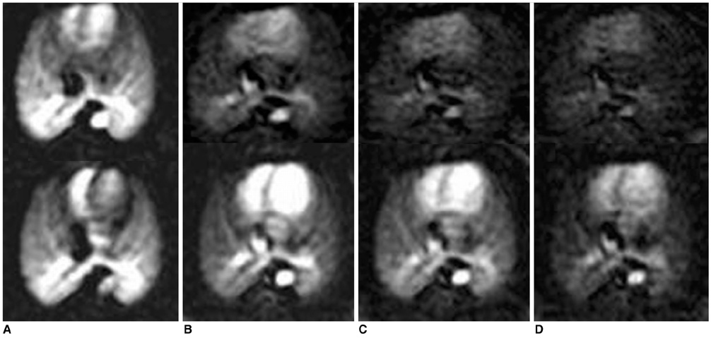
Fig. 1 Gd-DOTA (0.1 mmol·kg-1 bw) (top row) and P792-enhanced (0.013 mmol·kg-1 bw) (bottom row) images of normal rabbit lungs at time of peak contrast (A), and 1 minute (B), 5 minutes (C) and 10 minutes (D) after injection.
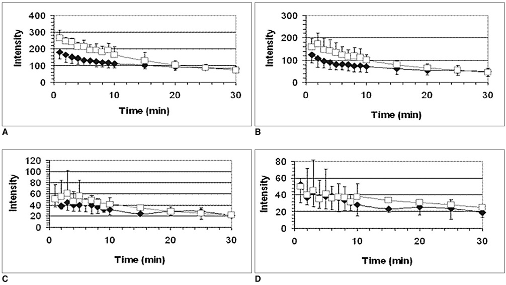
Fig. 2 Average washout curves for Gd-DOTA (diamonds) and P792 (squares) in aorta (A), pulmonary artery (B), right lung (C) and left lung (D). (Dose: Gd-DOTA 0.1 mmol·kg-1 bw, P792 0.013 mmol·kg-1 bw.) Data points for left and right lungs were obtained in rabbits without pulmonary artery occlusion.
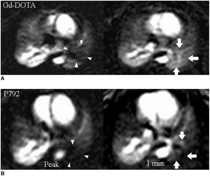
Fig. 3 Gd-DOTA (top row) and P792-enhanced (bottom row) perfusion images of rabbit with obstructed pulmonary artery at time of peak contrast (A) and one minute (B) after injection of contrast agent. Note that fillin of occluded lung occurs for both agents at one minute, likely by systemic collaterals. (Dose: Gd-DOTA 0.1 mmol·kg-1 bw, P792 0.013 mmol·kg-1 bw.)
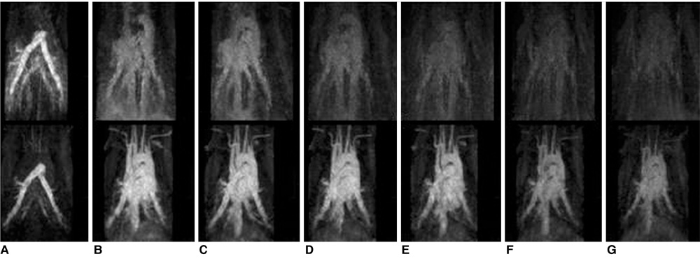
Fig. 4 Gd-DOTA (top row) and P792-enhanced (bottom row) images of two different rabbits, acquired at 3 seconds (A), 30 seconds (B), 1 (C), 3 (D), 5 (E), 10 (F) and 15 minutes (G) after contrast injection. (Dose: Gd-DOTA 0.3 mmol·kg-1 bw, P792 0.039 mmol·kg-1 bw.) All images are filmed at identical window settings.
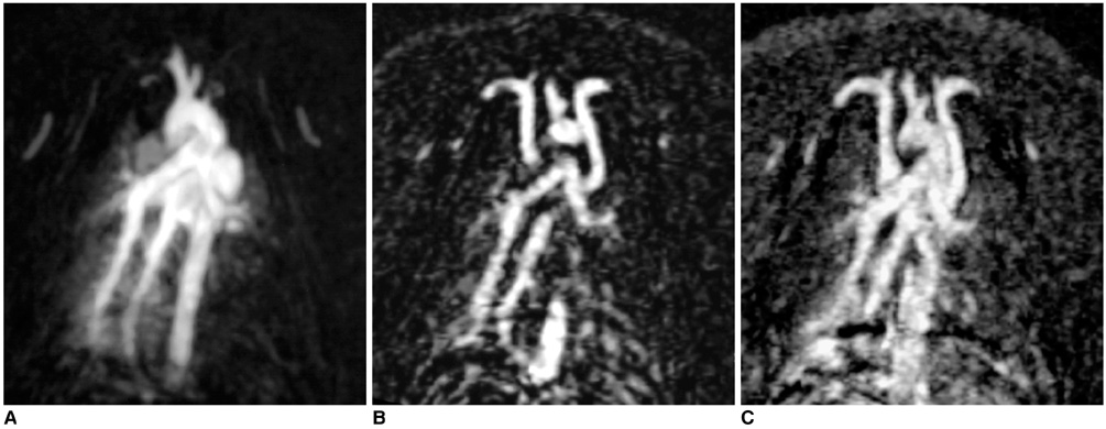
Fig. 5 P792-enhanced (0.013 mmol·kg-1 bw) images of rabbit during balloon occlusion of left pulmonary artery at time of first pass (A), as well as 5 minutes (B) and 15 minutes (C) after injection.
Reference
-
1. Carson JL, Kelley MA, Duff A, Weg JG, Fulkerson WJ, Palevsky HI, et al. The clinical course of pulmonary embolism. N Engl J Med. 1992. 326:1240–1245.2. Goldhaber SZ. Pulmonary embolism. N Engl J Med. 1998. 339:93–104.3. Barritt DW, Jordan SC. Anticoagulant drugs in the treatment of pulmonary embolism. A controlled trial. Lancet. 1960. 1:1309–1312.4. The PIOPED Investigators. Value of the ventilation/perfusion scan in acute pulmonary embolism: result of the Prospective Investigation of Pulmonary Embolism Diagnosis (PIOPED). JAMA. 1990. 263:2753–2759.5. Meaney JF, Weg JG, Chenevert TL, Stafford-Johnson D, Hamilton BH, Prince MR. Diagnosis of pulmonary embolism with magnetic resonance angiography. N Engl J Med. 1997. 336:1422–1427.6. Oudkerk M, van Beek EJ, Wielopolski P, van Ooijen PM, Brouwers-Kyuper EM, Bongaerts AH, et al. Comparison of contrast-enhanced magnetic resonance angiography and conventional pulmonary angiography for the diagnosis of pulmonary embolism: a prospective study. Lancet. 2002. 359:1643–1647.7. Pruessmann KP, Weiger M, Scheidegger MB, Boesiger P. SENSE: sensitivity encoding for fast MRI. Magn Reson Med. 1999. 42:952–962.8. Griswold MA, Jakob PM, Heidemann RM, Nittka M, Jellus V, Wang J, et al. Generalized autocalibrating partially parallel acquisitions (GRAPPA). Magn Reson Med. 2002. 47:1202–1210.9. Ohno Y, Higashino T, Takenaka D, Sugimoto K, Yoshikawa T, Kawai H, et al. MR angiography with sensitivity encoding (SENSE) for suspected pulmonary embolism: comparison with MDCT and ventilation-perfusion scintigraphy. AJR Am J Roentgenol. 2004. 183:91–98.10. Hatabu H, Tadamura E, Levin DL, Chen Q, Li W, Kim D, et al. Quantitative assessment of pulmonary perfusion with dynamic contrast-enhanced MRI. Magn Reson Med. 1999. 42:1033–1038.11. Fink C, Ley S, Puderbach M, Plathow C, Bock M, Kauczor HU. 3D pulmonary perfusion MRI and MR angiography of pulmonary embolism in pigs after a single injection of a blood pool MR contrast agent. Eur Radiol. 2004. 14:1291–1296.12. Port M, Corot C, Rousseaux O, Raynal I, Devoldere L, Idee JM, et al. P792: a rapid clearance blood pool agent for magnetic resonance imaging: preliminary results. MAGMA. 2001. 12:121–127.13. Port M, Corot C, Raynal I, Idee JM, Dencausse A, Lancelot E, et al. Physicochemical and biological evaluation of P792, a rapid clearance blood pool agent for magnetic resonance imaging. Invest Radiol. 2001. 36:445–454.14. Keilholz SD, Mai VM, Berr SS, Fujiwara N, Hagspiel KD. Comparison of first-pass Gd-DOTA and FAIRER MR perfusion imaging in a rabbit model of pulmonary embolism. J Magn Reson Imaging. 2002. 16:168–171.15. Corot C, Violas X, Robert P, Port M. Pharmacokinetics of three gadolinium chelates with different molecular sizes shortly after intravenous injection in rabbits: relevance to MR angiography. Invest Radiol. 2000. 35:213–218.16. Corot C, Violas X, Robert P, Gagneur G, Port M. Comparison of different types of blood pool agents (P792, MS325, USPIO) in a rabbit MR angiography-like protocol. Invest Radiol. 2003. 38:311–319.17. Kluge A, Muller C, Hansel J, Gerriets T, Bachmann G. Real-time MR with TrueFISP for the detection of acute pulmonary embolism: initial clinical experience. Eur Radiol. 2004. 14:709–718.18. Goyen M, Laub G, Ladd ME, Debatin JF, Barkhausen J, Truemmler KH, et al. Dynamic 3D MR angiography of the pulmonary arteries in under four seconds. J Magn Reson Imaging. 2001. 13:372–377.19. Dirksen MS, Lamb HJ, Kunz P, Robert P, Corot C, de Roos A. Improved MR coronary angiography with use of a new rapid clearance blood pool contrast agent in pigs. Radiology. 2003. 227:802–808.
- Full Text Links
-
- Actions
-
Cited
- CITED
-
- Close
- Share
-
- Similar articles
-
- Dynamic Contrast-Enhanced MRI Using a Macromolecular MR Contrast Agent (P792): Evaluation of Antivascular Drug Effect in a Rabbit VX2 Liver Tumor Model
- Contrast-enhanced Magnetic Resonance Imaging of Brain Metastases at 7.0T versus 1.5T: A Preliminary Result
- Strong Contrast Stagnation of Unilateral Vertebral Artery on Three-Dimensional Black Blood-Enhanced MRI Predicts Acute Medulla Infarction
- 3D Whole-Heart Coronary MR Angiography at 1.5T in Healthy Volunteers: Comparison between Unenhanced SSFP and Gd-Enhanced FLASH Sequences
- Comparison of Gadomer-17 and Gd-DTPA in image quality of contrast-enhanced MR angiographies using flow phantom model
