Culturing at Low Cell Density Delays Cellular Senescence of Human Bone Marrow-Derived Mesenchymal Stem Cells in Long-Term Cultures
- Affiliations
-
- 1SCM Lifesciences Co. Ltd., Incheon, Korea
- 2Department of Biomedical Sciences, Inha University College of Medicine, Incheon, Korea
- KMID: 2513087
- DOI: http://doi.org/10.15283/ijsc20078
Abstract
- Background and Objectives
Mesenchymal stem cells (MSCs) have immense therapeutic potential for treating intractable and immune diseases. They also have applications in regenerative medicine in which distinct treatments do not exist. Thus, MSCs are gaining attention as important raw materials in the field of cell therapy. Importantly, the number of MSCs in the bone marrow is limited and they are present only in small quantities. Therefore, mass production of MSCs through long-term culture is necessary to use them in cell therapy. However, MSCs undergo cellular senescence through repeated passages during mass production. In this study, we explored methods to prolong the limited lifetime of MSCs by culturing them with different seeding densities.
Methods and Results
We observed that in long-term cultures, low-density (LD, 50 cells/cm2) MSCs showed higher population doubling level, leading to greater fold increase, than high-density (HD, 4,000 cells/cm2) MSCs. LD-MSCs suppressed the expression of aging-related genes. We also showed that reactive oxygen species (ROS) were decreased in LD-MSCs compared to that in HD-MSCs. Further, proliferation potential increased when ROS were inhibited in HD-MSCs.
Conclusions
The results in this study suggest that MSC senescence can be delayed and that life span can be extended by controlling cell density in vitro. These results can be used as important data for the mass production of stem cell therapeutic products.
Keyword
Figure
-
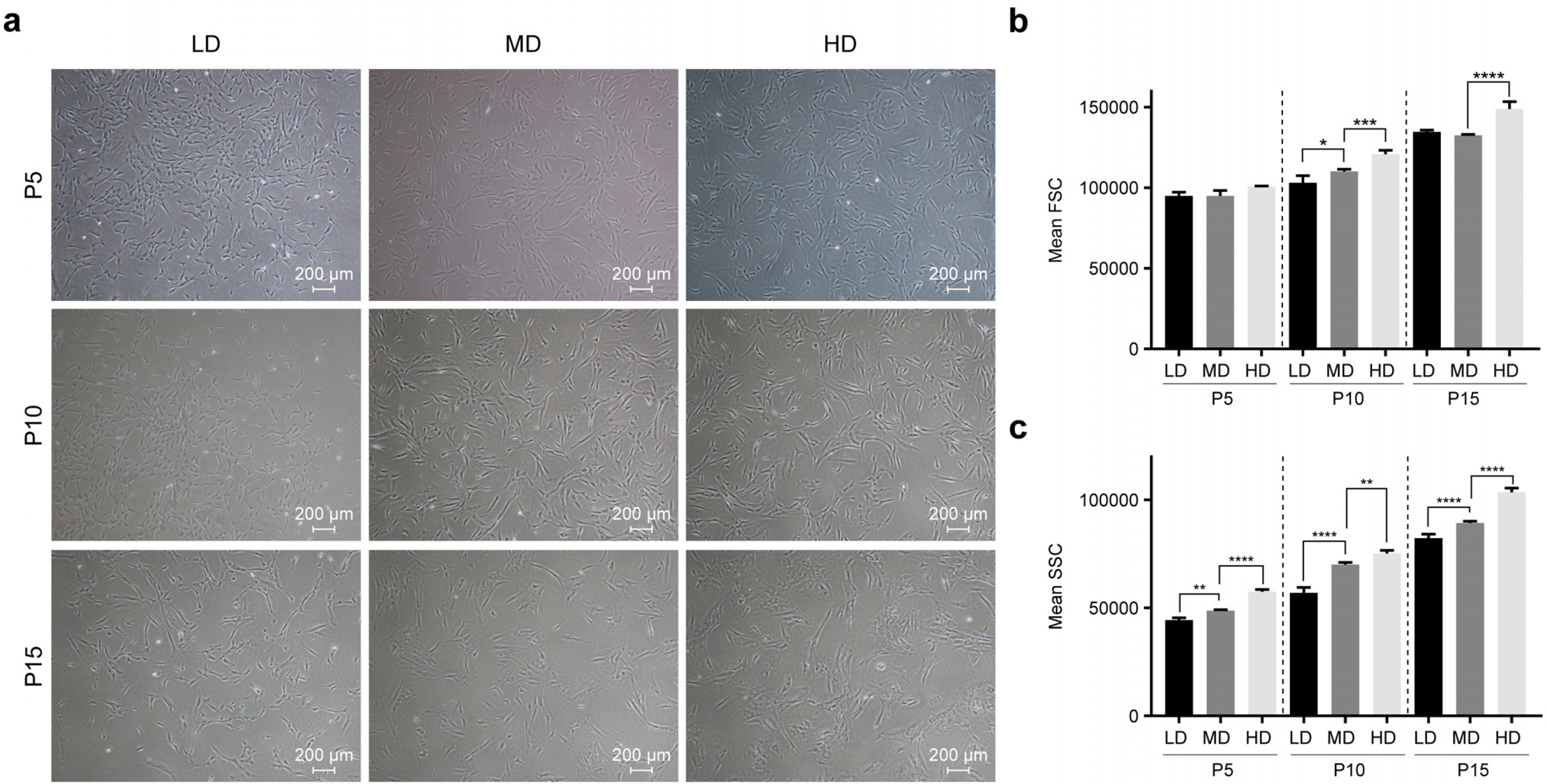
Fig. 1 Cell culturing density-dependent morphological changes in MSCs in long-term cultures. (a) At passages 5, 10, and 15, morphological changes were observed in MSCs under three different cell densities. Scale bar: 200 μm. LD=50 cells/cm2, MD=1000 cells/cm2, and HD=4000 cells/cm2. (b, c) Cell size and granularity were analyzed using flow cytometry. FSC=forward scatter and SSC=side scatter. Significance is *p<0.05, **p<0.01, ***p<0.001, ****p<0.0001.
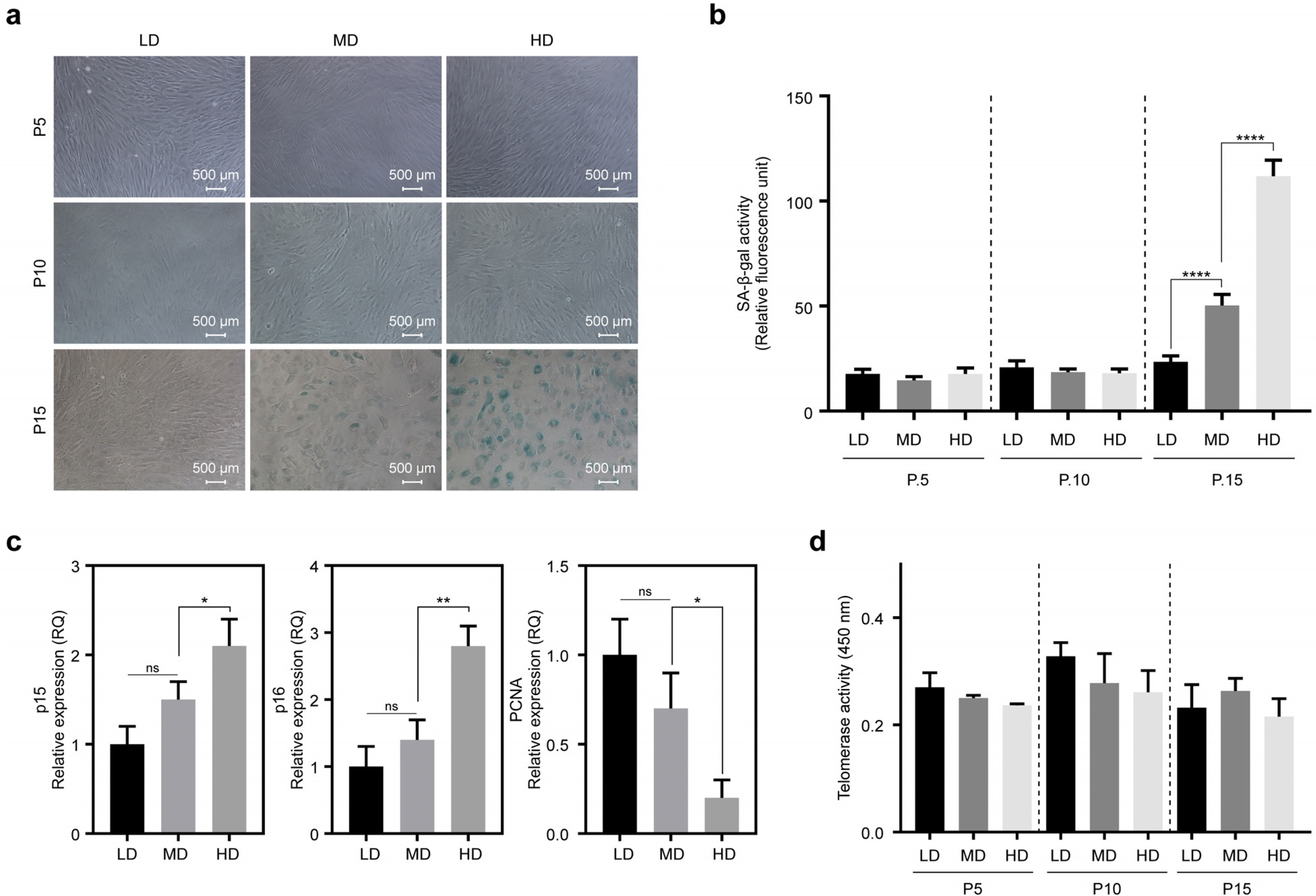
Fig. 2 Cell density-dependent changes in senescence of MSCs in long-term cultures. (a) At passages 5, 10, and 15, senescence changes were observed in LD, MD, and HD using β-galactosidase staining. Scale bar: 500 μm. (b) Quantification of SA-β-gal activity in LD, MD, and HD. Senescence change was measured using the quantitative cellular senescence assay kit and expressed as relative fluorescence unit (RFU). (c) At passage 15, transcriptional changes in p15 and p16 genes, which are associated with senescence, and proliferating cell nuclear antigen (PCNA), a marker for cell proliferation, were analyzed using quantitative real-time PCR in LD, MD, and HD. (d) Changes in telomerase activities were measured using a Telomerase PCR ELISA Kit in LD, MD, and HD. Significance is *p<0.05, **p<0.01, ****p<0.0001.
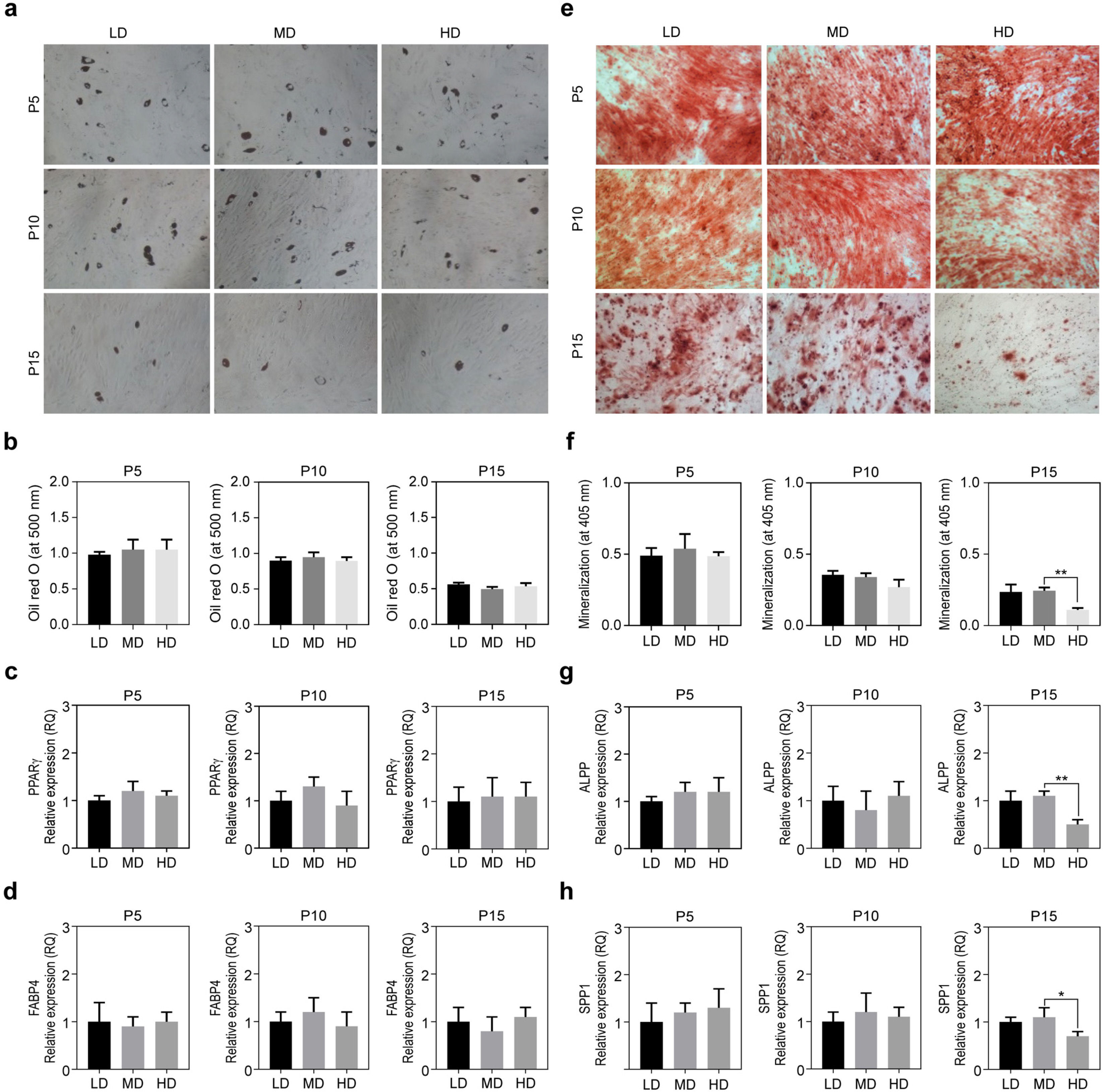
Fig. 3 Cell density-dependent changes in the lifespan of MSCs in long-term cultures. (a, b) Adipogenic differentiation potentials were compared among LD, MD, and HD. The cells were visualized by staining with Oil red O. After de-staining with 100% isopropanol, samples were quantified by measuring absorbance at 500 nm. (c, d) Quantitative PCR analysis of adipogenic differentiation markers (PPARγ, FABP4) in LD, MD, and HD. Graphs are represented as relative expression units compared with GAPDH. (e, f) Osteogenic differentiation potentials were compared among LD, MD, and HD. The cells were visualized by staining with Alizarin red S. After de-staining with 10% acetic acid, samples were quantified by measuring absorbance at 405 nm. (g, h) Quantitative PCR analysis of osteogenic differentiation markers (ALPP, SPP1) in LD, MD, and HD. Graphs are represented as relative expression units compared with GAPDH. Significance is *p<0.05, **p<0.01.

Fig. 4 Cell density-dependent production of total ROS and DNA damage in long-term cultures. (a) Total ROS were detected using 2’, 7’-dichlorofluorescein diacetate (DCFDA) assay. MSCs were treated with 10 μM DCFDA solution and fluorescence was quantified using a fluorescence reader (excitation=485 nm and emission=535 nm). (b) Oxidative DNA damage was analyzed by measuring 8-OHdG produced in LD, MD, and HD. (c) MSC proliferation was compared among HD at passages 11, 13, and 15, with and without AA (Ascorbic acid, 25 μg/ml) treatment. FI and relative proliferation were calculated by cell counting. (d) At passage 15, ROS generation was analyzed in HD treated with or without AA by staining with DCFDA. (e) At passage 15, p15, p16, and PCNA expressions were analyzed in HD treated with or without AA by Quantitative PCR. (f) At passage 15, Oxidative DNA damage was analyzed in HD treated with or without AA by measuring 8-OHdG production. Significance is *p<0.05, **p<0.01, ****p<0.0001.
Reference
-
References
1. Both SK, van der Muijsenberg AJ, van Blitterswijk CA, de Boer J, de Bruijn JD. 2007; A rapid and efficient method for expansion of human mesenchymal stem cells. Tissue Eng. 13:3–9. DOI: 10.1089/ten.2005.0513. PMID: 17518576.
Article2. Koller MR, Emerson SG, Palsson BO. 1993; Large-scale expansion of human stem and progenitor cells from bone marrow mononuclear cells in continuous perfusion cultures. Blood. 82:378–384. DOI: 10.1182/blood.V82.2.378.378. PMID: 8329697.
Article3. Wang LT, Ting CH, Yen ML, Liu KJ, Sytwu HK, Wu KK, Yen BL. 2016; Human mesenchymal stem cells (MSCs) for treatment towards immune- and inflammation-mediated diseases: review of current clinical trials. J Biomed Sci. 23:76. DOI: 10.1186/s12929-016-0289-5. PMID: 27809910. PMCID: PMC5095977.
Article4. Gharibi B, Farzadi S, Ghuman M, Hughes FJ. 2014; Inhibition of Akt/mTOR attenuates age-related changes in mesenchymal stem cells. Stem Cells. 32:2256–2266. DOI: 10.1002/stem.1709. PMID: 24659476.
Article5. de Witte SFH, Lambert EE, Merino A, Strini T, Douben HJCW, O'Flynn L, Elliman SJ, de Klein AJEMM, Newsome PN, Baan CC, Hoogduijn MJ. 2017; Aging of bone marrow- and umbilical cord-derived mesenchymal stromal cells during expansion. Cytotherapy. 19:798–807. DOI: 10.1016/j.jcyt.2017.03.071. PMID: 28462821.
Article6. Sethe S, Scutt A, Stolzing A. 2006; Aging of mesenchymal stem cells. Ageing Res Rev. 5:91–116. DOI: 10.1016/j.arr.2005.10.001. PMID: 16310414.
Article7. Basciano L, Nemos C, Foliguet B, de Isla N, de Carvalho M, Tran N, Dalloul A. 2011; Long term culture of mesenchymal stem cells in hypoxia promotes a genetic program maintaining their undifferentiated and multipotent status. BMC Cell Biol. 12:12. DOI: 10.1186/1471-2121-12-12. PMID: 21450070. PMCID: PMC3073900.
Article8. Ejtehadifar M, Shamsasenjan K, Movassaghpour A, Akbarzadehlaleh P, Dehdilani N, Abbasi P, Molaeipour Z, Saleh M. 2015; The effect of hypoxia on mesenchymal stem cell biology. Adv Pharm Bull. 5:141–149. DOI: 10.15171/apb.2015.021. PMID: 26236651. PMCID: PMC4517092.
Article9. Khan M, Akhtar S, Mohsin S, N Khan S, Riazuddin S. 2011; Growth factor preconditioning increases the function of diabetes-impaired mesenchymal stem cells. Stem Cells Dev. 20:67–75. DOI: 10.1089/scd.2009.0397. PMID: 20446810.
Article10. Kim JH, Kim WK, Sung YK, Kwack MH, Song SY, Choi JS, Park SG, Yi T, Lee HJ, Kim DD, Seo HM, Song SU, Sung JH. 2014; The molecular mechanism underlying the proliferating and preconditioning effect of vitamin C on adipose-derived stem cells. Stem Cells Dev. 23:1364–1376. DOI: 10.1089/scd.2013.0460. PMID: 24524758. PMCID: PMC4046194.
Article11. Rodrigues M, Griffith LG, Wells A. 2010; Growth factor regulation of proliferation and survival of multipotential stromal cells. Stem Cell Res Ther. 1:32. DOI: 10.1186/scrt32. PMID: 20977782. PMCID: PMC2983445.
Article12. Misra J, Mohanty ST, Madan S, Fernandes JA, Hal Ebetino F, Russell RG, Bellantuono I. 2016; Zoledronate attenuates accumulation of DNA damage in mesenchymal stem cells and protects their function. Stem Cells. 34:756–767. DOI: 10.1002/stem.2255. PMID: 26679354. PMCID: PMC4832316.
Article13. Oh J, Lee YD, Wagers AJ. 2014; Stem cell aging: mechanisms, regulators and therapeutic opportunities. Nat Med. 20:870–880. DOI: 10.1038/nm.3651. PMID: 25100532. PMCID: PMC4160113.
Article14. Schultz MB, Sinclair DA. 2016; When stem cells grow old: phenotypes and mechanisms of stem cell aging. Development. 143:3–14. DOI: 10.1242/dev.130633. PMID: 26732838. PMCID: PMC4725211.
Article15. Kim DS, Lee MW, Ko YJ, Chun YH, Kim HJ, Sung KW, Koo HH, Yoo KH. 2016; Cell culture density affects the proliferation activity of human adipose tissue stem cells. Cell Biochem Funct. 34:16–24. DOI: 10.1002/cbf.3158. PMID: 26778408.
Article16. Kim DS, Lee MW, Yoo KH, Lee TH, Kim HJ, Jang IK, Chun YH, Kim HJ, Park SJ, Lee SH, Son MH, Jung HL, Sung KW, Koo HH. 2014; Gene expression profiles of human adipose tissue-derived mesenchymal stem cells are modified by cell culture density. PLoS One. 9:e83363. DOI: 10.1371/journal.pone.0083363. PMID: 24400072. PMCID: PMC3882209.
Article17. Kim DS, Lee MW, Lee TH, Sung KW, Koo HH, Yoo KH. 2017; Cell culture density affects the stemness gene expression of adipose tissue-derived mesenchymal stem cells. Biomed Rep. 6:300–306. DOI: 10.3892/br.2017.845. PMID: 28451390. PMCID: PMC5403436.
Article18. Jeon MS, Yi TG, Lim HJ, Moon SH, Lee MH, Kang JS, Kim CS, Lee DH, Song SU. 2011; Characterization of mouse clonal mesenchymal stem cell lines established by subfrac-tionation culturing method. World J Stem Cells. 3:70–82. DOI: 10.4252/wjsc.v3.i8.70. PMID: 22007272. PMCID: PMC3192225.
Article19. Signer RA, Morrison SJ. 2013; Mechanisms that regulate stem cell aging and life span. Cell Stem Cell. 12:152–165. DOI: 10.1016/j.stem.2013.01.001. PMID: 23395443. PMCID: PMC3641677.
Article20. Zhao Q, Wang XY, Yu XX, Zhai YX, He X, Wu S, Shi YA. 2015; Expression of human telomerase reverse transcriptase mediates the senescence of mesenchymal stem cells through the PI3K/AKT signaling pathway. Int J Mol Med. 36:857–864. DOI: 10.3892/ijmm.2015.2284. PMID: 26178664.
Article21. Zimmermann S, Voss M, Kaiser S, Kapp U, Waller CF, Martens UM. 2003; Lack of telomerase activity in human mesenchymal stem cells. Leukemia. 17:1146–1149. DOI: 10.1038/sj.leu.2402962. PMID: 12764382.
Article22. Wagner W, Horn P, Castoldi M, Diehlmann A, Bork S, Saffrich R, Benes V, Blake J, Pfister S, Eckstein V, Ho AD. 2008; Replicative senescence of mesenchymal stem cells: a continuous and organized process. PLoS One. 3:e2213. DOI: 10.1371/journal.pone.0002213. PMID: 18493317. PMCID: PMC2374903.
Article23. Feng G, Tan W, Gu Z. 2013; Mesenchymal stem cells and senescence. Clon Transgen. 2:104. DOI: 10.4172/2168-9849.1000104.
Article24. Jacobs K, Zambelli F, Mertzanidou A, Smolders I, Geens M, Nguyen HT, Barbé L, Sermon K, Spits C. 2016; Higher-density culture in human embryonic stem cells results in DNA damage and genome instability. Stem Cell Reports. 6:330–341. DOI: 10.1016/j.stemcr.2016.01.015. PMID: 26923824. PMCID: PMC4788786.
Article25. Yang SR, Park JR, Kang KS. 2015; Reactive oxygen species in mesenchymal stem cell aging: implication to lung diseases. Oxid Med Cell Longev. 2015:486263. DOI: 10.1155/2015/486263. PMID: 26273422. PMCID: PMC4529978.
Article26. Li Y, Wu Q, Wang Y, Li L, Bu H, Bao J. 2017; Senescence of mesenchymal stem cells (Review). Int J Mol Med. 39:775–782. DOI: 10.3892/ijmm.2017.2912. PMID: 28290609.
Article27. Denu RA, Hematti P. 2016; Effects of oxidative stress on mesenchymal stem cell biology. Oxid Med Cell Longev. 2016:2989076. DOI: 10.1155/2016/2989076. PMID: 27413419. PMCID: PMC4928004.
Article28. Vono R, Jover Garcia E, Spinetti G, Madeddu P. 2018; Oxidative stress in mesenchymal stem cell senescence: regulation by coding and noncoding RNAs. Antioxid Redox Signal. 29:864–879. DOI: 10.1089/ars.2017.7294. PMID: 28762752. PMCID: PMC6080119.
Article29. Wei X, Yang X, Han ZP, Qu FF, Shao L, Shi YF. 2013; Mesenchymal stem cells: a new trend for cell therapy. Acta Pharmacol Sin. 34:747–754. DOI: 10.1038/aps.2013.50. PMID: 23736003. PMCID: PMC4002895.
Article30. Bartmann C, Rohde E, Schallmoser K, Pürstner P, Lanzer G, Linkesch W, Strunk D. 2007; Two steps to functional mesenchymal stromal cells for clinical application. Transfusion. 47:1426–1435. DOI: 10.1111/j.1537-2995.2007.01219.x. PMID: 17655587.
Article31. Colter DC, Class R, DiGirolamo CM, Prockop DJ. 2000; Rapid expansion of recycling stem cells in cultures of plastic-adherent cells from human bone marrow. Proc Natl Acad Sci U S A. 97:3213–3218. DOI: 10.1073/pnas.97.7.3213. PMID: 10725391. PMCID: PMC16218.
Article32. Fekete N, Rojewski MT, Fürst D, Kreja L, Ignatius A, Dausend J, Schrezenmeier H. 2012; GMP-compliant isolation and large-scale expansion of bone marrow-derived MSC. PLoS One. 7:e43255. DOI: 10.1371/journal.pone.0043255. PMID: 22905242. PMCID: PMC3419200.
Article
- Full Text Links
-
- Actions
-
Cited
- CITED
-
- Close
- Share
-
- Similar articles
-
- Nervonic Acid Inhibits Replicative Senescence of Human Wharton’s Jelly-Derived Mesenchymal Stem Cells
- Characterization of Senescence of Culture-expanded Human Adipose-derived Mesenchymal Stem Cells
- Clinical Safety and Efficacy of Autologous Bone Marrow-Derived Mesenchymal Stem Cell Transplantation in Sensorineural Hearing Loss Patients
- Concise Review: Differentiation of Human Adult Stem Cells Into Hepatocyte-like Cells In vitro
- Evaluation of Bone Marrow-derived Stem Cells and Adipose-derived Stem Cells Co-cultured on Human Nucleus Pulposus Cells: A Pilot Study
