Perirenal and epicardial fat and their association with carotid intima-media thickness in children
- Affiliations
-
- 1Pediatric Endocrinology, Girona Institute for Biomedical Research, Girona, Spain. alopezbermejo@idibgi.org
- 2Pediatrics, Dr. Josep Trueta Hospital, Girona, Spain.
- 3Department of Physical Therapy, EUSES University School, University of Girona, Salt, Spain.
- 4Pediatrics, Clinica Bofill, Girona, Spain.
- 5Institute of Legal Medicine of Catalonia, Girona, Spain.
- 6Maternal & Fetal Metabolic Research, Girona Institute for Biomedical Research, Salt, Spain.
- KMID: 2468722
- DOI: http://doi.org/10.6065/apem.2019.24.4.220
Abstract
- Recent data suggest that subclinical atherosclerosis is more related to visceral adipose tissue distribution than to overall fat mass. Both perirenal fat and epicardial fat are visceral fat depots surrounding the kidneys and the myocardium, respectively, which can be easily assessed by ultrasound. Their clinical relevance in children is largely unknown. This review describes studies relating perirenal and epicardial fat to cardiovascular disease or carotid intima-media thickness (cIMT), a well-established surrogate for subclinical atherosclerosis, and discusses this in context with our own data from children. In adults, both perirenal and epicardial fat are useful biological markers of visceral obesity. The former has been related to hypertension in overweight subjects and with atherosclerosis in patients with human immunodeficiency virus. The latter was associated with several metabolic syndrome components and with calcification of the carotid artery. In healthy prepubertal children, both epicardial and perirenal fat thickness, rather than total body fat mass, were related to cIMT. Ultrasonography measures of perirenal and epicardial fat are related to atherosclerosis in adults and may be convenient tools for the assessment of cardiometabolic risk in children.
Keyword
MeSH Terms
Figure
-
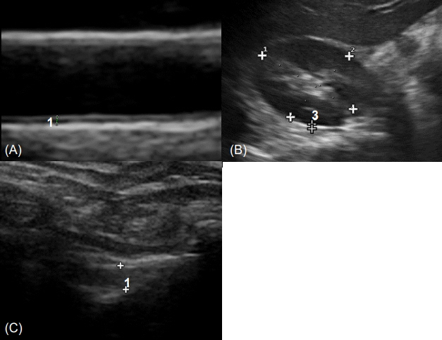
Fig. 1. (A) Echocardiographic carotid intima-media thickness. Diastolic images were obtained using a linear 12-MHz transducer at the level of the distal common carotid artery, 1 cm away from its bifurcation. The intima-media thickness in the image is indicated between the 2 cursors and marked using the number 1. (B) Echocardiographic perirenal fat thickness. Children were placed in the supine position and the probe was moved to find the position at which the surface of the kidney was almost parallel to the skin. Longitudinal scans were then performed using a linear 12-MHz transducer. The thickness from the inner side of the abdominal musculature to the surface of the kidney was measured. The perirenal fat thickness in the image is indicated between the 2 cursors and marked using the number 3. (C) Echocardiographic epicardial fat thickness. Children were placed in the left lateral decubitus position. Measurements of the fat thickness adjacent to the free wall of the right ventricle were taken from the parasternal long-axis views using a linear 12-MHz transducer. Epicardial adipose tissue appears as an echo-free space between the pericardium and myocardium indicated by the 2 cursors and marked using the number 1.

Fig. 2. Scatter plot of perirenal fat and the cIMT of children according to BMI categories. White dots and a dotted line depict lean children (BMI-SDS<1), gray dots and a dashed line depict overweight (OW) children (≤1 BMI-SDS <2), and black dots and a solid line depict obese children (BMI-SDS ≥2). cIMT, carotid intima-media thickness; BMI, body mass index; SDS, standard deviation score.
Reference
-
References
1. Must A, Jacques PF, Dallal GE, Bajema CJ, Dietz WH. Longterm morbidity and mortality of overweight adolescents. A follow-up of the Harvard Growth Study of 1922 to 1935. N Engl J Med. 1992; 327:1350–5.
Article2. Lorenz MW, Markus HS, Bots ML, Rosvall M, Sitzer M. Prediction of clinical cardiovascular events with carotid intima-media thickness: a systematic review and metaanalysis. Circulation. 2007; 115:459–67.
Article3. Brady TM, Schneider MF, Flynn JT, Cox C, Samuels J, Saland J, et al. Carotid intima-media thickness in children with CKD: results from the CKiD study. Clin J Am Soc Nephrol. 2012; 7:1930–7.4. Celermajer DS, Ayer JG. Childhood risk factors for adult cardiovascular disease and primary prevention in childhood. Heart. 2006; 92:1701–6.
Article5. Després JP. Body fat distribution and risk of cardiovascular disease: an update. Circulation. 2012; 126:1301–13.
Article6. Britton KA, Fox CS. Ectopic fat depots and cardiovascular disease. Circulation. 2011; 124:e837. –41.
Article7. Ma S, Zhu XY, Eirin A, Woollard JR, Jordan KL, Tang H, et al. Perirenal fat promotes renal arterial endothelial dysfunction in obese swine through tumor necrosis factor-α. J Urol. 2016; 195(4 Pt 1):1152–9.
Article8. Favre G, Grangeon-Chapon C, Raffaelli C, François-Chalmin F, Iannelli A, Esnault V. Perirenal fat thickness measured with computed tomography is a reliable estimate of perirenal fat mass. PLoS One. 2017; 12:e0175561.
Article9. De Pergola G, Campobasso N, Nardecchia A, Triggiani V, Caccavo D, Gesualdo L, et al. Para- and perirenal ultrasonographic fat thickness is associated with 24-hours mean diastolic blood pressure levels in overweight and obese subjects. BMC Cardiovasc Disord. 2015; 15:108.
Article10. Grima P, Guido M, Zizza A, Chiavaroli R. Sonographically measured perirenal fat thickness: an early predictor of atherosclerosis in HIV-1-infected patients receiving highly active antiretroviral therapy? J Clin Ultrasound. 2010; 38:190–5.
Article11. Iacobellis G, Bianco AC. Epicardial adipose tissue: emerging physiological, pathophysiological and clinical features. Trends Endocrinol Metab. 2011; 22:450–7.
Article12. Iacobellis G, Ribaudo MC, Assael F, Vecci E, Tiberti C, Zappaterreno A, et al. Echocardiographic epicardial adipose tissue is related to anthropometric and clinical parameters of metabolic syndrome: a new indicator of cardiovascular risk. J Clin Endocrinol Metab. 2003; 88:5163–8.
Article13. Djaberi R, Schuijf JD, van Werkhoven JM, Nucifora G, Jukema JW, Bax JJ. Relation of epicardial adipose tissue to coronary atherosclerosis. Am J Cardiol. 2008; 102:1602–7.
Article14. Iacobellis G, Lonn E, Lamy A, Singh N, Sharma AM. Epicardial fat thickness and coronary artery disease correlate independently of obesity. Int J Cardiol. 2011; 146:452–4.
Article15. Fitzgibbons TP, Czech MP. Epicardial and perivascular adipose tissues and their influence on cardiovascular disease: basic mechanisms and clinical associations. J Am Heart Assoc. 2014; 3:e000582.
Article16. Gray SL, Vidal-Puig AJ. Adipose tissue expandability in the maintenance of metabolic homeostasis. Nutr Rev. 2007; 65(6 Pt 2):S7–12.
Article17. Tokunaga K, Matsuzawa Y, Ishikawa K, Tarui S. A novel technique for the determination of body fat by computed tomography. Int J Obes. 1983; 7:437–45.18. Matsuzawa Y. Establishment of a concept of visceral fat syndrome and discovery of adiponectin. Proc Jpn Acad Ser B Phys Biol Sci. 2010; 86:131–41.
Article19. Fox CS, Massaro JM, Hoffmann U, Pou KM, Maurovich-Horvat P, Liu CY, et al. Abdominal visceral and subcutaneous adipose tissue compartments: association with metabolic risk factors in the Framingham Heart Study. Circulation. 2007; 116:39–48.
Article20. Czernichow S, Bertrais S, Oppert JM, Galan P, Blacher J, Ducimetière P, et al. Body composition and fat repartition in relation to structure and function of large arteries in middle-aged adults (the SU.VI.MAX study). Int J Obes (Lond). 2005; 29:826–32.
Article21. Park MH, Skow Á, De Matteis S, Kessel AS, Saxena S, Viner RM, et al. Adiposity and carotid-intima media thickness in children and adolescents: a systematic review. BMC Pediatr. 2015; 15:161.
Article22. Pou KM, Massaro JM, Hoffmann U, Vasan RS, Maurovich-Horvat P, Larson MG, et al. Visceral and subcutaneous adipose tissue volumes are cross-sectionally related to markers of inflammation and oxidative stress: the Framingham Heart Study. Circulation. 2007; 116:1234–41.
Article23. Geerts CC, Evelein AM, Bots ML, van der Ent CK, Grobbee DE, Uiterwaal CS. Body fat distribution and early arterial changes in healthy 5-year-old children. Ann Med. 2012; 44:350–9.
Article24. Epifanio M, Baldisserotto M, Sarria EE, Lazaretti A, Mattiello R. Ultrasound evaluation of carotid intima-media thickness in children. J Atheroscler Thromb. 2015; 22:1141–7.
Article25. Roever L, Resende ES, Veloso FC, Diniz AL, Penha-Silva N, Casella-Filho A, et al. Perirenal fat and association with metabolic risk factors: The Uberlândia Heart Study. Medicine (Baltimore). 2015; 94:e1105.26. Jung M, Volonté F, Buchs NC, Gayet-Ageron A, Pugin F, Gervaz P, et al. Perirenal fat surface area as a risk factor for morbidity after elective colorectal surgery. Dis Colon Rectum. 2014; 57:201–9.
Article27. Weisberg SP, McCann D, Desai M, Rosenbaum M, Leibel RL, Ferrante AW Jr. Obesity is associated with macrophage accumulation in adipose tissue. J Clin Invest. 2003; 112:1796–808.
Article28. Lamacchia O, Nicastro V, Camarchio D, Valente U, Grisorio R, Gesualdo L, et al. Para- and perirenal fat thickness is an independent predictor of chronic kidney disease, increased renal resistance index and hyperuricaemia in type-2 diabetic patients. Nephrol Dial Transplant. 2011; 26:892–8.
Article29. Chughtai HL, Morgan TM, Rocco M, Stacey B, Brinkley TE, Ding J, et al. Renal sinus fat and poor blood pressure control in middle-aged and elderly individuals at risk for cardiovascular events. Hypertension. 2010; 56:901–6.
Article30. Bassols J, Martínez-Calcerrada JM, Prats-Puig A, Carreras-Badosa G, Xargay-Torrent S, Lizarraga-Mollinedo E, et al. Perirenal fat is related to carotid intima-media thickness in children. Int J Obes (Lond). 2018; 42:641–7.
Article31. Rosito GA, Massaro JM, Hoffmann U, Ruberg FL, Mahabadi AA, Vasan RS, et al. Pericardial fat, visceral abdominal fat, cardiovascular disease risk factors, and vascular calcification in a community-based sample: the Framingham Heart Study. Circulation. 2008; 117:605–13.
Article32. Liu J, Fox CS, Hickson D, Sarpong D, Ekunwe L, May WD, et al. Pericardial adipose tissue, atherosclerosis, and cardiovascular disease risk factors: the Jackson heart study. Diabetes Care. 2010; 33:1635–9.
Article33. Yun CH, Lin TY, Wu YJ, Liu CC, Kuo JY, Yeh HI, et al. Pericardial and thoracic peri-aortic adipose tissues contribute to systemic inflammation and calcified coronary atherosclerosis independent of body fat composition, anthropometric measures and traditional cardiovascular risks. Eur J Radiol. 2012; 81:749–56.
Article34. Iacobellis G, Willens HJ. Echocardiographic epicardial fat: a review of research and clinical applications. J Am Soc Echocardiogr. 2009; 22:1311–9.
Article35. Neeland IJ, Ross R, Després JP, Matsuzawa Y, Yamashita S, Shai I, et al. Visceral and ectopic fat, atherosclerosis, and cardiometabolic disease: a position statement. Lancet Diabetes Endocrinol. 2019; 7:715–25.
Article36. Okada K, Ohshima S, Isobe S, Harada K, Hirashiki A, Funahashi H, et al. Epicardial fat volume correlates with severity of coronary artery disease in nonobese patients. J Cardiovasc Med (Hagerstown). 2014; 15:384–90.
Article37. Kim SJ, Kim HS, Jung JW, Kim NS, Noh CI, Hong YM. Correlation between epicardial fat thickness by echocardiography and other parameters in obese adolescents. Korean Circ J. 2012; 42:471–8.
Article38. Elshorbagy HH, Fouda ER, Kamal NM, Bassiouny MM, Fathi WM. Evaluation of epicardial fat and carotid intima-media thickness in obese children. Iran J Pediatr. 2016; 26:e2968.
Article39. Cabrera-Rego JO, Iacobellis G, Castillo-Herrera JA, Valiente-Mustelier J, Gandarilla-Sarmientos JC, Marín-Juliá SM, et al. Epicardial fat thickness correlates with carotid intima-media thickness, arterial stiffness, and cardiac geometry in children and adolescents. Pediatr Cardiol. 2014; 35:450–6.
Article40. Akyol B, Boyraz M, Aysoy C. Relationship of epicardial adipose tissue thickness with early indicators of atherosclerosis and cardiac functional changes in obese adolescents with metabolic syndrome. J Clin Res Pediatr Endocrinol. 2013; 5:156–63.
Article41. Schejbal V. Epicardial fatty tissue of the right ventricle--morphology, morphometry and functional significance. Pneumologie. 1989; 43:490–9.
- Full Text Links
-
- Actions
-
Cited
- CITED
-
- Close
- Share
-
- Similar articles
-
- Do we need individual measurement of carotid intima and media thickness?
- Carotid ultrasound in patients with coronary artery disease
- Gallstones are Associated with Intima-Media Thickness of Common Carotid Arteries in Men
- Response: Increased Carotid Intima-Media Thickness Is Associated with Progression of Diabetic Nephropathy in Patients with Type 2 Diabetes
- Letter: Increased Carotid Intima-Media Thickness Is Associated with Progression of Diabetic Nephropathy in Patients with Type 2 Diabetes
