The Effect of Different Routes of Injection of Bone Marrow Mesenchymal Stem Cells on Parotid Glands of Rats Receiving Cisplatin: A Comparative Study
- Affiliations
-
- 1Department of Oral Biology, Faculty of Dentistry, Mansoura University, Mansoura, Egypt. emy.hany11@gmail.com
- 2Urology and Nephrology Center, Mansoura University, Mansoura, Egypt.
- 3Department of Oral Biology, School of Dentistry, Badr University, Cairo, Egypt.
- KMID: 2400871
- DOI: http://doi.org/10.15283/ijsc17022
Abstract
- BACKGROUND AND OBJECTIVES
Cisplatin is a powerful antitumor chemotherapeutic agent that is widely used in the treatment of many cancers but it has many side effects on many organs including salivary glands. Bone marrow is considered to be a rich environment that comprises many types of stem cells of which BMSCs (Bone marrow mesenchymal stem cells) are the most studied with potentiality to differentiate into many cell types. This study was conducted to evaluate the effect of different routes of injection of BMSCs on parotid glands of rats receiving cisplatin.
METHODS AND RESULTS
Sprague-Dawley rats were divided into 3 groups: a negative control group receiving phosphate buffered saline, a positive control group receiving cisplatin, and an experimental group where rats received cisplatin and then received iron oxide-labeled BMSCs by either intravenous or intraparotid routes or both. Animals were sacrificed at periods of 3,6,10 and 15 days after cisplatin injection, then histological, ultrastructural and immunohistochemical studies were done. The experimental stem cell treated group showed better histological features and increased PCNA proliferation index when compared to the control. The systemic and combination groups showed better results than the local group. Iron oxide-labeled cells were detected with Prussian blue stain.
CONCLUSIONS
This study proved that BMSCs can improve cisplatin induced cytotoxicity in parotid glands. Systemic administration showed to have a better effect than local intraparotid administration and comparable effect to combined administration.
Keyword
MeSH Terms
Figure
-

Fig. 1 Photomicrographs of parotid glands at day 10. (A) Cisplatin group showing severe intracytoplasmic vacuolation with irregular nuclear and acinar outlines. (B) Local BMSCs administration group showing some vacuoles and some mitotic figures. (C) Systemic BMSCs administration group showing regular nuclei with some mitotic figures and normal ducts. (D) Combination BMSCs administration group showing normal ducts, regular acinar and nuclear outlines and some mitotic figures. (v) vacuoles, (arrows) mitotic figures, (d) ducts (H&E, 400×).

Fig. 2 Electron micrographs of parotid glands at day 10. (A) Cisplatin group showing irregular and atrophied nuclei, dilated rER and cytoplasmic vacuoles. (B) Local BMSCs administration group showing more regular nuclei but with condensed heterochromatin, dilated rER and many lysosomes. (C) Systemic BMSCs administration group showing regular euochromatic nuclei with homogenous secretory granules bordering a narrow lumen. (D) Combination BMSCs administration group showing euochromatic nucleus, regular rER and normal mitochondria. (N) nucleus, (rER) rough endoplasmic reticulum, (v) vacuoles, (L) lysosomes, (SG) secretory granules, (Lu) lumen, (M) mitochondria.

Fig. 3 Photomicrographs of immunostained parotid gland sections at day 10. (A) Cisplatin group. (B) Experimental local group. (C) Systemic group. (D) Combination group (anti PCNA antibody, 400×).

Fig. 4 Photomicrograph of the parotid gland showing cells with positive reaction to Prussian blue staining (arrows) (PB stain, 400×).
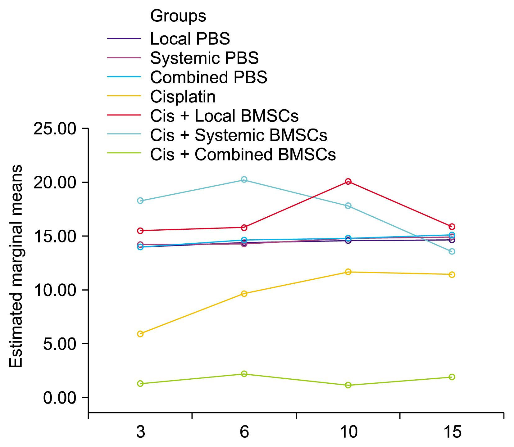
Fig. 5 Line graph showing the mean PCNA proliferation index for all groups at the different time periods.
Reference
-
References
1. Alcamo IE. Anatomy coloring workbook. 2nd ed. New York: Random House;2003. p. 222.2. Wang G, Reed E, Li QQ. Molecular basis of cellular response to cisplatin chemotherapy in non-small cell lung cancer (Review). Oncol Rep. 2004; 12:955–965. PMID: 15492778.
Article3. López BC, Esteve CG, Pérez GS. Dental treatment considerations in the chemotherapy patient. J Clin Exp Dent. 2011; 3:e31–e42. DOI: 10.4317/jced.3.e31.
Article4. Kitashima S. Morphological alterations of submandibular glands caused by cisplatin in the rat. Kurume Med J. 2005; 52:29–38. DOI: 10.2739/kurumemedj.52.29. PMID: 16119610.
Article5. Hey J, Setz J, Gerlach R, Vordermark D, Gernhardt CR, Kuhnt T. Effect of Cisplatin on parotid gland function in concomitant radiochemotherapy. Int J Radiat Oncol Biol Phys. 2009; 75:1475–1480. DOI: 10.1016/j.ijrobp.2008.12.071. PMID: 19515505.
Article6. Wexler SA, Donaldson C, Denning-Kendall P, Rice C, Bradley B, Hows JM. Adult bone marrow is a rich source of human mesenchymal ‘stem’ cells but umbilical cord and mobilized adult blood are not. Br J Haematol. 2003; 121:368–374. DOI: 10.1046/j.1365-2141.2003.04284.x. PMID: 12694261.
Article7. Yen MH, Wu YY, Liu YS, Rimando M, Ho JH, Lee OK. Efficient generation of hepatic cells from mesenchymal stromal cells by an innovative bio-microfluidic cell culture device. Stem Cell Res Ther. 2016; 7:120. DOI: 10.1186/s13287-016-0371-7. PMID: 27542358. PMCID: 4992324.
Article8. Woodbury D, Schwarz EJ, Prockop DJ, Black IB. Adult rat and human bone marrow stromal cells differentiate into neurons. J Neurosci Res. 2000; 61:364–370. DOI: 10.1002/1097-4547(20000815)61:4<364::AID-JNR2>3.0.CO;2-C. PMID: 10931522.
Article9. Qi S, Wu D. Bone marrow-derived mesenchymal stem cells protect against cisplatin-induced acute kidney injury in rats by inhibiting cell apoptosis. Int J Mol Med. 2013; 32:1262–1272. DOI: 10.3892/ijmm.2013.1517. PMID: 24126885. PMCID: 3829764.
Article10. Liu J, Zhang H, Zhang Y, Li N, Wen Y, Cao F, Ai H, Xue X. Homing and restorative effects of bone marrow-derived mesenchymal stem cells on cisplatin injured ovaries in rats. Mol Cells. 2014; 37:865–872. DOI: 10.14348/molcells.2014.0145. PMID: 25410907. PMCID: 4275703.
Article11. Weil BR, Markel TA, Herrmann JL, Abarbanell AM, Meldrum DR. Mesenchymal stem cells enhance the viability and proliferation of human fetal intestinal epithelial cells following hypoxic injury via paracrine mechanisms. Surgery. 2009; 146:190–197. DOI: 10.1016/j.surg.2009.03.031. PMID: 19628073.
Article12. Sumita Y, Liu Y, Khalili S, Maria OM, Xia D, Key S, Cotrim AP, Mezey E, Tran SD. Bone marrow-derived cells rescue salivary gland function in mice with head and neck irradiation. Int J Biochem Cell Biol. 2011; 43:80–87. DOI: 10.1016/j.biocel.2010.09.023.
Article13. Lim JY, Yi T, Choi JS, Jang YH, Lee S, Kim HJ, Song SU, Kim YM. Intraglandular transplantation of bone marrow-derived clonal mesenchymal stem cells for amelioration of post-irradiation salivary gland damage. Oral Oncol. 2013; 49:136–143. DOI: 10.1016/j.oraloncology.2012.08.010.
Article14. Lin CY, Chang FH, Chen CY, Huang CY, Hu FC, Huang WK, Ju SS, Chen MH. Cell therapy for salivary gland regeneration. J Dent Res. 2011; 90:341–346. DOI: 10.1177/0022034510386374. PMID: 21297017.
Article15. Tran SD, Liu Y, Xia D, Maria OM, Khalili S, Wang RW, Quan VH, Hu S, Seuntjens J. Paracrine effects of bone marrow soup restore organ function, regeneration, and repair in salivary glands damaged by irradiation. PLoS One. 2013; 8:e61632. DOI: 10.1371/journal.pone.0061632. PMID: 23637870. PMCID: 3634855.
Article16. Lotfy A, Salama M, Zahran F, Jones E, Badawy A, Sobh M. Characterization of mesenchymal stem cells derived from rat bone marrow and adipose tissue: a comparative study. Int J Stem Cells. 2014; 7:135–142. DOI: 10.15283/ijsc.2014.7.2.135. PMID: 25473451. PMCID: 4249896.
Article17. Roldán-Fidalgo A, Martín Saldaña S, Trinidad A, Olmedilla-Alonso B, Rodríguez-Valiente A, García-Berrocal JR, Ramírez-Camacho R. In vitro and in vivo effects of lutein against cisplatin-induced ototoxicity. Exp Toxicol Pathol. 2016; 68:197–204. DOI: 10.1016/j.etp.2016.01.003. PMID: 26850526.
Article18. Jeong J, Baek H, Kim YJ, Choi Y, Lee H, Lee E, Kim ES, Hah JH, Kwon TK, Choi IJ, Kwon H. Human salivary gland stem cells ameliorate hyposalivation of radiation-damaged rat salivary glands. Exp Mol Med. 2013; 45:e58. DOI: 10.1038/emm.2013.121. PMID: 24232257. PMCID: 3849572.
Article19. Huang S, Xu L, Zhang Y, Sun Y, Li G. Systemic and local administration of allogeneic bone marrow-derived mesenchymal stem cells promotes fracture healing in rats. Cell Transplant. 2015; 24:2643–2655. DOI: 10.3727/096368915X687219. PMID: 25647659.
Article20. Li XB, Schluesener HJ. Therapeutic effects of cisplatin on rat experimental autoimmune encephalomyelitis. Arch Immunol Ther Exp (Warsz). 2006; 54:51–53. DOI: 10.1007/s00005-006-0005-3.
Article21. Cepeda V, Fuertes MA, Castilla J, Alonso C, Quevedo C, Pérez JM. Biochemical mechanisms of cisplatin cytotoxicity. Anticancer Agents Med Chem. 2007; 7:3–18. DOI: 10.2174/187152007779314044. PMID: 17266502.
Article22. Fuertes MA, Castilla J, Alonso C, Pérez JM. Cisplatin biochemical mechanism of action: from cytotoxicity to induction of cell death through interconnections between apoptotic and necrotic pathways. Curr Med Chem. 2003; 10:257–266. DOI: 10.2174/0929867033368484. PMID: 12570712.
Article23. Casares C, Ramírez-Camacho R, Trinidad A, Roldán A, Jorge E, García-Berrocal JR. Reactive oxygen species in apoptosis induced by cisplatin: review of physiopathological mechanisms in animal models. Eur Arch Otorhinolaryngol. 2012; 269:2455–2459. DOI: 10.1007/s00405-012-2029-0. PMID: 22584749.
Article24. Izuwa Y, Kusaba J, Horiuchi M, Aiba T, Kawasaki H, Kurosaki Y. Comparative study of increased plasma quinidine concentration in rats with glycerol- and cisplatin-induced acute renal failure. Drug Metab Pharmacokinet. 2009; 24:451–457. DOI: 10.2133/dmpk.24.451. PMID: 19881257.
Article25. Lee JE, Nakagawa T, Kita T, Kim TS, Iguchi F, Endo T, Shiga A, Lee SH, Ito J. Mechanisms of apoptosis induced by cisplatin in marginal cells in mouse stria vascularis. ORL J Otorhinolaryngol Relat Spec. 2004; 66:111–118. DOI: 10.1159/000079329. PMID: 15316230.
Article26. Satoh M, Yoshihara T. Clinical and ultracytochemical investigation of sialadenosis. Acta Otolaryngol Suppl. 2004; (553):122–127. DOI: 10.1080/03655230410017814. PMID: 15277051.
Article27. McInnes EF. Background lesions in laboratory animals: a color atlas. China: Saunders Elsevier;2012. p. 49.28. Grove JE, Bruscia E, Krause DS. Plasticity of bone marrow-derived stem cells. Stem Cells. 2004; 22:487–500. DOI: 10.1634/stemcells.22-4-487. PMID: 15277695.
Article29. Quintana-Bustamante O, Alvarez-Barrientos A, Kofman AV, Fabregat I, Bueren JA, Theise ND, Segovia JC. Hematopoietic mobilization in mice increases the presence of bone marrow-derived hepatocytes via in vivo cell fusion. Hepatology. 2006; 43:108–116. DOI: 10.1002/hep.21005.
Article30. Rajantie I, Ilmonen M, Alminaite A, Ozerdem U, Alitalo K, Salven P. Adult bone marrow-derived cells recruited during angiogenesis comprise precursors for periendothelial vascular mural cells. Blood. 2004; 104:2084–2086. DOI: 10.1182/blood-2004-01-0336. PMID: 15191949. PMCID: 2698665.
Article31. Zarjou A, Kim J, Traylor AM, Sanders PW, Balla J, Agarwal A, Curtis LM. Paracrine effects of mesenchymal stem cells in cisplatin-induced renal injury require heme oxygenase-1. Am J Physiol Renal Physiol. 2011; 300:F254–F262. DOI: 10.1152/ajprenal.00594.2010. PMCID: 3023217.
Article32. Zhao W, Li JJ, Cao DY, Li X, Zhang LY, He Y, Yue SQ, Wang DS, Dou KF. Intravenous injection of mesenchymal stem cells is effective in treating liver fibrosis. World J Gastroenterol. 2012; 18:1048–1058. DOI: 10.3748/wjg.v18.i10.1048. PMID: 22416179. PMCID: 3296978.
Article33. Guo W, Wang H, Zou S, Gu M, Watanabe M, Wei F, Dubner R, Huang GT, Ren K. Bone marrow stromal cells produce long-term pain relief in rat models of persistent pain. Stem Cells. 2011; 29:1294–1303. DOI: 10.1002/stem.667. PMID: 21630378. PMCID: 3277433.
Article34. McFarlin K, Gao X, Liu YB, Dulchavsky DS, Kwon D, Arbab AS, Bansal M, Li Y, Chopp M, Dulchavsky SA, Gautam SC. Bone marrow-derived mesenchymal stromal cells accelerate wound healing in the rat. Wound Repair Regen. 2006; 14:471–478. DOI: 10.1111/j.1743-6109.2006.00153.x. PMID: 16939576.
Article35. Kwon DS, Gao X, Liu YB, Dulchavsky DS, Danyluk AL, Bansal M, Chopp M, McIntosh K, Arbab AS, Dulchavsky SA, Gautam SC. Treatment with bone marrow-derived stromal cells accelerates wound healing in diabetic rats. Int Wound J. 2008; 5:453–463. DOI: 10.1111/j.1742-481X.2007.00408.x. PMID: 18593394. PMCID: 3852907.
Article36. Aly LA, El-Menoufy H, Sadeq HS, Ragae A, Sabry D. Efficiency of systemic versus intralesional bone marrow-derived stem cells in regeneration of oral mucosa after induction of formocresol induced ulcers in dogs. Dent Res J (Isfahan). 2014; 11:212–221.37. Muschler GF, Nakamoto C, Griffith LG. Engineering principles of clinical cell-based tissue engineering. J Bone Joint Surg Am. 2004; 86-A:1541–1558. DOI: 10.2106/00004623-200407000-00029. PMID: 15252108.
Article38. Abbah SA, Spanoudes K, O’Brien T, Pandit A, Zeugolis DI. Assessment of stem cell carriers for tendon tissue engineering in pre-clinical models. Stem Cell Res Ther. 2014; 5:38. DOI: 10.1186/scrt426. PMID: 25157898. PMCID: 4056691.
Article39. Schwarz S, Huss R, Schulz-Siegmund M, Vogel B, Brandau S, Lang S, Rotter N. Bone marrow-derived mesenchymal stem cells migrate to healthy and damaged salivary glands following stem cell infusion. Int J Oral Sci. 2014; 6:154–161. DOI: 10.1038/ijos.2014.23. PMID: 24810808. PMCID: 4170149.
Article40. Papadimitriou N, Li S, Barreto Henriksson H. Iron sucrose-labeled human mesenchymal stem cells: in vitro multilineage capability and in vivo traceability in a lapine xenotransplantation model. Stem Cells Dev. 2015; 24:2403–2412. DOI: 10.1089/scd.2015.0140. PMID: 26076769.
Article
- Full Text Links
-
- Actions
-
Cited
- CITED
-
- Close
- Share
-
- Similar articles
-
- Study of the Effect of Route of Administration of Mesenchymal Stem Cells on Cisplatin-Induced Acute Kidney Injury in Sprague Dawley Rats
- Comparison of the Outcomes after Intralesional, Intracisternal, and Intravenous Transplantation of Human Bone Marrow Derived Mesenchymal Stem Cells for Spinal Cord Injured Rat
- Clinical Use of Mesenchymal Stem Cells in Bone Regeneration
- Fate of Transplanted Bone Marrow Derived Mesenchymal Stem Cells Following Spinal Cord Injury in Rats by Transplantation Routes
- Stem Cells and Niemann Pick Disease
