Comparison of Anterior Segment Parameters Obtained by Anterior Segment Optical Coherence Tomography and Dual Rotating Scheimpflug Camera
- Affiliations
-
- 1Department of Ophthalmology, Inha University School of Medicine, Incheon, Korea. panch325@hanmail.net
- KMID: 2397848
- DOI: http://doi.org/10.3341/jkos.2017.58.12.1341
Abstract
- PURPOSE
To compare the anterior segment measurements with a Galilei® dual Scheimpflug analyzer and anterior segment optical coherence tomography (Cirrus OCT®).
METHODS
Forty-eight eyes of 24 normal young adults were assessed for repeatability with two identical measurements of the central corneal thickness, minimum corneal thickness, anterior chamber depth, and anterior chamber angle using the Galilei® dual-Scheimpflug analyzer and Cirrus OCT®.
RESULTS
The central corneal thickness, anterior chamber depth, and anterior chamber angle were highly reproducible and repeatable (intraclass correlation coefficient ≥ 0.90). Repeatability of the minimum corneal thickness was slightly lower (intraclass correlation coefficient ≥ 0.69). The mean corneal thickness measured using the Galilei® dual Scheimpflug analyzer was 0.26 ± 7.11 µm thinner than that measured using the Cirrus OCT®, and the mean corneal thickness was 0.37 ± 7.35 µm thicker, but was not statistically significant. The anterior chamber depth was 0.22 ± 0.08 mm deeper than the Cirrus OCT® (p < 0.007), and the anterior chamber angle was 7.87°± 1.32° larger than the Cirrus OCT® (p = 0.04). The 95% agreements of the central corneal thickness, anterior chamber depth, and anterior chamber angle between instruments were 85.30 µm, 1.43 mm, and 27.90°, respectively, and showed a high correlation (r ≥ 0.90; p < 0.001). The repeatability of the minimum corneal thickness was slightly low (r = 0.69; p < 0.001), and the range of agreement was larger (109.58 µm).
CONCLUSIONS
The anterior segment measurements obtained with the dual rotating Scheimpflug camera and new anterior segment OCT in normal eyes was comparable and reproducible. However, the agreement ranges of the measured values were relatively large, so it was difficult to exchange values between instruments.
Keyword
Figure
-

Figure 1 An example of the pachymetry mode of the Cirrus OCT®. Every sector has three values, which indicates the minimum, average, and maximum corneal thicknesses. The average value is representative of each sector.

Figure 2 The angle opening distance 500 was measured on a line perpendicular to the trabecular meshwork at points 500 µm from the scleral spur. The anterior chamber angle (ACA) was measured on this image (white dotted line). AOD = angle opening distance.

Figure 3 Bland Altman plots between the parameters of the two instruments. The middle line is the mean and the lines on the side represent the upper and lower 95% limits of agreement. (A) The average central corneal thickness using the Galilei® dual Scheimpflug analyzer and Cirrus OCT®. (B) The average minimum corneal thickness using the Galilei® dual Scheimpflug analyzer and Cirrus OCT®. (C) The average anterior chamber depth using the Galilei® dual-Scheimpflug analyzer and Cirrus OCT®. (D) The average anterior chamber angle using the Galilei® dual Scheimpflug analyzer and Cirrus OCT®. CCT = central corneal thickness; MCT = minimum corneal thickness; ACD = anterior chamber depth; ACA = anterior chamber angle.
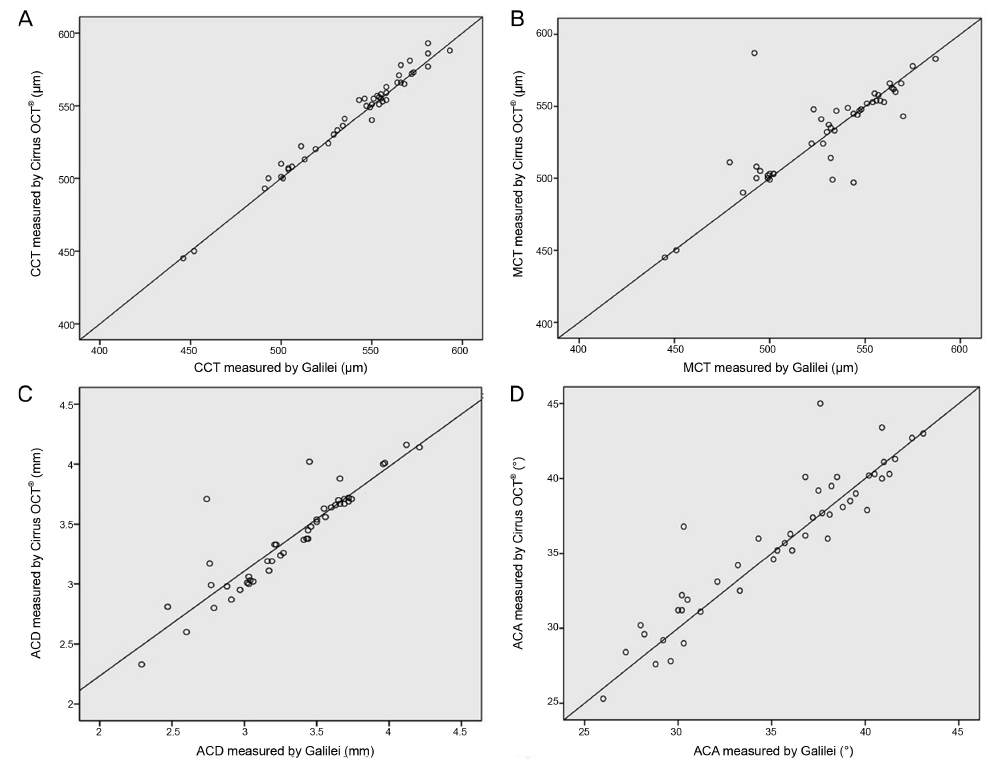
Figure 4 Scattergram showing correlations of the anterior chamber depth, central corneal thickness, minimum corneal thickness, and anterior chamber angle using the Galilei® dual Scheimpflug analyzer and Cirrus OCT®. (A) The correlation of central corneal thicknesses between the Galilei® dual Scheimpflug analyzer and Cirrus OCT® (r = 0.99; p < 0.01). (B) The correlation of the minimum corneal thickness between the Galilei® dual Scheimpflug analyzer and Cirrus OCT® (r = 0.69; p < 0.01). (C) The correlation of the anterior chamber depth between the Galilei® dual Scheimpflug analyzer and Cirrus OCT® (r = 0.90; p < 0.01). (D) The correlation of the anterior chamber angle between the Galilei® dual Scheimpflug analyzer and Cirrus OCT® (r = 0.92; p < 0.01). CCT = central corneal thickness; MCT = minimum corneal thickness; ACD = anterior chamber depth; ACA = anterior chamber angle.
Reference
-
1. Wang Z, Chen J, Yang B. Posterior corneal surface topographic changes after laser in situ keratomileusis are related to residual corneal bed thickness. Ophthalmology. 1999; 106:406–409.2. Yau CW, Cheng HC. Microkeratome blades and corneal flap thickness in LASIK. Ophthalmic Surg Lasers Imaging. 2008; 39:471–475.3. Huang J, Pesudovs K, Wen D, et al. Comparison of anterior segment measurements with rotating Scheimpflug photography and partial coherence reflectometry. J Cataract Refract Surg. 2011; 37:341–348.4. Coskunseven E, Jankov MR 2nd, Hafezi F. Contralateral eye study of corneal collagen cross-linking with riboflavin and UVA irradiation in patients with keratoconus. J Refract Surg. 2009; 25:371–376.5. Ryu HW, Kim KR, Chung SK. Comparison of A-scan, Scheimpflug camera, and Orbscan for measurement of anterior chamber depth. J Korean Ophthalmol Soc. 2006; 47:1287–1291.6. Giers U, Epple C. Comparison of A-scan device accuracy. J Cataract Refract Surg. 1990; 16:235–242.7. Wang L, Shirayama M, Koch DD. Repeatability of corneal power and wavefront aberration measurements with a dual-Scheimpflug Placido corneal topographer. J Cataract Refract Surg. 2010; 36:425–430.8. Patel RP, Pandit RT. Comparison of anterior chamber depth measurements from the Galilei Dual Scheimpflug Analyzer with IOLMaster. J Ophthalmol. 2012; 2012:430249.9. Lee YE, Jun RM. The intra and inter-examiner repeatability of corneal parameters obtained by GALILEI(TM) in normal subjects. J Korean Ophthalmol Soc. 2009; 50:1611–1616.10. Han KE, Jun RM. Measurement of white-to-white diameter and anterior chamber depth by dual Scheimpflug camera. J Korean Ophthalmol Soc. 2010; 51:169–174.11. Bland JM, Altman DG. Statistical methods for assessing agreement between two methods of clinical measurement. Lancet. 1986; 1:307–310.12. Bland JM, Altman DG. Measurement error. BMJ. 1996; 313:744.13. Olsen T, Thorwest M. Calibration of axial length measurements with the Zeiss IOLMaster. J Cataract Refract Surg. 2005; 31:1345–1350.14. Alsbirk PH. Primary angle-closure glaucoma. Oculometry, epidemiology, and genetics in a high risk population. Acta Ophthalmol Suppl. 1976; 54:5–31.15. Shaffer RN. A suggested anatomic classification to define the pupillary block glaucomas. Invest Ophthalmol. 1973; 12:540–542.16. Pereira FA, Cronemberger S. Ultrasound biomicroscopic study of anterior segment changes after phacoemulsification and foldable intraocular lens implantation. Ophthalmology. 2003; 110:1799–1806.17. Cosar CB, Sener AB. Orbscan corneal topography system in evaluating the anterior structures of the human eye. Cornea. 2003; 22:118–121.18. Prospero Ponce CM, Rocha KM, Smith SD, Krueger RR. Central and peripheral corneal thickness measured with optical coherence tomography, Scheimpflug imaging, and ultrasound pachymetry in normal, keratoconus-suspect, and post-laser in situ keratomileusis eyes. J Cataract Refract Surg. 2009; 35:1055–1062.19. Amano S, Honda N, Amano Y, et al. Comparison of central corneal thickness measurements by rotating Scheimpflug camera, ultrasonic pachymetry, and scanning-slit corneal topography. Ophthalmology. 2006; 113:937–941.20. Lieberman DM, Grierson JW. The lids influence on corneal shape. Cornea. 2000; 19:336–342.21. Marsich MW, Bullimore MA. The repeatability of corneal thickness measures. Cornea. 2000; 19:792–795.22. Khoramnia R, Rabsilber TM, Auffarth GU. Central and peripheral pachymetry measurements according to age using the Pentacam rotating Scheimpflug camera. J Cataract Refract Surg. 2007; 33:830–836.23. Lee DG, Choi SH. Measurement of anterior segment using Visante OCT in Koreans. J Korean Ophthalmol Soc. 2009; 50:542–550.24. Grewal DS, Brar GS, Jain R, Grewal SP. Comparison of Scheimpflug imaging and spectral domain anterior segment optical coherence tomography for detection of narrow anterior chamber angles. Eye (Lond). 2011; 25:603–611.25. Kim TG, Moon SW, Yang JH, Jin KH. Clinical usefulness of UBM in the sitting position in anterior chamber depth and angle measurements. J Korean Ophthalmol Soc. 2014; 55:1007–1016.
- Full Text Links
-
- Actions
-
Cited
- CITED
-
- Close
- Share
-
- Similar articles
-
- Corneal Thickness Measured by Dual Scheimpflug, Anterior Segment Optical Coherence Tomography, and Ultrasound Pachymetry
- A Comparison of Central Corneal Thickness Measurements and Measurement Repeatability Using Three Imaging Modalities
- Comparison of Anterior Segment Measurements between Scheimpflug-Placido Camera and New Swept-source Optical Coherence Tomography
- Comparison of Anterior Segment Measurements between Scheimpflug Camera and New Module of Optical Coherence Tomography
- Central Corneal Thickness Measured by Noncontact Specular Microscopy, Dual Rotating Scheimpflug Camera and Ultrasound Pachymetry
