J Korean Ophthalmol Soc.
2012 Oct;53(10):1412-1418.
Corneal Thickness Measured by Dual Scheimpflug, Anterior Segment Optical Coherence Tomography, and Ultrasound Pachymetry
- Affiliations
-
- 1Department of Ophthalmology, Kangnam Sacred Heart Hospital, Hallym University College of Medicine, Seoul, Korea. schinn@hanmail.net
Abstract
- PURPOSE
To compare central corneal thickness (CCT) as measured by dual rotating Scheimpflug camera (Galilei), anterior segment optical coherence tomography (AS-OCT), and ultrasound pachymetry (USP).
METHODS
The measurements of CCT using a dual rotating Scheimpflug camera, AS-OCT, and USP in 40 eyes of 20 healthy subjects were compared.
RESULTS
The average measurements of CCT by dual rotating Scheimpflug camera, AS-OCT, and USP were 538.10 +/- 31.36 microm, 536.20 +/- 31.21 microm, and 541.93 +/- 34.93 microm, respectively. The CCT measurement by USP was statistically significantly thicker than by the dual rotating Scheimpflug camera and AS-OCT (p = 0.017, p = 0.001, respectively). There was no significant difference between the dual rotating Scheimpflug camera and AS-OCT (p = 0.054). A significant linear correlation was observed between the dual rotating Scheimpflug camera, the AS-OCT, and the USP (r > 0.900, p < 0.001).
CONCLUSIONS
The results of the 3 methods have significant correlation with each other, but the measurement by USP was significantly thicker than the dual rotating Scheimpflug camera and AS-OCT. Therefore, CCT should be interpreted in the context of the instrument used.
Keyword
MeSH Terms
Figure
-
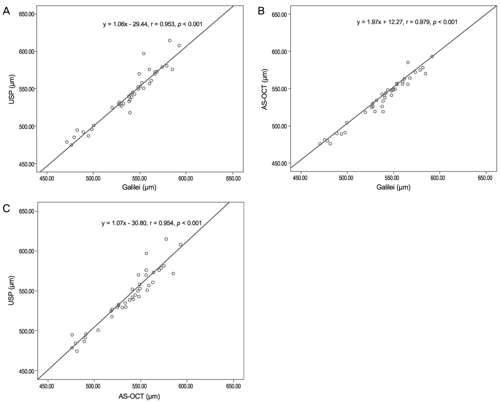
Figure 1 Scattergram showing the correlation of central corneal thickness measured by Galilei, anterior segment optical coherence tomography (AS-OCT), and ultrasound pachtmetry (USP). (A) Correlation between Galilei and USP. (B) Correlation between Galilei and AS-OCT. (C) Correlation between AS-OCT and USP.
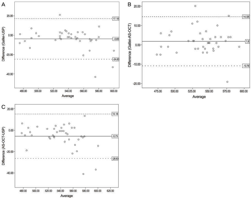
Figure 2 Bland Altman plots between the 2 methods. The middle line is the mean and the lines on the side represent the upper and lower 95% limits of agreement (LoA). (A) Galilei and ultrasound pachymetry (USP). (B) Galilei and anterior segment optical coherence tomography (AS-OCT). (C) AS-OCT and USP.
Reference
-
1. Yau CW, Cheng HC. Microkeratome blades and corneal flap thickness in LASIK. Ophthalmic Surg Lasers Imaging. 2008. 39:471–475.2. Whitacre MM, Stein RA, Hassanein K. The effect of corneal thickness on applanation tonometry. Am J Ophthalmol. 1993. 115:592–596.3. Kim DH, Kim MS, Kim JH. Early corneal-thickness changes after penetrating keratoplasty. J Korean Ophthalmol Soc. 1997. 38:1355–1361.4. Wang Z, Chen J, Yang B. Posterior corneal surface topographic changes after laser in situ keratomileusis are related to residual corneal bed thickness. Ophthalmology. 1999. 106:406–409.5. Doughty MJ, Zaman ML. Human corneal thickness and its impact on intraocular pressure measures: a review and meta-analysis approach. Surv Ophthalmol. 2000. 44:367–408.6. Realini T, Lovelace K. Measuring central corneal thickness with ultrasound pachymetry. Optom Vis Sci. 2003. 80:437–439.7. Sanchis-Gimeno JA, Lleo-Perez A, Casanova J, et al. Inter-observer variability of central corneal thickness measurements using non-contact specular microscopy after laser in situ keratomileusis. Clin Exp Optom. 2004. 87:15–18.8. Yoo C, Eom YS, Suh YW, Kim YY. Central corneal thickness and anterior scleral thickness in Korean patients with open-angle glaucoma: an anterior segment optical coherence tomography study. J Glaucoma. 2011. 20:95–99.9. Hashemi H, Roshani M, Mehravaran S, et al. Effect of corneal thickness on the agreement between ultrasound and Orbscan II pachymetry. J Cataract Refract Surg. 2007. 33:1694–1700.10. Ceylan OM, Turk A, Erdurman C, et al. Comparison of Oculus Pentacam and Stratus optical coherence tomography for measurement of central corneal thickness. Cornea. 2011. 30:670–674.11. Li EY, Mohamed S, Leung CK, et al. Agreement among 3 methods to measure corneal thickness: ultrasound pachymetry, Orbscan II, and Visante anterior segment optical coherence tomography. Ophthalmology. 2007. 114:1842–1847.12. Wang L, Shirayama M, Koch DD. Repeatability of corneal power and wavefront aberration measurements with a dual-Scheimpflug Placido corneal topographer. J Cataract Refract Surg. 2010. 36:425–430.13. Zhao MH, Zou J, Wang WQ, Li J. Comparison of central corneal thickness as measured by non-contact specular microscopy and ultrasound pachymetry before and post LASIK. Clin Experiment Ophthalmol. 2007. 35:818–823.14. Doughty MJ, Jonuscheit S. An assessment of regional differences in corneal thickness in normal human eyes, using the Orbscan II or ultrasound pachymetry. Optometry. 2007. 78:181–190.15. Al-Mezaine HS, Al-Amro SA, Kangave D, et al. Comparison of central corneal thickness measurements using Pentacam and ultrasonic pachymetry in post-LASIK eyes for myopia. Eur J Ophthalmol. 2010. 20:852–857.16. Kim HY, Budenz DL, Lee PS, et al. Comparison of central corneal thickness using anterior segment optical coherence tomography vs ultrasound pachymetry. Am J Ophthalmol. 2008. 145:228–232.17. Thomas J, Wang J, Rollins AM, Sturm J. Comparison of corneal thickness measured with optical coherence tomography, ultrasonic pachymetry, and a scanning slit method. J Refract Surg. 2006. 22:671–678.18. Ling T, Ho A, Holden BA. Method of evaluating ultrasonic pachometers. Am J Optom Physiol Opt. 1986. 63:462–466.19. Copt RP, Thomas R, Mermoud A. Corneal thickness in ocular hypertension, primary open-angle glaucoma, and normal tension glaucoma. Arch Ophthalmol. 1999. 117:14–16.20. Harper CL, Boulton ME, Bennett D, et al. Diurnal variations in human corneal thickness. Br J Ophthalmol. 1996. 80:1068–1072.21. Rodrigues EB, Johanson M, Penha FM. Anterior segment tomography with the cirrus optical coherence tomography. J Ophthalmol. 2012. 2012:806989. Epub 2012 Jan 24.22. Savini G, Carbonelli M, Barboni P, Hoffer KJ. Repeatability of automatic measurements performed by a dual Scheimpflug analyzer in unoperated and post-refractive surgery eyes. J Cataract Refract Surg. 2011. 37:302–309.23. Yeter V, Sönmez B, Beden U. Comparison of central corneal thickness measurements by Galilei Dual-Scheimpflug analyzer® and ultrasound pachymeter in myopic eyes. Ophthalmic Surg Lasers Imaging. 2012. 43:128–134.24. Ladi JS, Shah NA. Comparison of central corneal thickness measurements with the Galilei dual Scheimpflug analyzer and ultrasound pachymetry. Indian J Ophthalmol. 2010. 58:385–388.25. Bechmann M, Thiel MJ, Neubauer AS, et al. Central corneal thickness measurement with a retinal optical coherence tomography device versus standard ultrasonic pachymetry. Cornea. 2001. 20:50–54.26. Leung DY, Lam DK, Yeung BY, Lam DS. Comparison between central corneal thickness measurements by ultrasound pachymetry and optical coherence tomography. Clin Experiment Ophthalmol. 2006. 34:751–754.27. Azen SP, Burg KA, Smith RE, Maguen E. A comparison of three methods for the measurement of corneal thickness. Invest Ophthalmol Vis Sci. 1979. 18:535–538.28. Lee YE, Jun RM. The intra and inter-examiner repeatability of corneal parameters obtained by GALILEI(TM) in normal subjects. J Korean Ophthalmol Soc. 2009. 50:1611–1616.29. Menassa N, Kaufmann C, Goggin M, et al. Comparison and reproducibility of corneal thickness and curvature readings obtained by the Galilei and the Orbscan II analysis systems. J Cataract Refract Surg. 2008. 34:1742–1747.
- Full Text Links
-
- Actions
-
Cited
- CITED
-
- Close
- Share
-
- Similar articles
-
- A Comparison of Central Corneal Thickness Measurements and Measurement Repeatability Using Three Imaging Modalities
- Comparison of Anterior Segment Parameters Obtained by Anterior Segment Optical Coherence Tomography and Dual Rotating Scheimpflug Camera
- Utility of the Swept Source Optical Coherence Tomography for Measurements of Central Corneal Thickness
- Utility of the Anterior Segment Optical Coherence Tomography for Measurements of Central Corneal Thickness
- Reproducibility of IntraLASIK Flap Thickness Measured with Optical Coherence Tomography
