Transcription Factors Regulating Inflammatory Cytokine Production Are Differentially Expressed in Peripheral Blood Mononuclear Cells of Behçet Disease Depending on Disease Activity
- Affiliations
-
- 1Department of Microbiology, Ajou University School of Medicine, Suwon, Korea. sinsun@ajou.ac.kr
- 2Department of Biomedical Sciences, The Graduate School, Ajou University, Suwon, Korea.
- 3Department of Dermatology, Ajou University School of Medicine, Suwon, Korea. esl@ajou.ac.kr
- KMID: 2394840
- DOI: http://doi.org/10.5021/ad.2017.29.2.173
Abstract
- BACKGROUND
Behçet disease (BD) is a relapsing inflammatory disease with increased production of inflammatory cytokines in peripheral blood mononuclear cells (PBMCs); however, the underlying molecular mechanisms are not well known.
OBJECTIVE
To analyze whether the differential expression of transcription factors is involved in the increased tumor necrosis factor (TNF)-α and interleukin (IL)-6 production by PBMCs of BD patients compared to healthy controls (HCs).
METHODS
Expression of transcription factors was examined by real-time reverse transcriptase-polymerase chain reaction and western blotting. Cytokine production by CD11b+ cells transfected with siRNAs against transcription factors was measured by enzyme-linked immunosorbent assay.
RESULTS
In the absence of lipopolysaccharide stimulation, the transcript level of CCAAT-enhancer-binding proteins (C/EBP) β was increased in PBMCs from patients with active BD compared to that in PBMCs from patients with stable BD. The C/EBPδ transcript level was higher in PBMCs from patients with active BD than in those from HCs. The activating transcription factor 3 (ATF3) transcript level was increased in PBMCs from patients with stable BD compared to that in PBMCs from HCs. siRNAs targeting C/EBPβ and C/EBPδ significantly reduced the production of IL-6 and TNF-α in lipopolysaccharide-stimulated CD11b+ cells from patients with BD as well as from HCs.
CONCLUSION
We found differential expression of C/EBPβ, C/EBPδ, and ATF3 in PBMCs from patients with BD depending on disease activity, indicating the involvement of these molecules in BD pathogenesis.
Keyword
MeSH Terms
-
Activating Transcription Factor 3
Behcet Syndrome*
Blotting, Western
CCAAT-Enhancer-Binding Proteins
Cytokines
Enzyme-Linked Immunosorbent Assay
Gene Expression
Humans
Interleukin-6
Interleukins
RNA, Small Interfering
Transcription Factors*
Tumor Necrosis Factor-alpha
Activating Transcription Factor 3
CCAAT-Enhancer-Binding Proteins
Cytokines
Interleukin-6
Interleukins
RNA, Small Interfering
Transcription Factors
Tumor Necrosis Factor-alpha
Figure
-
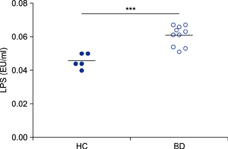
Fig. 1 Increased lipopolysaccharide (LPS) concentration in sera of patients with Behçet disease (BD). LPS concentration was analyzed in the sera of five healthy controls (HCs) and 10 patients with BD (BD), using a Chromogenic LAL Endotoxin Assay Kit. The Mann-Whitney test was conducted. ***p<0.005.
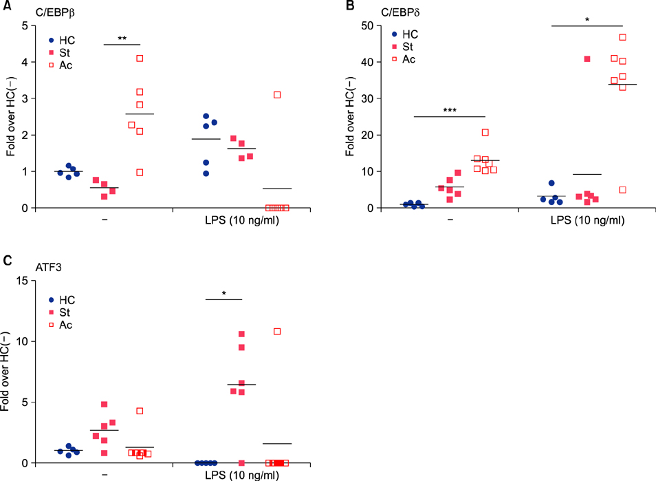
Fig. 2 Differential mRNA expression of CCAAT-enhancer-binding proteins (C/EBP) β (A), C/EBPδ (B), and activating transcription factor 3 (ATF3) (C) in peripheral blood mononuclear cells (PBMCs) from Behçet disease (BD) patients. PBMCs isolated from 5 healthy controls (HCs), 4 to 6 stable BD patients (St) and 6 to 7 active BD patients (Ac) were cultured with or without lipopolysaccharide (LPS) for 3 hours. mRNA levels of C/EBPβ, C/EBPδ, and ATF3 were analyzed by real-time reverse transcription-polymerase chain reaction. Fold over HC(–): Relative mRNA level versus the average mRNA level in HC without LPS stimulation. Each symbol represents a single subject. Bars represent the mean of each group. The Kruskal-Wallis test with Dunn's procedure was conducted. *p <0.05, **p<0.01, ***p<0.005.
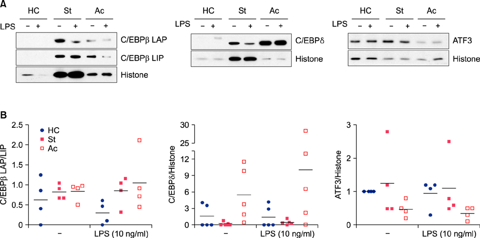
Fig. 3 Protein levels of CCAAT-enhancer-binding proteins (C/EBPβ), C/EBPδ, and activating transcription factor 3 (ATF3) in peripheral blood mononuclear cells (PBMCs) from Behçet disease (BD) patients. PBMCs isolated from healthy controls (HCs), stable BD patients (St), or active BD patients (Ac) were cultured with or without lipopolysaccharide (LPS) for 3 hours. Cell lysates were subjected to western blotting. Representative Western blots of nuclear lysates of 4 or 5 independent experiments (A). Relative band intensity to the indicated protein was compared between groups (B). Each symbol represents a subject and the bars represent the mean.
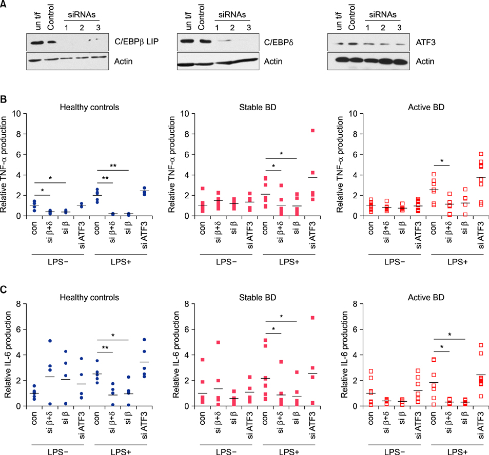
Fig. 4 Suppressive effect of siRNA targeting CCAAT-enhancer-binding proteins (C/EBP) β and C/EBPδ on the production of tumor necrosis factor (TNF)-α and interleukin (IL)-6. (A) Knockdown of the indicated transcription factors using specific siRNA. THP-1 cells were transfected with control siRNA (control) or siRNA specific for C/EBPβ, C/EBPδ, or ATF3. After 24 hours of transfection, protein levels were determined by Western blotting. un t/f: untransfected. (B, C) CD11b+ cells isolated from 5 healthy controls (HCs), 9 stable Behçet disease (BD) patients (St), and 10 active BD patients (Ac) were transfected with the indicated siRNA. After 24 hours, culture media was replaced with fresh media with or without lipopolysaccharide (LPS) (10 ng/ml). After 3 hours, the concentration of TNF-α and IL-6 in the media was measured. Relative production is the ratio of cytokine concentration in each culture condition relative to the average cytokine concentration in the control siRNA (con)-transfected culture without LPS stimulation. Each symbol represents a subject and the bars represent the mean of each group. Con, siRNA for C/EBPβ (siβ), siRNA for ATF3 (siATF3), a combination of siβ and C/EBPδ (siβ+δ). The Mann-Whitney test was conducted. *p<0.05, **p<0.01. un t/f: untransfection.
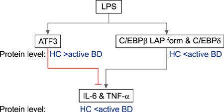
Fig. 5 Graphical summary. LPS: lipopolysccharide, ATF3: activating transcription factor 3, C/EBPβ LAP: CCAAT-enhancerbinding proteins β liver-enriched transcriptional-activator protein, HC: healthy controls, BD: Behcet disease, IL-6: interleukin-6, TNF-α: tumor necrosis factor-α.
Reference
-
1. Pineton de Chambrun M, Wechsler B, Geri G, Cacoub P, Saadoun D. New insights into the pathogenesis of Behçet's disease. Autoimmun Rev. 2012; 11:687–698.
Article2. Sim JH, Park MJ, Park S, Lee ES. Altered expression of costimulatory molecules in Behçet's disease according to clinical activity. Br J Dermatol. 2011; 164:1285–1291.
Article3. Mege JL, Dilsen N, Sanguedolce V, Gul A, Bongrand P, Roux H, et al. Overproduction of monocyte derived tumor necrosis factor alpha, interleukin (IL) 6, IL-8 and increased neutrophil superoxide generation in Behçet's disease. A comparative study with familial Mediterranean fever and healthy subjects. J Rheumatol. 1993; 20:1544–1549.4. Akman A, Sallakci N, Coskun M, Bacanli A, Yavuzer U, Alpsoy E, et al. TNF-alpha gene 1031 T/C polymorphism in Turkish patients with Behçet's disease. Br J Dermatol. 2006; 155:350–356.5. Medzhitov R, Horng T. Transcriptional control of the inflammatory response. Nat Rev Immunol. 2009; 9:692–703.
Article6. Lu YC, Kim I, Lye E, Shen F, Suzuki N, Suzuki S, et al. Differential role for c-Rel and C/EBPbeta/delta in TLR-mediated induction of proinflammatory cytokines. J Immunol. 2009; 182:7212–7221.
Article7. Whitmore MM, Iparraguirre A, Kubelka L, Weninger W, Hai T, Williams BR. Negative regulation of TLR-signaling pathways by activating transcription factor-3. J Immunol. 2007; 179:3622–3630.
Article8. Cangemi R, Pignatelli P, Carnevale R, Bartimoccia S, Nocella C, Falcone M, et al. Low-grade endotoxemia, gut permeability and platelet activation in community-acquired pneumonia. J Infect. 2016; 73:107–114.
Article9. Calkhoven CF, Müller C, Leutz A. Translational control of C/EBPalpha and C/EBPbeta isoform expression. Genes Dev. 2000; 14:1920–1932.10. Tsai VW, Mohammad MG, Tolhurst O, Breit SN, Sawchenko PE, Brown DA. CCAAT/enhancer binding protein-δ expression by dendritic cells regulates CNS autoimmune inflammatory disease. J Neurosci. 2011; 31:17612–17621.
Article11. Dasgupta S, Jana M, Liu X, Pahan K. Role of very-late antigen-4 (VLA-4) in myelin basic protein-primed T cell contact-induced expression of proinflammatory cytokines in microglial cells. J Biol Chem. 2003; 278:22424–22431.
Article12. Tsushima H, Okazaki K, Hayashida M, Ushijima T, Iwamoto Y. CCAAT/enhancer binding protein β regulates expression of matrix metalloproteinase-3 in arthritis. Ann Rheum Dis. 2012; 71:99–107.
Article13. Lu D, Chen J, Hai T. The regulation of ATF3 gene expression by mitogen-activated protein kinases. Biochem J. 2007; 401:559–567.
Article14. Pan YX, Chen H, Kilberg MS. Interaction of RNA-binding proteins HuR and AUF1 with the human ATF3 mRNA 3'-untranslated region regulates its amino acid limitation-induced stabilization. J Biol Chem. 2005; 280:34609–34616.
Article
- Full Text Links
-
- Actions
-
Cited
- CITED
-
- Close
- Share
-
- Similar articles
-
- The Suppressive Effect of Butyrate and Bromopyruvate on Inflammatory Cytokine Production and Short Chain Fatty Acid Receptor Expression by Blood Mononuclear Cells in Patients with Behçet's Disease
- Analysis of Cytokine Gene Expression in Thyroid Aspirates and Peripheral Blood Mononuclear Cell and in vitro Production of Interferon - Gamma by Peripheral Blood Mononuclear Cell Culture
- Serum Tumor Necrosis Factor,Interleukin-1β and Interleukin-6 Levels in Behçet's Disease
- Cytokine production of peripheral blood mononuclear cells from atopic asthmatics
- Expression of mRNA of GATA-3 and T-bet in the peripheral blood mononuclear cells of atopic asthmatics
