Accuracy of several implant bite registration techniques: an in-vitro pilot study
- Affiliations
-
- 1Department of Prosthodontics, Dental Research Institute, Institute of Translation Dental Science, School of Dentistry, Pusan National University, Yangsan, Republic of Korea. huhjb@pusan.ac.kr
- 2Department of Prosthodontics, Seoul National University Gwanak Dental Hospital, Seoul, Republic of Korea.
- 3BK21 PLUS Project, School of Dentistry, Pusan National University, Yangsan, Republic of Korea.
- 4Department of Prosthodontics, In-Je University Haeundae Paik Hospital, Busan, Republic of Korea.
- KMID: 2393219
- DOI: http://doi.org/10.4047/jap.2017.9.5.341
Abstract
- PURPOSE
This study evaluated the accuracies of different bite registration techniques for implant-fixed prostheses using three dimensional file analysis.
MATERIALS AND METHODS
Implant fixtures were placed on the mandibular right second premolar, and the first and second molar in a polyurethane model. Aluwax (A), Pattern Resin (P), and Blu-Mousse (B) were used as the bite registration materials on the healing abutments (H) or temporary abutments (T). The groups were classified into HA, HP, HB, TA, TP, and TB according to each combination. The group using the bite impression coping was the BC group; impression taking and bite registration were performed simultaneously. After impression and bite taking, the scan bodies were connected to the lab analogs of the casts. These casts were scanned using a model scanner. The distances between two reference points in three-dimensional files were measured in each group. One-way ANOVA and Duncan's test were used at the 5% significance level.
RESULTS
The smallest distance discrepancy was observed in the TB group using the temporary abutments. The Blu-Mousse and HP groups showed the largest distance discrepancy. The TB and BC groups showed a lower distance discrepancy than the HP group (P=.001), and there was no significant difference between the groups using the temporary abutments and healing abutments (P>.05).
CONCLUSION
Although this study has limitations as an in-vitro investigation, the groups using the temporary abutments to hold the Blu-Mousse record and bite impression coping showed greater accuracy than the group using the healing abutments to hold the pattern resin record.
Keyword
MeSH Terms
Figure
-
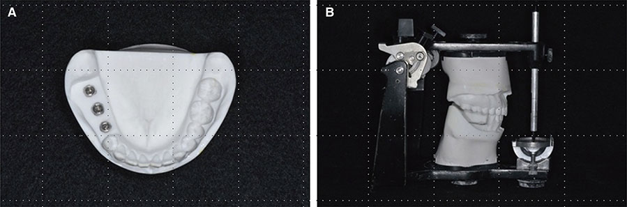
Fig. 1 Fabricated reference model. (A) Top view of the mandibular polyurethane cast with the implant fixtures. (B) Mounting of the polyurethane casts on a semi-adjustable dental articulator.
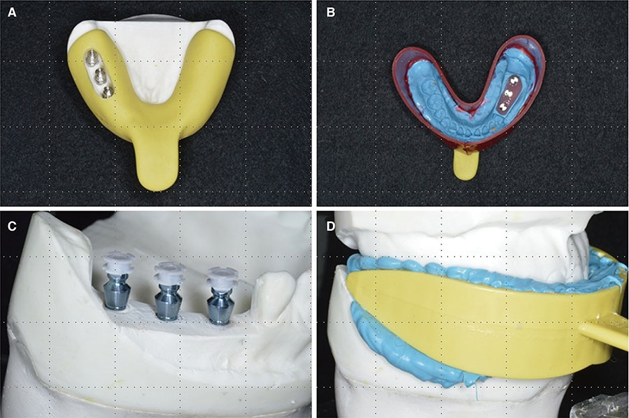
Fig. 2 Final impression taking. (A) Individual tray for groups using the healing abutment and temporary abutment. (B) Final impression with an individual tray. (C) Connection of the Pick Cap coping for the group with a bite impression coping. (D) Final impression with a bite tray for the group with a bite impression coping.
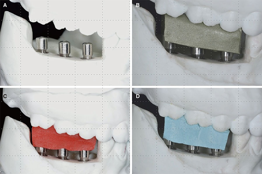
Fig. 3 Bite registration for groups with the healing abutments. (A) Connection of the healing abutments, (B) Group HA: Aluwax, (C) Group HP: Pattern Resin, (D) Group HB: Blu-Mousse.
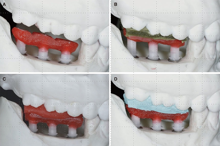
Fig. 4 Bite registration for groups using the temporary abutments. (A) Connection of the temporary abutments, (B) Group TA: Aluwax, (C) Group TP: Pattern Resin, (D) Group TB: Blu-Mousse.
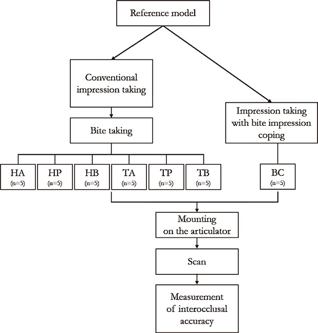
Fig. 5 Schematic diagram of this study. HA = using healing abutments to hold the Aluwax record; HP = using healing abutments to hold the Pattern Resin record; HB = using healing abutments to hold the Blu-Mousse record; TA = using temporary abutments to hold the Aluwax record; TP = using temporary abutments to hold the Pattern Resin record; TB = using temporary abutments to hold Blu-Mousse record; BC = using bite impression copings and a bite tray.
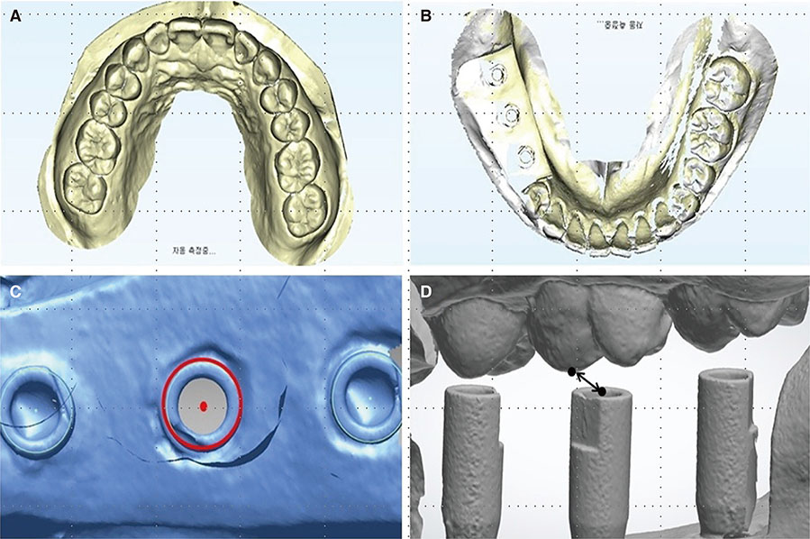
Fig. 6 Measurements for the evaluation of interocclusal accuracy. (A) Maxillary arch 3D file, (B) Mandibular arch 3D file, (C) Mandibular reference point: the center of the superior surface of the right mandibular first molar site scan body, (D) Distance between the center of the superior surface of the right mandibular first molar site scan body and the mesiopalatal cusp tip of the maxillary first molar.
Reference
-
1. Freilich MA, Altieri JV, Wahle JJ. Principles for selecting interocclusal records for articulation of dentate and partially dentate casts. J Prosthet Dent. 1992; 68:361–367.2. Ockert-Eriksson G, Eriksson A, Lockowandt P, Eriksson O. Materials for interocclusal records and their ability to reproduce a 3-dimensional jaw relationship. Int J Prosthodont. 2000; 13:152–158.3. Anup G, Ahila SC, Vasanthakumar M. Evaluation of dimensional stability, accuracy and surface hardness of interocclusal recording materials at various time intervals: an in vitro study. J Indian Prosthodont Soc. 2011; 11:26–31.4. Mullick SC, Stackhouse JA Jr, Vincent GR. A study of interocclusal record materials. J Prosthet Dent. 1981; 46:304–307.5. Savabi O, Nejatidanesh F. Interocclusal record for fixed implant-supported prosthesis. J Prosthet Dent. 2004; 92:602–603.6. Wicks R, Ahuja S, Jain V, Ferreira CF. A technique to create an interocclusal bite registration using in situ implant healing abutments. J Tenn Dent Assoc. 2013; 93:47–49.7. Monzavi A, Alikhasi M, Taghavi F. Occlusal recording components for dental implant-supported prostheses. J Dent (Tehran). 2012; 9:76–78.8. Tejo SK, Kumar AG, Kattimani VS, Desai PD, Nalla S, Chaitanya KK. A comparative evaluation of dimensional stability of three types of interocclusal recording materials-an in-vitro multi-centre study. Head Face Med. 2012; 8:27.9. Chun JH, Pae A, Kim SH. Polymerization shrinkage strain of interocclusal recording materials. Dent Mater. 2009; 25:115–120.10. Roh JY, Kamg MK, Shim SY, Kim KN, Kim KM. Evaluation of physical properties of polyvinylsiloxane interocclusal recording materials. Res Soc Dent Mater. 2006; 33:301–307.11. Ghazal M, Ludwig K, Habil RN, Kern M. Evaluation of vertical accuracy of interocclusal recording materials. Quintessence Int. 2008; 39:727–732.12. Gibbs SB, Versluis A, Tantbirojn D, Ahuja S. Comparison of polymerization shrinkage of pattern resins. J Prosthet Dent. 2014; 112:293–298.13. Müller J, Götz G, Hörz W, Kraft E. An experimental study on the influence of the derived casts on the accuracy of different recording materials. Part I: Plaster, impression compound, and wax. J Prosthet Dent. 1990; 63:263–269.14. Park HY, Chang IT, Kim KN. An experimental study on the accuracy of interocclusal recording materials. J Korean Acad Prosthodont. 1990; 28:91–108.15. Breeding LC, Dixon DL, Kinderknecht KE. Accuracy of three interocclusal recording materials used to mount a working cast. J Prosthet Dent. 1994; 71:265–270.16. Park JY, Kim HY, Kim JH, Kim JH, Kim WC. Comparison of prosthetic models produced by traditional and additive manufacturing methods. J Adv Prosthodont. 2015; 7:294–302.17. Müller J, Götz G, Hörz W, Kraft E. Study of the accuracy of different recording materials. J Prosthet Dent. 1990; 63:41–46.18. Iwaki Y, Wakabayashi N, Igarashi Y. Dimensional accuracy of optical bite registration in single and multiple unit restorations. Oper Dent. 2013; 38:309–315.19. Tripodakis AP, Vergos VK, Tsoutsos AG. Evaluation of the accuracy of interocclusal records in relation to two recording techniques. J Prosthet Dent. 1997; 77:141–146.20. Lee H, So JS, Hochstedler JL, Ercoli C. The accuracy of implant impressions: a systematic review. J Prosthet Dent. 2008; 100:285–291.21. Parker MH, Cameron SM, Hughbanks JC, Reid DE. Comparison of occlusal contacts in maximum intercuspation for two impression techniques. J Prosthet Dent. 1997; 78:255–259.22. Lassila V. Comparison of five interocclusal recording materials. J Prosthet Dent. 1986; 55:215–218.23. Michalakis KX, Pissiotis A, Anastasiadou V, Kapari D. An experimental study on particular physical properties of several interocclusal recording media. Part II: Linear dimensional change and accompanying weight change. J Prosthodont. 2004; 13:150–159.24. Millstein PL, Kronman JH, Clark RE. Determination of the accuracy of wax interocclusal registrations. J Prosthet Dent. 1971; 25:189–196.25. Vergos VK, Tripodakis AP. Evaluation of vertical accuracy of interocclusal records. Int J Prosthodont. 2003; 16:365–368.26. Ghazal M, Albashaireh ZS, Kern M. The ability of different materials to reproduce accurate records of interocclusal relationships in the vertical dimension. J Oral Rehabil. 2008; 35:816–820.27. Mojon P, Oberholzer JP, Meyer JM, Belser UC. Polymerization shrinkage of index and pattern acrylic resins. J Prosthet Dent. 1990; 64:684–688.28. Müller J, Götz G, Hörz W, Kraft E. An experimental study on the influence of the derived casts on the accuracy of different recording materials. Part II: Polyether, acrylic resin, and corrected wax wafer. J Prosthet Dent. 1990; 63:389–395.29. Millstein PL, Hsu CC. Differential accuracy of elastomeric recording materials and associated weight change. J Prosthet Dent. 1994; 71:400–403.30. Breeding LC, Dixon DL. Compression resistance of four interocclusal recording materials. J Prosthet Dent. 1992; 68:876–878.31. Odman PA, Jemt TM. Accuracy of impression materials in a semi-clinical model. Dent Mater. 1988; 4:64–67.32. Klineberg I, Jagger R. Occlusion and clinical practice: an evidence-based approach. Edinburgh: Wright/Elsevier;2004. p. 83–89.33. Davies SJ, Gray RJ, Linden GJ, James JA. Occlusal considerations in periodontics. Br Dent J. 2001; 191:597–604.
- Full Text Links
-
- Actions
-
Cited
- CITED
-
- Close
- Share
-
- Similar articles
-
- A novel method for testing accuracy of bite registration using intraoral scanners
- Accuracy of new implant impression technique using dual arch tray and bite impression coping
- Accuracy of the Point-Based Image Registration Method in Integrating Radiographic and Optical Scan Images: A Pilot Study
- Accuracy of five implant impression technique: effect of splinting materials and methods
- Effect of image matching experience on the accuracy and working time for 3D image registration between radiographic and optical scan images
