Effects of Selenium on Colon Carcinogenesis Induced by Azoxymethane and Dextran Sodium Sulfate in Mouse Model with High-Iron Diet
- Affiliations
-
- 1Department of Veterinary Medicine, College of Veterinary Medicine and Research Institute of Veterinary Medicine, Chungbuk National University, Cheongju, Republic of Korea. beomjun@cbu.ac.kr
- KMID: 2391852
- DOI: http://doi.org/10.5625/lar.2011.27.1.9
Abstract
- Selenium (Se) is known to prevent several cancers while the relationship between high iron and the risk of colorectal cancer is controversial. To investigate the effects of Se in colon carcinogenesis, we subjected three different levels of Se and high-iron diet to a mouse model of colon cancer in which animals were treated with three azoxymethane (AOM) injections followed by dextran sodium sulfate (DSS) administration. There were five experimental groups including vehicle group [normal-Fe (NFe, 45 ppm)+medium-Se (MSe, 0.1 ppm)], positive control group (AOM/DSS+NFe+MSe), AOM/DSS+high-Fe (HFe, 450 ppm)+low-Se (LSe, 0.02 ppm), AOM/DSS+HFe+MSe, and AOM/DSS+HFe+high-Se (HSe, 0.5 ppm). The animals were fed on the three different Se diets for 24 weeks. The incidence of colon tumor in the high-Se diet group (AOM/DSS+HFe+HSe) showed 19.4% lower than positive control group, 5.9% lower than AOM/DSS+HFe+MSe diet group, and 11.1% lower than AOM/DSS+HFe+LSe group. The tumor multiplicity was significantly higher in the low-Se diet group (AOM/DSS+HFe+LSe) compare to all other AOM/DSS treated groups. In the high-Se diet group, the activity of hepatic GPx was comparable to that of positive control group, and significantly higher than those of low-Se or medium-Se diet groups. Expression level of hepatic GPx-1 showed similar results. Hepatic malondialdehyde (MDA) level (indicator of oxidative stress) in the low-Se diet group showed the highest compared to the other groups, and it was significantly higher than positive control group. In the high-Se diet group the level of MDA in the liver was significantly lower than all other AOM/DSS treated groups. High-Se diet group showed significantly lower proliferative index than low-Se and medium-Se groups. The apoptotic indices in low-Se group and medium-Se group were significantly lower than positive control group. However, apoptotic index of high-Se diet group was significantly higher than all other AOM/DSS treated groups. These findings suggest that dietary Se supplement may have protective effect against colon cancer by decreasing proliferation, increasing apoptosis of tumor cells, and reducing oxidative stress in mice with high iron diet.
MeSH Terms
Figure
-
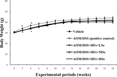
Figure 1 Change in the body weights of mice fed the high Fe diet. All AOM/DSS-treated groups showed a low body weight compared with the vehicle group during experimental periods. AOM: azoxymethane, DSS: dextran sodium sulfate, HFe: high-Fe diet (450 ppm), LSe: low-Se diet (0.02 ppm), MSe: medium-Se diet (0.1 ppm), HSe: high-Se diet (0.5 ppm). Data were represented as mean±SE. abMeans not sharing common superscript letters are significantly different at P<0.05.
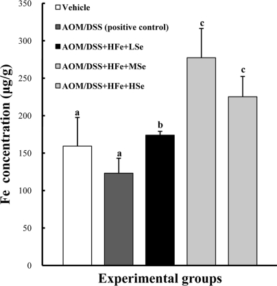
Figure 2 Hepatic Fe levels in mice fed the high Fe diet. Fe concentration was determined using an inductively coupled plasma spectrophotometer. High Fe diet significantly increased hepatic Fe levels compared with the control group. AOM: azoxymethane, DSS: dextran sodium sulfate, HFe: high-Fe diet (450 ppm), LSe: low-Se diet (0.02 ppm), MSe: medium-Se diet (0.1 ppm), HSe: high-Se diet (0.5 ppm). Data were represented as mean±SE. abcMeans not sharing common superscript letters are significantly different at P<0.05.
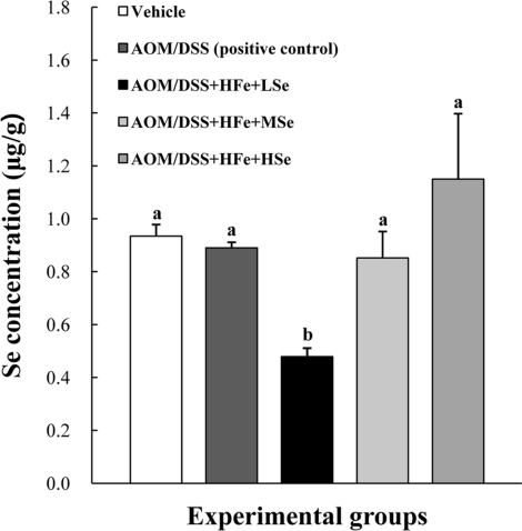
Figure 3 Hepatic Se levels in mice fed the high Fe diet with different Se levels. The Se concentration was determined using an inductively coupled plasma mass spectrophotometer. The hepatic Se levels were dependent on dietary Se levels. AOM: azoxymethane, DSS: dextran sodium sulfate, HFe: high-Fe diet (450 ppm), LSe: low-Se diet (0.02 ppm), MSe: medium-Se diet (0.1 ppm), HSe: high-Se diet (0.5 ppm). Data were represented as mean±SE. abMeans not sharing common superscript letters are significantly different at P<0.05.
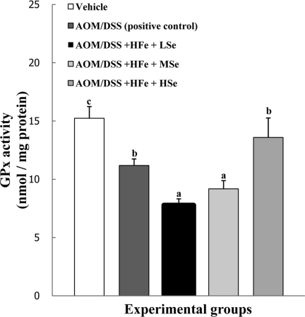
Figure 4 Effect of Se on hepatic GPx activity in mice fed the high Fe diet with different Se levels. The activity of GPx was dependent on dietary Se levels. AOM: azoxymethane, DSS: dextran sodium sulfate, HFe: high-Fe diet (450 ppm), LSe: low-Se diet (0.02 ppm), MSe: medium-Se diet (0.1 ppm), HSe: high-Se diet (0.5 ppm). Data were represented as mean±SE. abcMeans not sharing common superscript letters are significantly different at P<0.05.
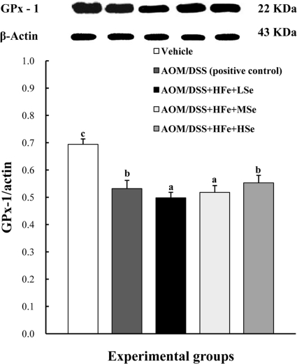
Figure 5 Effect of Se on hepatic GPx-1 by western blot in mice fed the high Fe diet with different Se levels. The expression of GPx-1 was up-regulated in the high-Se group compared with the low-Se group. AOM: azoxymethane, DSS: dextran sodium sulfate, HFe: high-Fe diet (450 ppm), LSe: low-Se diet (0.02 ppm), MSe: medium-Se diet (0.1 ppm), HSe: high-Se diet (0.5 ppm). Data were represented as mean±SE. abcMeans not sharing common superscript letters are significantly different at P<0.05.
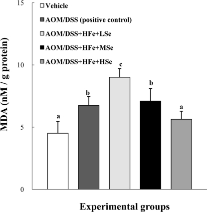
Figure 6 Effect of Se on hepatic MDA levels in mice fed the high Fe diet with different Se levels. The MDA levels were decreased by dietary Se in a dose-dependent manner. AOM: azoxymethane, DSS: dextran sodium sulfate, HFe: high-Fe diet (450 ppm), LSe: low-Se diet (0.02 ppm), MSe: medium-Se diet (0.1 ppm), HSe: high-Se diet (0.5 ppm). Data were represented as mean±SE. abcdMeans not sharing common superscript letters are significantly different at P<0.05.
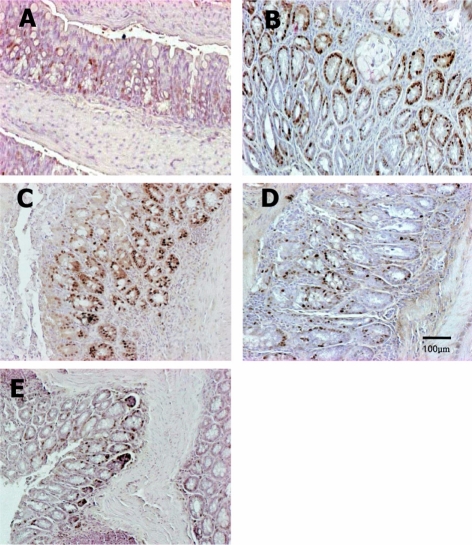
Figure 7 Immunohistochemistry of PCNA in the colon of mice fed the low Fe diet with different Se levels. The PCNA-positive cells were increased by treatment with AOM/DSS, but they were reduced by co-administration of a high concentration of Se. Vehicle (A), AOM/DSS+NFe+MSe (positive control, B), AOM/DSS+HFe+LSe (C), AOM/DSS+HFe+MSe (D), AOM/DSS+HFe+HSe (E).
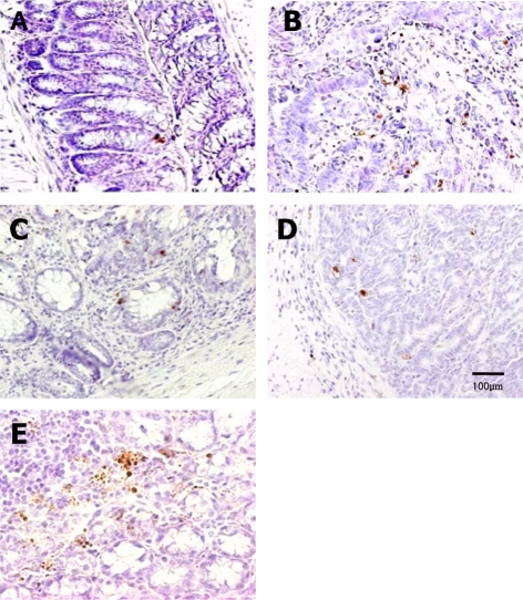
Figure 8 TUNEL assay for apoptotic nuclei in distal colon sections of mice fed high-Fe diet with different Se levels. The TUNEL-positive cells were increased by dietary Se in a dose-dependent manner. Vehicle (A), AOM/DSS+NFe+MSe (positive control, B), AOM/DSS+HFe+LSe (C), AOM/DSS+HFe+MSe (D), AOM/DSS+HFe+HSe (E).
Reference
-
1. Beckman KB, Ames BN. The free radical theory of aging matures. Physiol Rev. 1998; 78(2):547–581. PMID: 9562038.
Article2. Beno I, Klvanova J, Magalova T, Brtkova A. Blood levels of natural antioxidants in gastric and colorectal precancerous lesions and cancers in Slovakia. Neoplasma. 2000; 47(1):37–40. PMID: 10870685.3. Britton RS. Metal-induced hepatotoxicity. Semin Liver Dis. 1996; 16(1):3–12. PMID: 8723319.
Article4. Burk RF, Hill KE. Regulation of selenoproteins. Annu Rev Nutr. 1993; 13:65–81. PMID: 8369160.
Article5. Butler JA, Thomson CD, Whanger PD, Robinson MF. Selenium distribution in blood fractions of New Zealand women taking organic or inorganic selenium. Am J Clin Nutr. 1991; 53(3):748–754. PMID: 2000831.
Article6. Clark LC, Combs GF Jr, Turnbull BW, Slate EH, Chalker DK, Chow J, Davis LS, Glover RA, Graham GF, Gross EG, Krongrad A, Lesher JL Jr, Park HK, Sanders BB Jr, Smith CL, Taylor JR. Nutritional Prevention of Cancer Study Group. Effects of selenium supplementation for cancer prevention in patients with carcinoma of the skin. A randomized controlled trial. JAMA. 1996; 276(24):1957–1963. PMID: 8971064.7. Combs GF Jr, Clark LC, Turnbull BW. An analysis of cancer prevention by selenium. Biofactors. 2001; 14(1-4):153–159. PMID: 11568452.
Article8. Connelly-Frost A, Poole C, Satia JA, Kupper LL, Millikan RC, Sandler RS. Selenium, apoptosis, and colorectal adenomas. Cancer Epidemiol Biomarkers Prev. 2006; 15(3):486–493. PMID: 16537706.
Article9. Evans P, Halliwell B. Micronutrients: oxidant/antioxidant status. Br J Nutr. 2001; 85:S67–S74. PMID: 11509092.
Article10. Feng Y, Finley JW, Davis CD, Becker WK, Fretland AJ, Hein DW. Dietary selenium reduces the formation of aberrant crypts in rats administered 3,2'-dimethyl-4-aminobiphenyl. Toxicol Appl Pharmacol. 1999; 157(1):36–42. PMID: 10329505.
Article11. Finley JW, Davis CD. Selenium (Se) from high-selenium broccoli is utilized differently than selenite, selenate and selenomethionine, but is more effective in inhibiting colon carcinogenesis. Biofactors. 2001; 14(1-4):191–196. PMID: 11568456.
Article12. Ilsley JN, Belinsky GS, Guda K, Zhang Q, Huang X, Blumberg JB, Milbury PE, Roberts LJ 2nd, Stevens RG, Rosenberg DW. Dietary iron promotes azoxymethane-induced colon tumors in mice. Nutr Cancer. 2004; 49(2):162–169. PMID: 15489209.
Article13. Khanna SS, Karjodkar FR. Circulating immune complexes and trace elements (Copper, Iron and Selenium) as markers in oral precancer and cancer: a randomised, controlled clinical trial. Head Face Med. 2006; 2:33–39. PMID: 17040577.
Article14. Korea National Statistical Office. Death and cause of death statistics 2006. 2007. Daejeon: KNSO;p. 35–120.15. Larsson SC, Wolk A. Meat consumption and risk of colorectal cancer: a meta-analysis of prospective studies. Int J Cancer. 2006; 119(11):2657–2664. PMID: 16991129.
Article16. Lund EK, Wharf SG, Fairweather-Tait SJ, Johnson IT. Increases in the concentrations of available iron in response to dietary iron supplementation are associated with changes in crypt cell proliferation in rat large intestine. J Nutr. 1998; 128(2):175–179. PMID: 9446839.
Article17. National Research Council. Selenium in Nutrition. 1983. revised edition. Washington DC: National Academy Press;p. 13–25.18. Norat T, Riboli E. Meat consumption and colorectal cancer: a review of epidemiologic evidence. Nutr Rev. 2001; 59(2):37–47. PMID: 11310774.
Article19. Okayasu I, Hatakeyama S, Yamada M, Ohkusa T, Inagaki Y, Nakaya R. A novel method in the induction of reliable experimental acute and chronic ulcerative colitis in mice. Gastroenterology. 1990; 98(3):694–702. PMID: 1688816.
Article20. Ravn-Haren G, Bugel S, Krath BN, Hoac T, Stagsted J, Jorgensen K, Bresson JR, Larsen EH, Dragsted LO. A short-term intervention trial with selenate, selenium-enriched yeast and selenium-enriched milk: effects on oxidative defence regulation. Br J Nutr. 2008; 99(4):883–892. PMID: 17888202.
Article21. Rosenberg DW, Giardina C, Tanaka T. Mouse models for the study of colon carcinogenesis. Carcinogenesis. 2009; 30(2):183–196. PMID: 19037092.
Article22. Saito Y, Sato N, Hirashima M, Takebe G, Nagasawa S, Takahashi K. Domain structure of bi-functional selenoprotein P. Biochem J. 2004; 381(Pt 3):841–846. PMID: 15117283.
Article23. Sohn OS, Fiala ES, Requeijo SP, Weisburger JH, Gonzalez FJ. Differential effects of CYP2E1 status on the metabolic activation of the colon carcinogens azoxymethane and methylazoxymethanol. Cancer Res. 2001; 61(23):8435–8440. PMID: 11731424.24. Soyars KE, Fischer JG. Iron supplementation does not affect cell proliferation or aberrant crypt foci development in the colon of sprague-dawley rats. J Nutr. 1998; 128(4):764–770. PMID: 9521641.
Article25. Stevens RG, Kalkwarf DR. Iron, radiation, and cancer. Environ Health Perspect. 1990; 87:291–300. PMID: 2269234.
Article26. Tanaka T, de Azevedo MB, Duran N, Alderete JB, Epifano F, Genovese S, Tanaka M, Curini M. Colorectal cancer chemoprevention by 2 beta-cyclodextrin inclusion compounds of auraptene and 4'-geranyloxyferulic acid. Int J Cancer. 2010; 126(4):830–840. PMID: 19688830.27. Tanaka T, Kohno H, Suzuki R, Yamada Y, Sugie S, Mori H. A novel inflammation-related mouse colon carcinogenesis model induced by azoxymethane and dextran sodium sulfate. Cancer Sci. 2003; 94(11):965–973. PMID: 14611673.
Article
- Full Text Links
-
- Actions
-
Cited
- CITED
-
- Close
- Share
-
- Similar articles
-
- Dietary Selenium Supplement Prevents Colon Carcinogenesis Induced by Azoxymethane and Dextran Sodium Sulfate in ICR Mice
- Epithelial Cell-specific Deletion of Microsomal Prostaglandin E Synthase-1 Does Not Influence Colon Tumor Development in Mice
- Effects of luteolin on chemical induced colon carcinogenesis in high fat diet-fed obese mouse
- Increase in dietary protein content exacerbates colonic inflammation and tumorigenesis in azoxymethane-induced mouse colon carcinogenesis
- Effect of iron overload with ascorbic acid on experimental colon carcinogenesis in mice
