Difference in Spinal Fusion Process in Osteopenic and Nonosteopenic Living Rat Models Using Serial Microcomputed Tomography
- Affiliations
-
- 1Department of Neurosurgery, Seoul National University Boramae Medical Center, Seoul, Korea.
- 2Department of Neurosurgery, Seoul National University College of Medicine, Seoul, Korea. chungc@snu.ac.kr
- 3Clinical Research Institute, Seoul National University Hospital, Seoul, Korea.
- 4Department of Brain and Cognitive Sciences, Seoul National University College of Natural Sciences, Seoul, Korea.
- KMID: 2382770
- DOI: http://doi.org/10.3340/jkns.2016.0707.002
Abstract
OBJECTIVE
To identify and investigate differences in spinal fusion between the normal and osteopenic spine in a rat model.
METHODS
Female Sprague Dawley rats underwent either an ovariectomy (OVX) or sham operation and were randomized into two groups: non-OVX group and OVX group. Eight weeks after OVX, unilateral lumbar spinal fusion was performed using autologous iliac bone. Bone density (BD) was measured 2 days and 8 weeks after fusion surgery. Microcomputed tomography was used to evaluate the process of bone fusion every two weeks for 8 weeks after fusion surgery. The fusion rate, fusion process, and bone volume parameters of fusion bed were compared between the two groups.
RESULTS
BD was significantly higher in the non-OVX group than in the OVX group 2 days and 8 weeks after fusion surgery. The fusion rate in the non-OVX group was higher than that in the OVX group 8 weeks after surgery (p=0.044). The bony connection of bone fragments with transverse processes and bone formation between transverse processes in non-OVX group were significantly superior to those of OVX group from 6 weeks after fusion surgery. The compactness and bone maturation of fusion bed in non-OVX were prominent compared with the non-OVX group.
CONCLUSION
The fusion rate in OVX group was inferior to non-OVX group at late stage after fusion surgery. Bone maturation of fusion bed in the OVX group was inferior compared with the non-OVX group. Fusion enhancement strategies at early stage may be needed to patients with osteoporosis who need spine fusion surgery.
Keyword
MeSH Terms
Figure
-
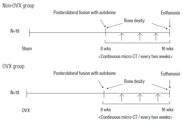
Fig. 1 Experimental groups and time schedule. Eighteen rats underwent bilateral OVX (OVX group), and another 18 rats underwent a sham operation (non-OVX group). Eight weeks after OVX, all rats underwent unilateral spinal fusion using autologous iliac bone. Sequential micro-CT evaluations in all rats were obtained every 2 weeks for 8 weeks after surgery. Bone density of all rats was assessed at 2 days and 8 weeks after fusion surgery. OVX: ovariectomy, micro-CT: microcomputed tomography.
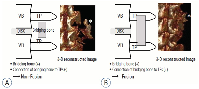
Fig. 2 Assessment of fusion processes using three-dimensional (3D) images. The scheme and 3D reconstructed images show nonfusion (A) and fusion (B). Fusion was defined as the simultaneous presence of bridging bone and the connection of bridging bone to both upper and lower TPs. The shape of the bone fragments in 3D reconstructed image A (asterisk) does not appear as compact as that in 3D reconstructed image B (asterisk). VB: vertebral body, TP: transverse process.
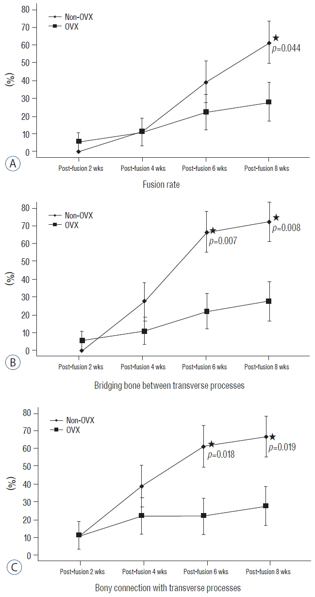
Fig. 3 Comparison of the fusion process between two groups. This graph shows the difference between non-OVX and OVX groups in fusion rate (A), and the ratios of the presence of a bony connection with the TP (B) and bridging bone between TPs (C) at postoperative weeks 2, 4, 6 and 8 (asterisk, p<0.05). OVX: ovariectomy, TP: transverse process.
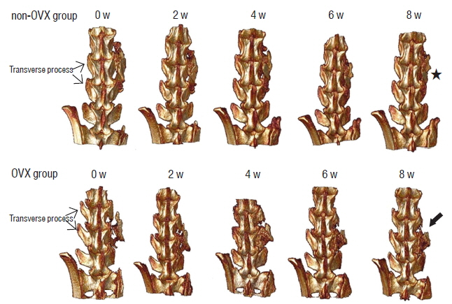
Fig. 4 Micro-CT scanning of fusion masses. The figures show 3D reconstructed images from both groups after surgery. The picture in the upper row shows serial 3D reconstructed images from a rat in the non-OVX group. The grafted bone materials were inserted on the fusion bed between the L4 and L5 TPs. The shape of the bridging bone 8 weeks after surgery (asterisk) was more compact than that in the early period after surgery. The picture in the lower row is a 3D reconstructed image from a rat in the OVX group. The shape of the grafted bone at 8 weeks was less compact in the OVX group (arrow) than in the non-OVX group. Micro-CT: microcomputed tomography, OVX: ovariectomy, TP: transverse process.
Reference
-
References
1. Aldini NN, Fini M, Giavaresi G, Giardino R, Greggi T, Parisini P. Pedicular fixation in the osteoporotic spine: a pilot in vivo study on long-term ovariectomized sheep. J Orthop Res. 20:1217–1224. 2002.
Article2. Bae HW, Zhao L, Kanim LE, Wong P, Marshall D, Delamarter RB. Bone marrow enhances the performance of rhBMP-2 in spinal fusion: a rodent model. J Bone Joint Surg Am. 95:338–347. 2013.3. Boden SD. Biology of lumbar spine fusion and use of bone graft substitutes: present, future, and next generation. Tissue Eng. 6:383–399. 2000.
Article4. Boden SD, Schimandle JH, Hutton WC. An experimental lumbar intertransverse process spinal fusion model. Radiographic, histologic, and biomechanical healing characteristics. Spine (Phila Pa 1976). 20:412–420. 1995.5. Boden SD, Schimandle JH, Hutton WC, Chen MI. 1995 Volvo Award in basic sciences. The use of an osteoinductive growth factor for lumbar spinal fusion Part I: Biology of spinal fusion. Spine (Phila Pa 1976). 20:2626–2632. 1995.6. Bouxsein ML, Boyd SK, Christiansen BA, Guldberg RE, Jepsen KJ, Müller R. Guidelines for assessment of bone microstructure in rodents using micro-computed tomography. J Bone Miner Res. 25:1468–1486. 2010.
Article7. Bridwell KH, Sedgewick TA, O’Brien MF, Lenke LG, Baldus C. The role of fusion and instrumentation in the treatment of degenerative spondylolisthesis with spinal stenosis. J Spinal Disord. 6:461–472. 1993.
Article8. Coe JD, Warden KE, Herzig MA, McAfee PC. Influence of bone mineral density on the fixation of thoracolumbar implants. A comparative study of transpedicular screws, laminar hooks, and spinous process wires. Spine (Phila Pa 1976). 15:902–907. 1990.
Article9. Kamoda H, Ohtori S, Ishikawa T, Miyagi M, Arai G, Suzuki M, et al. The effect of platelet-rich plasma on posterolateral lumbar fusion in a rat model. J Bone Joint Surg Am. 95:1109–1116. 2013.
Article10. Moazzaz P, Gupta MC, Gilotra MM, Gilotra MN, Maitra S, Theerajunyaporn T, et al. Estrogen-dependent actions of bone morphogenetic protein-7 on spine fusion in rats. Spine (Phila Pa 1976). 30:1706–1711. 2005.
Article11. Nakao S, Minamide A, Kawakami M, Boden SD, Yoshida M. The influence of alendronate on spine fusion in an osteoporotic animal model. Spine (Phila Pa 1976). 36:1446–1452. 2011.
Article12. Namkung-Matthai H, Appleyard R, Jansen J, Hao Lin J, Maastricht S, Swain M, et al. Osteoporosis influences the early period of fracture healing in a rat osteoporotic model. Bone. 28:80–86. 2001.
Article13. Omi N, Ezawa I. Animal models for bone and joint disease. Low calcium diet-induced rat model of osteoporosis. Clin Calcium. 21:173–180. 2011.14. Park SB, Chung CK. Strategies of spinal fusion on osteoporotic spine. J Korean Neurosurg Soc. 49:317–322. 2011.
Article15. Park SB, Kim CH, Hong M, Yang HJ, Chung CK. Effect of a selective estrogen receptor modulator on bone formation in osteoporotic spine fusion using an ovariectomized rat model. Spine J. 16:72–81. 2016.
Article16. Schindeler A, McDonald MM, Bokko P, Little DG. Bone remodeling during fracture repair: the cellular picture. Semin Cell Dev Biol. 19:459–466. 2008.
Article17. Takahata M, Ito M, Abe Y, Abumi K, Minami A. The effect of anti-resorptive therapies on bone graft healing in an ovariectomized rat spinal arthrodesis model. Bone. 43:1057–1066. 2008.
Article18. Toribatake Y, Hutton WC, Tomita K, Boden SD. Vascularization of the fusion mass in a posterolateral intertransverse process fusion. Spine (Phila Pa 1976). 23:1149–1154. 1998.
Article19. Wiltse LL, Spencer CW. New uses and refinements of the paraspinal approach to the lumbar spine. Spine (Phila Pa 1976). 13:696–706. 1988.
Article20. Xu SW, Yu R, Zhao GF, Wang JW. Early period of fracture healing in ovariectomized rats. Chin J Traumatol. 6:160–166. 2003.21. Zipfel GJ, Guiot BH, Fessler RG. Bone grafting. Neurosurg Focus. 14:e8. 2003.
Article
- Full Text Links
-
- Actions
-
Cited
- CITED
-
- Close
- Share
-
- Similar articles
-
- The Effect of Hyaluronate-Carboxymethyl Cellulose on Bone Graft Substitute Healing in a Rat Spinal Fusion Model
- Animal Models of Orthopedic Research: A Spinal Fusion Model
- Influence of Alendronate and Endplate Degeneration to Single Level Posterior Lumbar Spinal Interbody Fusion
- The Effect of Risedronate on Posterior Lateral Spinal Fusion in a Rat Model
- Clinical and Radiological Outcomes of Segmental Spinal Fusion in Transforaminal Lumbar Interbody Fusion with Spinous Process Tricortical Autograft
