Inhibiting Cytochrome C Oxidase Leads to Alleviated Ischemia Reperfusion Injury
- Affiliations
-
- 1Department of Anesthesia, Xiang-Ya Second Hospital, Central South University, Changsha, China. lei_liull@sina.com
- 2Department of Anesthesiology, the Second Affiliated Hospital, University of South China, Hengyang, China.
- 3Department of Anesthesiology, Zunyi Medical College, Zunyi, Guizhou, China.
- KMID: 2377459
- DOI: http://doi.org/10.4070/kcj.2016.0137
Abstract
- BACKGROUND AND OBJECTIVES
The overall purpose of this study was to investigate the role of cytochrome C oxidase (CcO) in preventing ischemia reperfusion-induced cardiac injury through gaseous signaling molecule pathways.
MATERIALS AND METHODS
We used CcO inhibitor, potassium cyanide (KCN) to mimic the pre-treatment of gaseous signaling molecules in a global ischemia/reperfusion (IR) injury model in rats. Intracellular reactive oxygen species (ROS) was determined by measuring mitochondrial H2O2 and mitochondrial complex activity.
RESULTS
KCN pre-treatment led to decreased infarction area after IR injury and improved cardiac function. KCN pre-treated group challenged with IR injury was associated with reduced ROS production through inhibition of activity and not downregulation of CcO expression. In addition, KCN pre-treatment was associated with enhanced expression and activity of mitochondrial antioxidase, suggesting the role of CcO in regulating IR injury through oxidative stress.
CONCLUSION
KCN pre-treatment reduced the severity of IR injury. The potential mechanism could be increased endogenous anti-oxidase activity and consequently, the enhanced clearance of ROS.
MeSH Terms
-
Animals
Cytochromes c*
Cytochromes*
Down-Regulation
Electron Transport Complex IV*
Infarction
Ischemia*
Mitochondria
Myocardial Infarction
Oxidative Stress
Potassium Cyanide
Rats
Reactive Oxygen Species
Reperfusion Injury*
Cytochromes
Cytochromes c
Electron Transport Complex IV
Potassium Cyanide
Reactive Oxygen Species
Figure
-

Fig. 1 Experimental scheme. K-S: Krebs Henseleit, C: control group, KCN: potassium cyanide pre-treatment group, C-IR: control+ischemia reperfusion group, KCN-IR: potassium cyanide pre-treatment+ischemia reperfusion group.
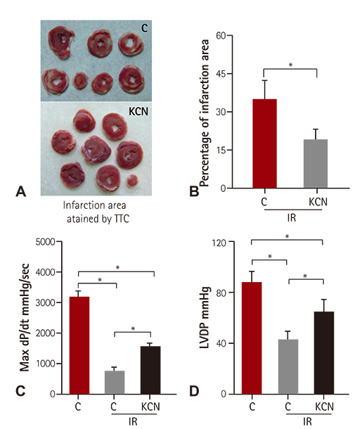
Fig. 2 Pre-treatment of IR resulted in decreased infarction area and improved cardiac function. (A, B) Myocardium infarct size (n=8) with and without KCN (10 uM) pre-treatment including representative images (A) and quantification (B). (C,D) Cardiac function of heart pre-treatment with and without KCN reflected by (C) max dP/dt and (D) LVDP (n=8). C: control group, KCN: potassium cyanide pre-treatment group, TTC: triphenyl tetrazolium chloride, C-IR: control+ischemia reperfusion group, KCN-IR: potassium cyanide pre-treatment+ischemia reperfusion group, LVDP: left ventricular developed pressure, dP/dt: the rate of rise of left ventricular pressure. *p<0.05.
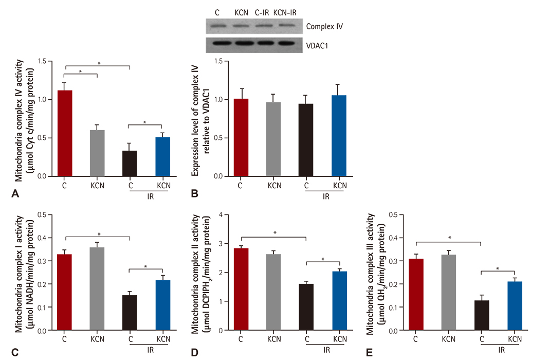
Fig. 3 KCN pre-treatment was associated with inhibited activity of CcO. Mitochondrial complex I, II, III, activity in cardiac tissue of rats challenged with and without IR injury in both control and KCN pre-treated groups. Cardiac (A) mitochondrial complex IV (n=6), (B) complex IV protein expression level (n=6), (C) complex I activity (n=6), (D) complex II activity (n=6) and (E) III (n=6) activity. C: control group, KCN: potassium cyanide pre-treatment group, VDAC1: voltage-dependent anion-selective channel 1, NADH: reduced form of nicotinamide-adenine dinucleotid, DCPIPH2: reduced form of dichlorophenalindophenol, QH2: reduced form of coenzyme Q10, C-IR: control+ischemia reperfusion group, KCN-IR: potassium cyanide pre-treatment+ischemia reperfusion group. *p<0.05.
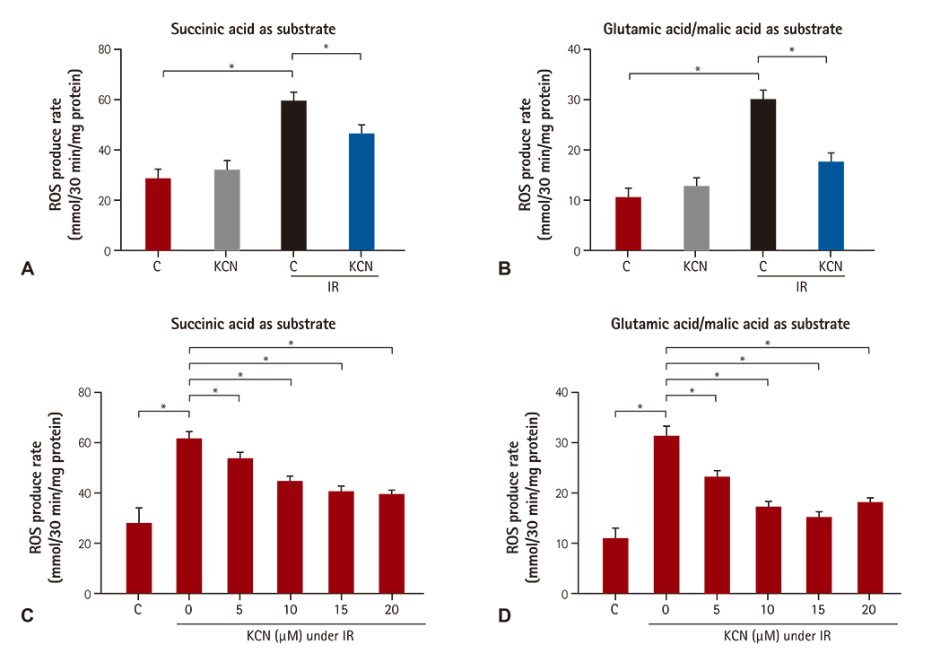
Fig. 4 KCN pre-treated group challenged with IR injury was associated with reduced ROS production. ROS production of cardiac tissue challenged with and without IR injury in both control and KCN pre-treated groups. H2O2 production by heart mitochondria oxidizing complex I (A) and (B) complex II substrates in the four groups. Effect of different concentrations of KCN pre-treatment (5, 10, 15 and 20 uM) on ROS production levels by complex I (C) and complex II (D) induced by IR treatment (both n=6). ROS: reactive oxygen species, C: control group, KCN: potassium cyanide pre-treatment group, C-IR: control+ischemia reperfusion group, KCN-IR: potassium cyanide pre-treatment+ischemia reperfusion group. *p<0.05.
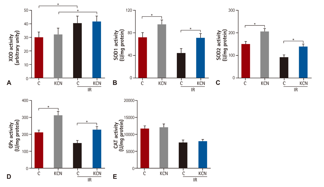
Fig. 5 Pre-treatment was associated with enhanced activity of mitochondrial antioxidase. ROS-related enzyme activity (XOD and antioxidase activities) in cardiac mitochondria of rats challenged with and without IR injury in both control and KCN pre-treated groups. The activity of (A) SOD1 (n=8), (B) SOD2 (n=8), (C) GPx (n=8), (D) CAT (n=8) and (E) XOD (n=8) in cardiac mitochondria. *p<0.05 between indicated groups. XOD: xanthine oxidase activity, SOD1: superoxide dismutases 1, SOD2: superoxide dismutases 2, GPx: glutathione peroxidase, CAT: catalase, C: control group, C: control group, KCN: potassium cyanide pre-treatment group, C-IR: control+ischemia reperfusion group, KCN-IR: potassium cyanide pre-treatment+ischemia reperfusion group.
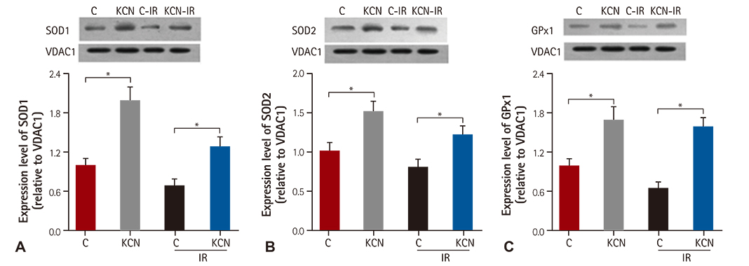
Fig. 6 Pre-treatment was associated with increased expression levels of mitochondrial antioxidase. ROS-related enzyme expression levels. The expression of antioxidase including (A) SOD1 (n=3), (B) SOD2 (n=3) and (C) GPx1(n=3) in cardiac mitochondria of rats challenged with and without IR injury in both control and KCN pre-treated groups. SOD1: superoxide dismutases 1, SOD2: superoxide dismutases 2, GPx: glutathione peroxidase, VDAC1: voltage dependent anion channel 1, C: control group, KCN: potassium cyanide pre-treatment group, C-IR: control+ischemia reperfusion group, KCN-IR: potassium cyanide pre-treatment+ischemia reperfusion group. *p<0.05.
Reference
-
1. Tsukihara T, Aoyama H, Yamashita E, et al. Structures of metal sites of oxidized bovine heart cytochrome c oxidase at 2.8 A. Science. 1995; 269:1069–1074.2. Hüttemann M, Helling S, Sanderson TH, et al. Regulation of mitochondrial respiration and apoptosis through cell signaling: cytochrome c oxidase and cytochrome c in ischemia/reperfusion injury and inflammation. Biochim Biophys Acta. 2012; 1817:598–609.3. Groening P, Huang Z, La Gamma EF, Levy RJ. Glutamine restores myocardial cytochrome C oxidase activity and improves cardiac function during experimental sepsis. JPEN J Parenter Enteral Nutr. 2011; 35:249–254.4. Bruno C, Martinuzzi A, Tang Y, et al. A stop-codon mutation in the human mtDNA cytochrome c oxidase I gene disrupts the functional structure of complex IV. Am J Hum Genet. 1999; 65:611–620.5. Comi GP, Bordoni A, Salani S, et al. Cytochrome c oxidase subunit I microdeletion in a patient with motor neuron disease. Ann Neurol. 1998; 43:110–116.6. Muller-Hocker J. Cytochrome-c-oxidase deficient cardiomyocytes in the human heart--an age-related phenomenon. A histochemical ultracytochemical study. Am J Pathol. 1989; 134:1167–1173.7. Antonicka H, Mattman A, Carlson CG, et al. Mutations in COX15 produce a defect in the mitochondrial heme biosynthetic pathway, causing early-onset fatal hypertrophic cardiomyopathy. Am J Hum Genet. 2003; 72:101–114.8. Papadopoulou LC, Sue CM, Davidson MM, et al. Fatal infantile cardioencephalomyopathy with COX deficiency and mutations in SCO2, a COX assembly gene. Nat Genet. 1999; 23:333–337.9. Prabu SK, Anandatheerthavarada HK, Raza H, Srinivasan S, Spear JF, Avadhani NG. Protein kinase A-mediated phosphorylation modulates cytochrome c oxidase function and augments hypoxia and myocardial ischemia-related injury. J Biol Chem. 2006; 281:2061–2070.10. Booth EA, Flint RR, Lucas KL, Knittel AK, Lucchesi BR. Estrogen protects the heart from ischemia-reperfusion injury via COX-2-derived PGI2. J Cardiovasc Pharmacol. 2008; 52:228–235.11. Vogt S, Ramzan R, Weber P, et al. Ischemic preconditioning results in an ATP-dependent inhibition of cytochrome C oxidase. Shock. 2013; 40:407–413.12. Sun WH, Liu F, Chen Y, Zhu YC. Hydrogen sulfide decreases the levels of ROS by inhibiting mitochondrial complex IV and increasing SOD activities in cardiomyocytes under ischemia/reperfusion. Biochem Biophys Res Commun. 2012; 421:164–169.13. Whittington HJ, Hall AR, McLaughlin CP, Hausenloy DJ, Yellon DM, Mocanu MM. Chronic metformin associated cardioprotection against infarction: not just a glucose lowering phenomenon. Cardiovasc Drugs Ther. 2013; 27:5–16.14. Burkard N, Williams T, Czolbe M, et al. Conditional overexpression of neuronal nitric oxide synthase is cardioprotective in ischemia/reperfusion. Circulation. 2010; 122:1588–1603.15. Guo W, Cheng ZY, Zhu YZ. Hydrogen sulfide and translational medicine. Acta Pharmacol Sin. 2013; 34:1284–1291.16. Zuckerbraun BS, Chin BY, Bilban M, et al. Carbon monoxide signals via inhibition of cytochrome c oxidase and generation of mitochondrial reactive oxygen species. FASEB J. 2007; 21:1099–1106.17. Jain M, Rivera S, Monclus EA, et al. Mitochondrial reactive oxygen species regulate transforming growth factor-beta signaling. J Biol Chem. 2013; 288:770–777.18. Diaz F. Cytochrome c oxidase deficiency: patients and animal models. Biochim Biophys Acta. 2010; 1802:100–110.19. Nicholson CK, Calvert JW. Hydrogen sulfide and ischemia-reperfusion injury. Pharmacol Res. 2010; 62:289–297.20. Chen Q, Camara AK, Stowe DF, Hoppel CL, Lesnefsky EJ. Modulation of electron transport protects cardiac mitochondria and decreases myocardial injury during ischemia and reperfusion. Am J Physiol Cell Physiol. 2007; 292:C137–C147.21. Tanaka-Esposito C, Chen Q, Lesnefsky EJ. Blockade of electron transport before ischemia protects mitochondria and decreases myocardial injury during reperfusion in aged rat hearts. Transl Res. 2012; 160:207–216.
- Full Text Links
-
- Actions
-
Cited
- CITED
-
- Close
- Share
-
- Similar articles
-
- The Effects of Gabexate Mesilate on the Ishemia-Reperfusion Injury in the Rabbit Liver
- Change of Plasma Xanthine Oxidase Activity by Intermittent Hepatic Ischemia-Reperfusion
- Effects of Renal Ischemia/Reperfusion on Renal Function in Rats
- Effect of Methylprednisolone on Cytochrome Oxidase and Lipid Peroxidation of the Contused Spinal Cord
- Reactive oxygen species and N-methyl-D-aspartate receptor-mediated central sensitization in hindlimb ischemia/reperfusion injury-induced neuropathic pain rats
