Ethanol Extract of Perillae Herba Enhances Pentobarbital-Induced Sleep and Non-Rapid Eye Movement (NREM) Sleep through GABA(A)-ergic Systems
- Affiliations
-
- 1College of Pharmacy, Chungbuk National University, Cheongju 361-763, Republic of Korea. kiwan@chungbuk.ac.kr
- KMID: 2376500
- DOI: http://doi.org/10.20307/nps.2017.23.1.53
Abstract
- Perillae Herba has been traditionally used for the sedation in the oriental countries. Therefore, this study was conducted to determine whether Perillae Herba ethanol extract (PHEE) enhances pentobarbital-induced sleeping behaviors in animals. In addition, the possible mechanisms are demonstrated. PHEE (12.5, 25 and 50 mg/kg. p.o.) reduced the locomotor activity in mice. PHEE reduced sleep latency and augmented the total sleep time in pentobarbital (42 mg/kg, i.p.)-induced sleep in mice. Furthermore, the number of sleeping mice treated with sub-hypnotic pentobarbital (28 mg/kg, i.p.) increased. PHEE (50 mg/kg. p.o.) decreased the sleep/wake cycles and wakefulness, and increased total sleeping time and NREM sleep in electroencephalogram (EEG) of rats. In addition, PHEE (0.1, 1.0 and 10 µg/ml) increased the intracellular Clâ» level through the GABA receptors in the hypothalamus of rats. Moreover, the protein of glutamate decarboxylase (GAD) was overexpressed by PFEE. It was found that PHEE enhanced pentobarbital-induced sleeping behaviors through GABA(A)-ergic transmissions.
Keyword
MeSH Terms
Figure
-
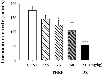
Fig. 1 Effects of PHEE and diazepam (DZ) on locomotor activity test in mice. DZ and PHEE were orally administrated, respectively 30 min and 1 hour before the testing. The measurement of ambulation activity was carried out for 1 hour. Each bar represents the mean with the mean ± S.E.M. The significance was evaluated by using Dunnettest. *P<0.05, **P<0.01, ***P<0.005, compared to the control (CONT).
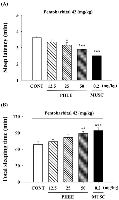
Fig. 2 Effects of muscimol (MUSC) and PHEE on onset and duration of sleep in mice treated with pentobarbital. Mice were starved from 24 hours prior to theexperiment. Before pentobarbital injection, muscimol and PHEE weretreated by i.p. respectively. (A) The sleep latency and (B) Total sleeping time were measured. Each bar represents the mean ± S.E.M. The significance was evaluated by using Dunnett-test. *P<0.05, **P<0.01, ***P<0.005, compared to the control (CONT).

Fig. 3 Effects of PHEE (50 mg/kg) on counts of sleep-wake cycles in rats. Each bar represents the mean ± S.E.M. The significance was evaluated by using Dunnet test. ***P<0.005, compared to the control (CONT).
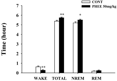
Fig. 4 Effects of PHEE on sleep architectures in rats. It was separated the wakefulness and sleep (NREM and REM) state. Each bar represents the mean ± S.E.M. The significance was evaluated by using Dunnet test. **P<0.01, compared to the control.
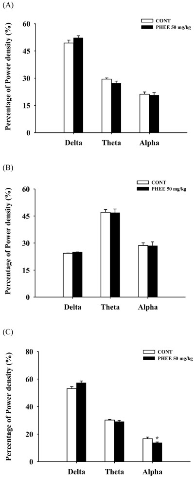
Fig. 5 Effects of PHEE on EEG power density of wakefulness (A), REM sleep (B) and NREM sleep (C).The power density was divided with delta-wave, theta-wave and alpha-wave. Each bar represents the mean ± S.E.M. The significance was evaluated by using Dunnet test. *P<0.05, compared to the control.

Fig. 6 Effects of pentobarbital and PHEE on Cl− influx in primary cultured hypothalamic neuron cells.The hypothalamic neuron cells were cultured for 8 days, and then Cl− influx of the cells was incubated with MQAE. After pentobarbital (PENT, 10 µM) and PHEE (0.1, 1 and 10 µg/ml) were treated for 1 hour, the measurement was carried out. Each bar represents the mean ± S.E.M. The significance was evaluated by using Dunnet t-test. ***P<0.005, compared to the control (CONT).
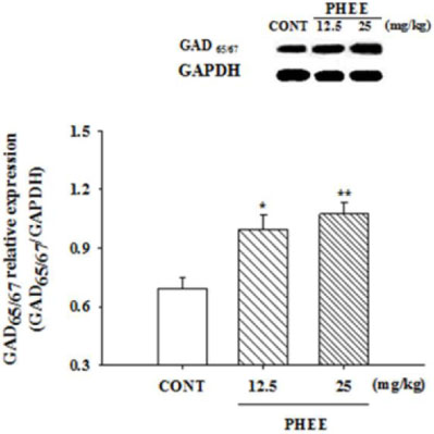
Fig. 7 Effects of PHEE on the expression of GAD. After PHEE (12.5 and 24 mg/kg) oral administration, theexpression of GAD was measured with the expression of GAPDH. GAPDH was needed equally to compare with the expression of the proteins. Each bar represents the mean ± S.E.M. The significance was evaluated by using Dunnet test. *P<0.05, **P<0.01, compared to the control (CONT).
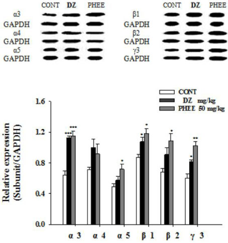
Fig. 8 Effects of PHEE on the expression of GABAA receptors subunits.After PHEE (50mg/kg) oral administration, theexpression of GABAA receptors subunits was measured with the expression of GAPDH. GAPDH was needed equally to compare with the expression of the proteins. Each bar represents the mean ± S.E.M. The significance was evaluated by using Dunnet test. *P<0.05, **P<0.01, ***P<0.005, compared to the control.
Reference
-
1. Pilcher JJ, Ginter DR, Sadowsky B. J Psychosom Res. 1997; 42:583–596.2. Doi Y, Minowa M, Tango T. Sleep. 2003; 26:467–471.3. Iliescu EA, Coo H, McMurray MH. Nephrol Dial Transplant. 2003; 18:126–132.4. Phillips KD, Sowell RL, Boyd M, Dudgeon WD, Hand GA. Mind-Body Research Group. Qual Life Res. 2005; 14:959–970.5. Joanna MW, Stachowicz K, Nowak G, Pilc A. The Loss of Glutamate-GABA Harmony in Anxiety Disorders. USA: InTech;2011. p. 24.6. Olsen RW, Sieghart W. Pharmacol Rev. 2008; 60:243–260.7. Whiting PJ. Drug Discov Today. 2003; 8:445–450.8. Ahn H. Korean J Food Preserv. 2006; 13:703–707.9. Honda G, Koezuka Y, Tabata M. Chem Pharm Bull. 1988; 36:3153–3155.10. Yi LT, Li J, Geng D. J Ethnopharmacol. 2013; 147:245–253.11. Takeda H, Tsuji M, Inazu M, Egashira T, Matsumiya T. Eur J Pharmacol. 2002; 449:261–267.12. Takeda H, Tsuji M, Miyamoto J, Matsumiya T. Psychopharmacology (Berl). 2002; 164:233–235.13. Morton GJ, Kaiyala KJ, Fisher JD, Ogimoto K, Schwartz MW, Wisse BE. Am J Physiol Endocrinol Metab. 2011; 300:E392–E401.14. Wolfman C, Viola H, Marder M, Wasowski C, Ardenghi P, Izquierdo I, Paladini AC, Medina JH. Eur J Pharmacol. 1996; 318:23–30.15. Sanford LD, Yang L, Liu X, Tang X. Brain Res. 2006; 1084:80–88.16. Tokunaga S, Takeda Y, Niimoto T, Nishida N, Kubo T, Ohno T, Matsuura Y, Kawahara Y, Shinomiya K, Kamei C. Biol Pharm Bull. 2007; 30:363–366.17. Ma Y, Han H, Eun JS, Kim HC, Hong JT, Oh KW. Biol Pharm Bull. 2007; 30:1748–1753.18. West MR, Molloy CR. Anal Biochem. 1996; 241:51–58.19. Wagner C, Vargas AP, Roos DH, Morel AF, Farina M, Nogueira CW, Aschner M, Rocha JB. Arch Toxicol. 2010; 84:89–97.20. Fanger BO. Anal Biochem. 1987; 162:11–17.21. Igarashi M, Miyazaki Y. Evid Based Complement Alternat Med. 2013; 2013:925342.22. Ito N, Nagai T, Oikawa T, Yamada H, Hanawa T. Evid Based Complement Alternat Med. 2011; 2011:512697.23. Hu HZ, Li ZW. J Physiol. 1997; 501(Pt 1):67–75.24. Möhler H. J Recept Signal Transduct Res. 2006; 26:731–740.25. Miller MA. Front Neurol. 2015; 6:1–9.26. Liu Z, Xu XH, Liu TY, Hong ZY, Urade Y, Huang ZL, Qu WM. CNS Neurosci Ther. 2012; 18:623–630.
Article27. Mehta AK, Ticku MK. Brain Res Brain Res Rev. 1999; 29:196–217.28. Lambert JJ, Belelli D, Harney SC, Peters JA, Frenguelli BG. Brain Res Brain Res Rev. 2001; 37:68–80.29. Choi JJ, Kim YS, Kwon YO, Yoo JH, Chong MS, Lee MK, Hong JT, Oh KW. Nat Prod Sci. 2015; 21:219–225.
- Full Text Links
-
- Actions
-
Cited
- CITED
-
- Close
- Share
-
- Similar articles
-
- 4-Hydroxybenzaldehyde, One of Constituents from Gastrodiae Rhizoma Augments Pentobarbital-induced Sleeping Behaviors and Non-rapid Eye Movement (NREM) Sleep in Rodents
- Sinomenine, an Alkaloid Derived from Sinomenium acutum Potentiates Pentobarbital-Induced Sleep Behaviors and Non-Rapid Eye Movement (NREM) Sleep in Rodents
- Rosmarinic Acid Potentiates Pentobarbital-Induced Sleep Behaviors and Non-Rapid Eye Movement (NREM) Sleep through the Activation of GABA(A)-ergic Systems
- Poria cocos ethanol extract and its active constituent, pachymic acid, modulate sleep architectures via activation of GABA(A)-ergic transmission in rats
- Potentiation of decursinol angelate on pentobarbital-induced sleeping behaviors via the activation of GABA(A)-ergic systems in rodents
