Two Case Reports of Calcified Spinal Meningioma and a Literature Review
- Affiliations
-
- 1Department of Orthopedic Surgery, Kyungpook National University Hospital, Daegu, South Korea.
- 2Department of Orthopedic Surgery, Changwon Gyeongsang National University Hospital, Changwon, South Korea. whiteugi@naver.com
- KMID: 2365083
- DOI: http://doi.org/10.4184/jkss.2016.23.4.227
Abstract
- STUDY DESIGN: Case Report.
OBJECTIVES
The aim of this study was to report 2 cases of calcified spinal meningioma that displayed differences in appearance during resection, and to review the current literature on calcified and ossified spinal meningiomas. SUMMARY OF LITERATURE REVIEW: Calcified and ossified spinal meningiomas are rare, and tumor calcification is a risk factor for poor neurological outcomes resulting from the additional manipulations required to dissect the tumor.
MATERIALS AND METHODS
We describe the clinical course and intraoperative findings of 2 female patients who presented with symptoms of myelopathy. Magnetic resonance imaging showed calcified spinal meningiomas of the thoracic spine. The type of tumor resection performed was dependent on the solidity and texture of the individual tumors.
RESULTS
Pathologic evaluation revealed psammoma bodies, which suggested calcified meningioma. The patients' neurologic symptoms resolved with no neurologic sequelae.
CONCLUSIONS
Although there are a few pathologic differences regarding the main type and pathogenesis of ossified and calcified meningioma, both are thought to have a poor prognosis. For these tumors, adequately accounting for the expected poor prognosis and performing a wide laminectomy in order to ensure an adequate surgical margin are important factors for achieving a favorable outcome.
MeSH Terms
Figure
-
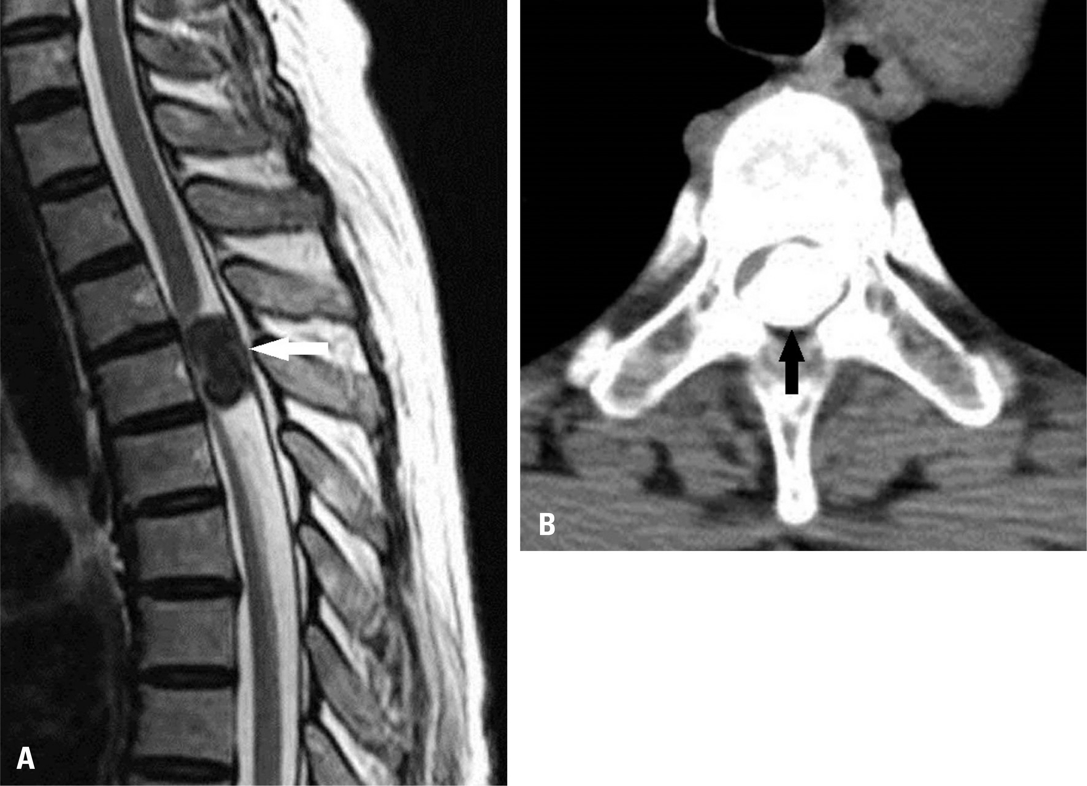
Fig. 1. A sagittal plane T2-weighted magnetic resonance imaging scan exhibited a very low signal (white arrow) in an intradural ex-tramedullary tumor occupying the spinal canal at T4 (A). Computed tomography without enhancement depicted a completely calcified intradural mass (black arrow) occupying the entire spinal canal at the T4 level. The axial view is shown in (B).
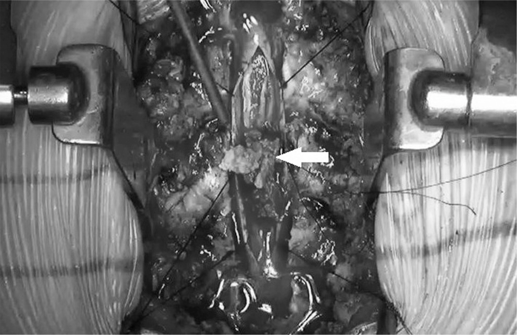
Fig. 2. Intraoperative image showing a tumor (white arrow) at the T4 level. The tumor had a friable solidity and was dissected in a piecemeal manner.
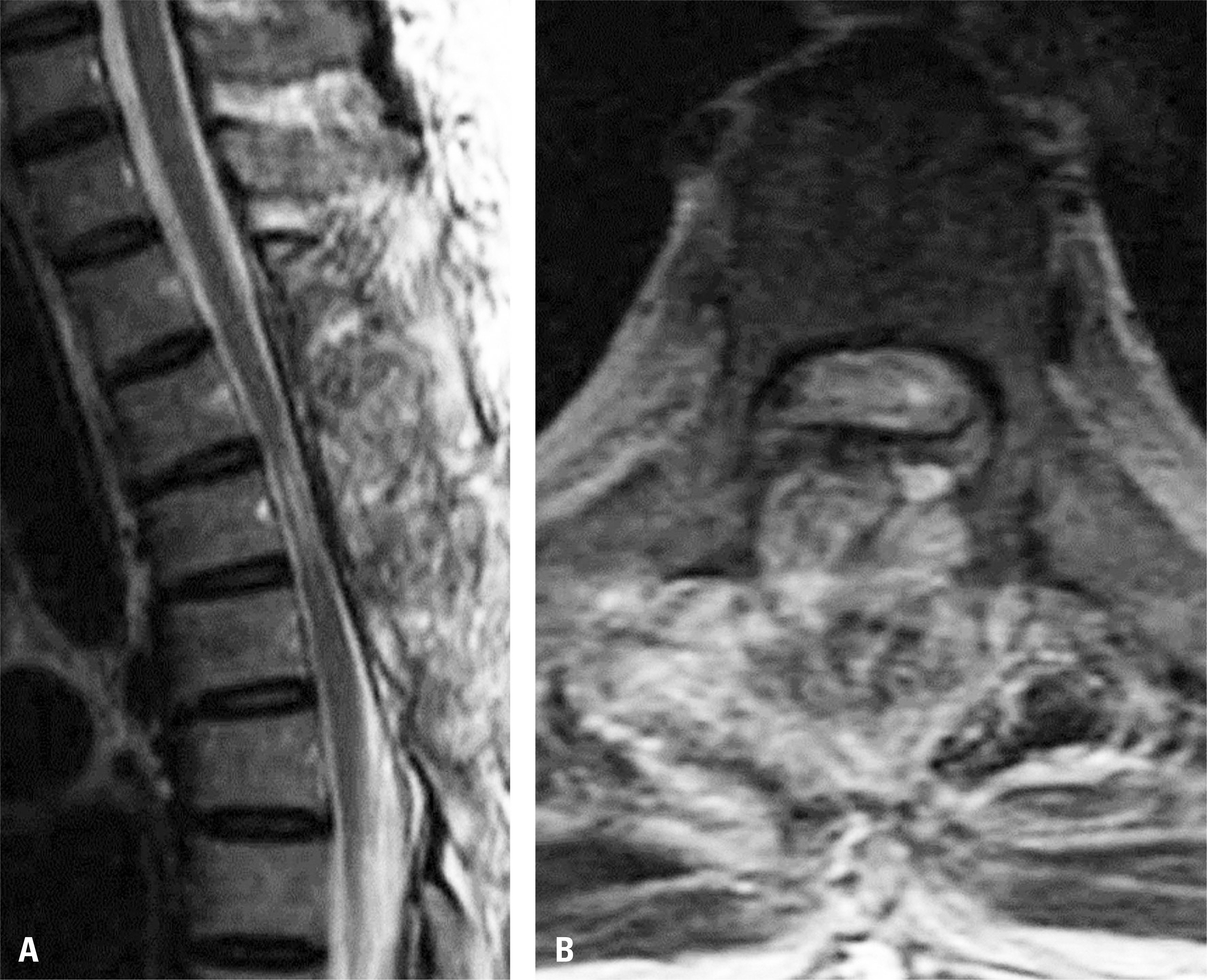
Fig. 3. Postoperative T2-weighted magnetic resonance imaging scan revealing decompression of the flattened cord and recovery of the cord diameter in the thoracic spine. The sagittal view is shown in (A), and the axial view in (B).
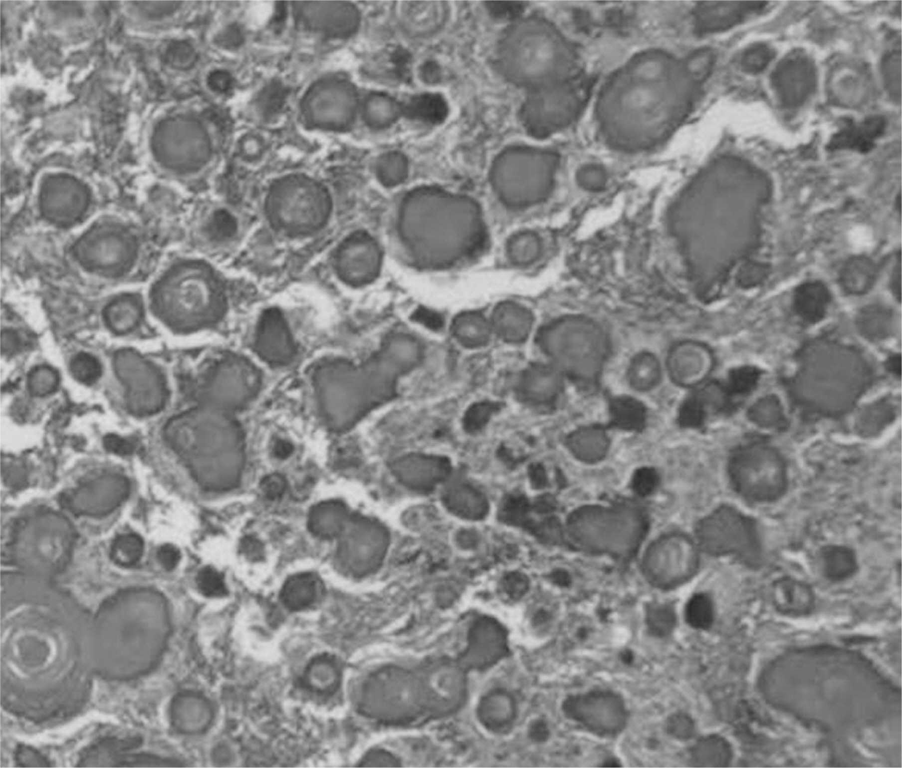
Fig. 4. Microscopy of the specimen demonstrating a psammomatous subtype that was composed of numerous calcified psammoma bodies, suggestive of a calcified meningioma (hematoxylin and eosin staining, ×200 magnification).
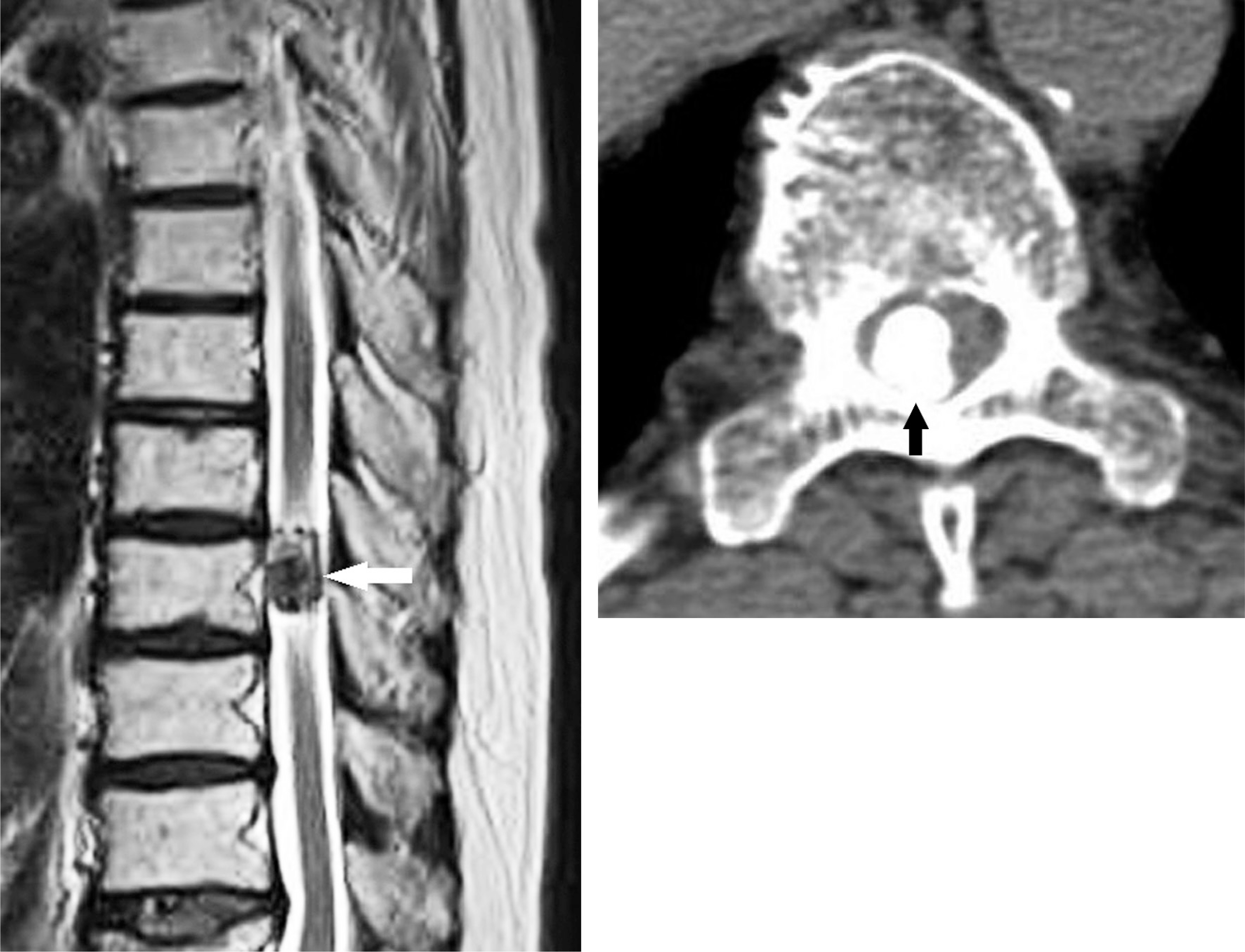
Fig. 5. T2-weighted magnetic resonance imaging scan showing an oval-shaped mass (white arrow) clearly demarcated by a hypodense signal. The sagittal view shown in (A). Computed tomography image showing a calcified mass (black arrow) occupying the spinal canal at the level of the T9 vertebra. The axial view is shown in (B).
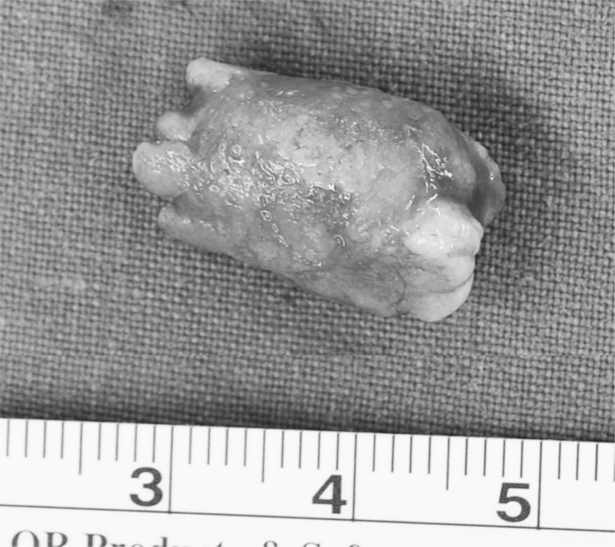
Fig. 6. Gross photography showing en bloc dissection of the firm, solid tumor.
Reference
-
1. Gamache FW Jr, Wang JC, Deck M, et al. Unusual appearance of an en plaque meningioma of the cervical spinal canal. A case report and literature review. Spine (Phila Pa 1976). 2001; 26:E87–9.2. Doita M, Harada T, Nishida K, et al. Recurrent calcified spinal meningioma detected by plain radiograph. Spine(Phila Pa 1976). 2001; 26:E249–52.
Article3. Lee JW, Lee IS, Choi KU, et al. CT and MRI findings of calcified spinal meningiomas: correlation with pathological findings. Skeletal radiology. 2010; 39:345–52.
Article4. Zhu Q, Qian M, Xiao J, et al. Myelopathy due to calcified meningiomas of the thoracic spine: minimum 3-year follow-up after surgical treatment. J Neurosurg Spine. 2013; 18:436–42.
Article5. Levy WJ Jr, Bay J, et al. Spinal cord meningioma. J Neurosurg. 1982; 57:804–12.
Article6. Niijima K, Huang YP, Malis LI, et al. Ossified spinal meningioma en plaque. Spine. 1993; 18:2340–3.
Article7. Huang TY, Kochi M, Kuratsu J, et al. Intraspinal osteo-genic meningioma: report of a case. J Formos Med Assoc. 1999; 98:218–21.8. Uchida K, Nakajima H, Yayama T, et al. Immunohisto-chemical findings of multiple ossified en plaque meningiomas in the thoracic spine. J Clin Neurosci. 2009; 16:1660–2.
Article9. Kubota T, Sato K, Yamamoto S, et al. Ultrastructural study of the formation of psammoma bodies in fibroblastic me-ningioma. J Neurosurg. 1984; 60:512–7.
Article10. Chotai SP, Mrak RE, Mutgi SA, et al. Ossification in an extra-intradural spinal meningioma-pathologic and surgical vistas. Spine J. 2013; 13:E21–6.
Article
- Full Text Links
-
- Actions
-
Cited
- CITED
-
- Close
- Share
-
- Similar articles
-
- Synchronous Development of Spinal Cord Tumor: Meningioma and Schwannoma: A Case Report
- A Case of Totally Calcified Meningioma
- Intraventricular Malignant Meningioma with CSF-Disseminated Spinal Metastasis : Case Report and Literature Review
- Recurrent Spinal Meningioma: A Case Report
- Extradural Spinal Lymphoplasmacyte-Rich Meningioma in the Thoracic Spine: A Case Report and Literature Review
