Three Cases of Focal Choroidal Excavation in the Macula Detected by Spectral-Domain Optical Coherence Tomography
- Affiliations
-
- 1Department of Ophthalmology, Yeungnam University College of Medicine, Daegu, Korea. changwh@ynu.ac.kr
Abstract
- PURPOSE
To report the clinical finding of 3 patients with focal choroidal excavation in the macula detected by spectral-domain optical coherence tomography (SD-OCT).
CASE SUMMARY
Five eyes of 3 patients with focal choroidal excavation detected by SD-OCT were enrolled in the present study. All patients had myopia (average refractive power -5.60 diopter). Two of the 3 patients had focal choroidal excavation in both eyes. All 5 eyes revealed foveal pigmentary changes on fundus examination. The excavation area in the autofluorescence image was hypofluorescent. Fluorescein angiographic finding was normal to various degrees of hyperfluoresence. Indocyanine green angiography revealed hypofluoresence at the excavation area. The excavation involoved from the retinal pigment epithelium layer to the external limiting membrane or outer nuclear layer and average choroidal thickness at excavation were statistically thinner than the uninvolved area based on SD-OCT (p = 0.002). Retinoschisis, serous pigment epithelial detachment and choroidal neovascularziation (CNV) were detected individually in 3 eyes. The other 2 eyes had no specific abnormalities.
CONCLUSIONS
During the follow-up period, the choroidal excavation remained relatively stable in 4 of 5 eyes, but CNV developed in 1 eye. Therefore, intravitreal bevacizumab injection was performed. Longer follow-up periods are necessary to determine the etiology, clinical course and visual prognosis of eyes with focal choroidal excavation.
Keyword
MeSH Terms
Figure
-

Figure 1. A 55-year-old man with high myopia. Color photographs of the right (A) and left (B) eyes show mild foveal retinal pig-ment epithelial changes. Spectral domain optical coherence tomographic scans through the fovea shows retinoschisis with two cho-roidal excavation of the right eye (C) and focal mild choroidal excavation of the left eye (D).
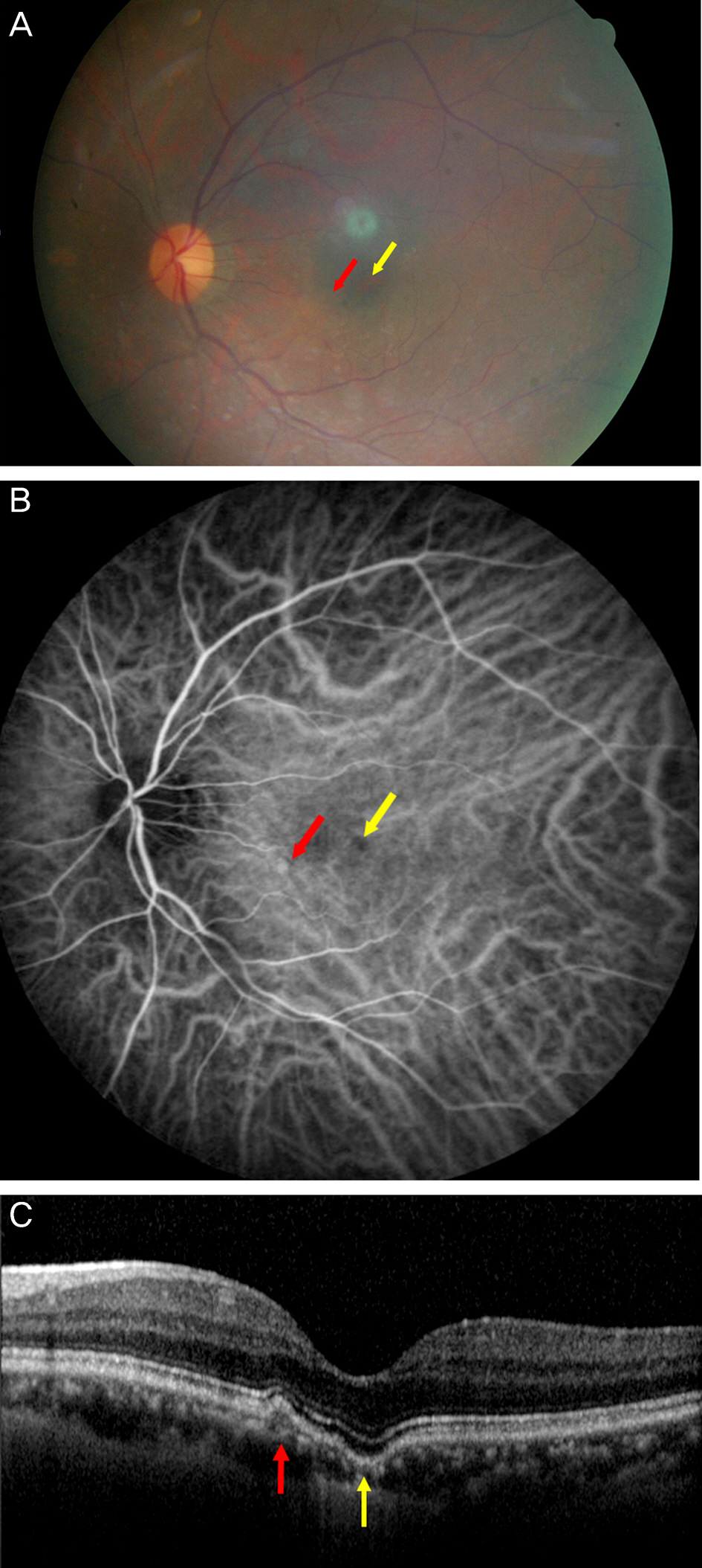
Figure 2. Fundus photography, indocyanine green angiography (ICGA), spectral domain optical coherence tomography (SD-OCT) images of a 56-year-old woman. (A) Fundus photograph at the first examination showed foveal (yellow arrow) and paraf-oveal depigmentation (red arrow) in the left eye. (B) Foveal hy-pofluoresence (yellow arrow) and parafoveal hyperfluoresence (red arrow) are observed on ICGA. (C) Focal foveal choroidal excavation (yellow arrow) and parafoveal serous pigment epi-thelial detachment (red arrow) are seen on SD-OCT.
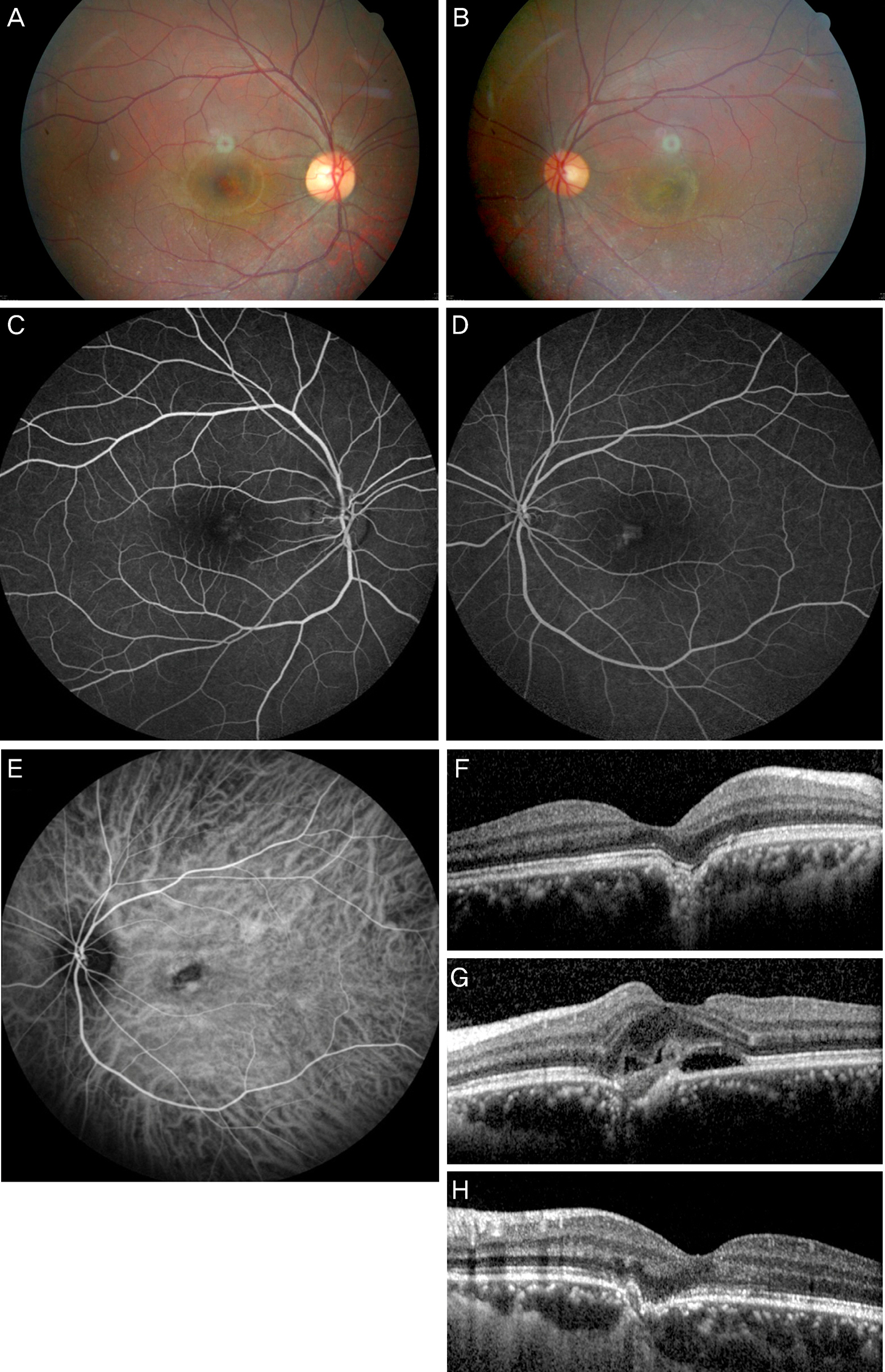
Figure 3. Findings in a 27-year-old man with bilateral focal choroidal excavation. Fundus photographs on the initial presentation show pigmentary change at the parafovea in the right eye (A) and yellowish exudates lesion at the parafovea in the left eye (B). Fluorescein angiography shows window defect in both eyes (C) and weak leakage in the left eye (D). Indocyanine green angiography shows hypofluoresence and choroidal neovascularization at the parafovea in the left eye (E). Spectral domain optical coherence to-mographic shows focal choroidal excavation at the parafovea in the right eye (F) and Bruch's membrane disruption with subretinal fluid and choroidal excavation at the parafovea in the left eye (G). One month after three times of intravitreal bevacizumab in-jections, the subretinal fluid was absorbed but focal choroidal excavation was unchanged in the left eye (H).
Reference
-
References
1. Jampol LM, Shankle J, Schroeder R, et al. Diagnostic and ther-apeutic challenges. Retina. 2006; 26:1072–6.
Article2. Wakabayashi Y, Nishimura A, Higashide T, et al. Unilateral choroi-dal excavation in the macula detected by spectral-domain optical coherence tomography. Acta Ophthalmol. 2010; 88:e87–91.
Article3. Margolis R, Mukkamala SK, Jampol LM, et al. The expanded spectrum of focal choroidal excavation. Arch Ophthalmol. 2011; 129:1320–5.
Article4. Kobayashi W, Abe T, Tamai H, Nakazawa T. Choroidal excavation with polypoidal choroidal vasculopathy: a case report. Clin Ophthalmol. 2012; 6:1373–6.
Article5. Katome T, Mitamura Y, Hotta F, et al. Two cases of focal choroidal excavation detected by spectral-domain optical coherence tomography. Case Rep Ophthalmol. 2012; 3:96–103.
Article
- Full Text Links
-
- Actions
-
Cited
- CITED
-
- Close
- Share
-
- Similar articles
-
- A Case of Focal Choroidal Excavation Associated with Chronic Central Serous Chorioretinopathy
- Choroidal Thickness at the Outside of Fovea in Diabetic Retinopathy Using Spectral-Domain Optical Coherence Tomography
- A Case of Ocular Toxoplasmosis Imaged with Spectral Domain Optical Coherence Tomography
- Availability of Optical Coherence Tomography in Diagnosis and Classification of Choroidal Neovascularization
- Choroidal Thickness in Primary Open-Angle Glaucoma Using Spectral-Domain Optical Coherence Tomography
