J Korean Ophthalmol Soc.
2010 Oct;51(10):1414-1418.
Case of Tsutsugamushi Disease With Anterior Uveitis
- Affiliations
-
- 1Department of Ophthalmology, Sungmo Eye Hospital, Busan, Korea. hippo007@dreamwiz.com
Abstract
- PURPOSE
To report a single case of tsutsugamushi disease with anterior uveitis and eschars on the upper eyelid.
CASE SUMMARY
A 56-year-old female patient complained of ocular pain and gradually decreasing visual acuity in her right eye. On physical examination, lymphadenopathy was palpable on the right side of the neck, and eschars were observed on the forehead and upper eyelid. On slit lamp examination, conjunctival injection, episcleral vessel dilations and severe intraocular inflammatory reaction were observed. Fundus examination showed no abnormal findings. A blood test was submitted for analysis, and tsutsugamushi disease was diagnosed. The patient's ocular manifestations responded well to treatment with steroids. After three weeks, the patient showed improvement on ocular examination, and no problems were observed at the six-month follow-up.
CONCLUSIONS
Although ocular manifestation of tsutusgamushi disease with conjunctivitis and limbitis has previously been reported, ocular manifestation of tsutusgamushi disease with anterior uveitis has not been reported. Anterior uveitis may respond satisfactorily to steroid treatment, along with improvement in systemic conditions.
Keyword
MeSH Terms
Figure
-
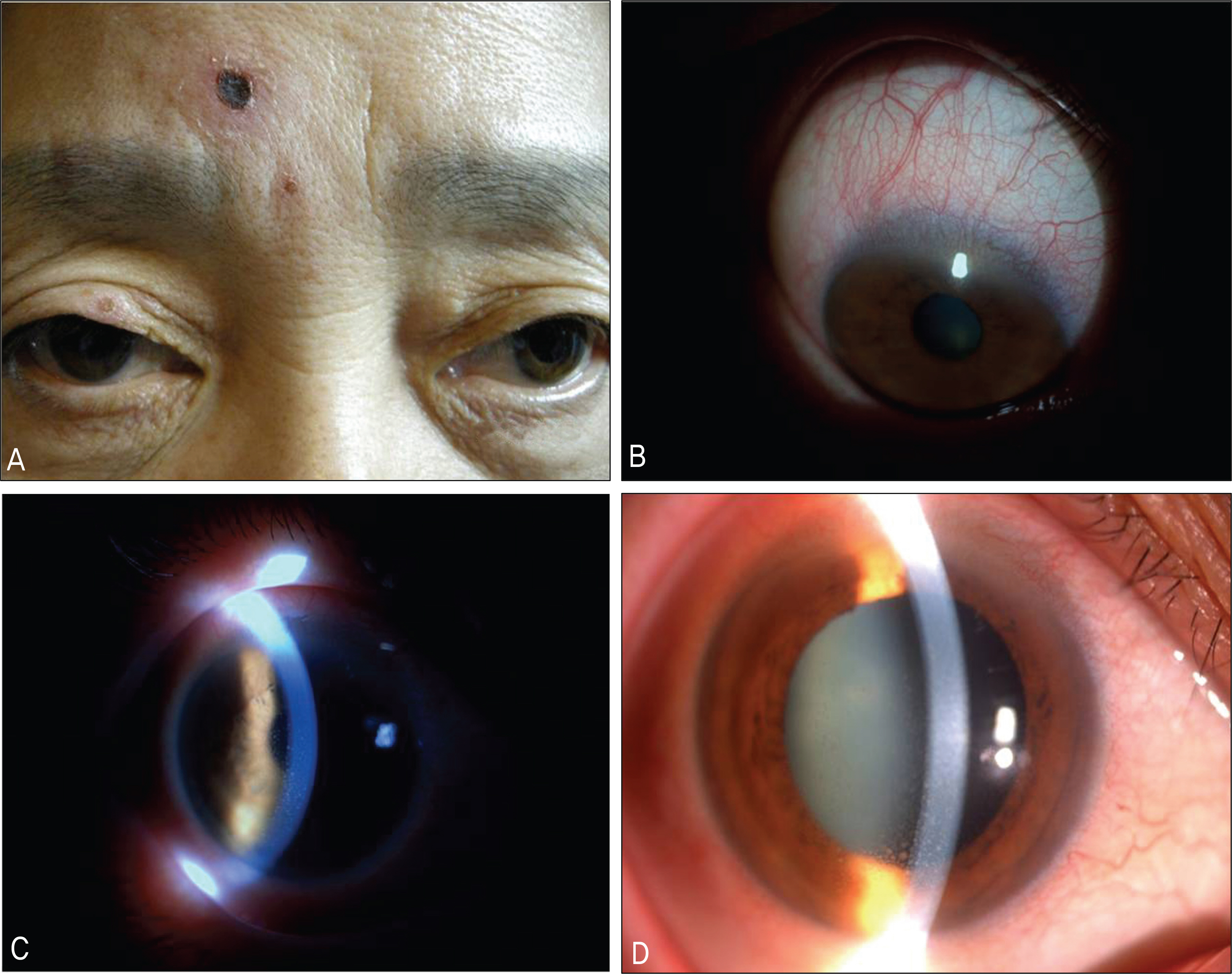
Figure 1. Photographs of a 56-year-old woman with pain sensation in her right eye. (A) The photograph shows eschars on the right upper lid and forehead. (B) Dilated episcleral vessels and conjuctival injection. (C), (D) Anterior chamber inflammation and keratic precipitates located in the inner endothelial layer of the cornea.

Figure 2. Three weeks after treatment. (A) Eschar on the right upper lid disappeared. (B) Eschar remained ulcer on her frontal head. (C) Dilated conjunctival and episcleral vessels were restored. (D) Anterior chamber inflammation and keratic precipitates resolved.
Reference
-
References
1. Tachibana N. Tsutsugamushi disease. Jpn J Clin Med. 1985; 43:728–32.2. Kim YW, Cho MK, Kim HS, et al. Patterns of Acute Febrile Illness (Murine Typhus, Scrub Typhus, Leptospirosis and Hemorrhagic Fever with Renal Syndrome) from 1986 to 1990 in Korea. J Korean Soc Microbiol. 1991; 26:431–41.3. La Scola B, Raoult D. Laboratory diagnosis of rickettsioses: current approaches to diagnosis of old and new rickettsial diseases. J Clin Microbiol. 1997; 35:2715–27.
Article4. Suto T. Rapid serologic diagnosis of tsutsugamushi disease employing the immunoperoxidase reaction with cell cultured rickettsia. Clin Virol. 1980; 8:425–9.5. Seong SY, Choi MS, Kim IS. Orientia tsutsugamushi infection: overview and immune responses. Microbes Infect. 2001; 3:11–21.6. Ogawa M, Hagiwara T, Kishimoto T, et al. Scrub typhus in Japan: epidemiology and clinical features of cases reported in 1998. Am J Trop Med Hyg. 2002; 67:162–5.
Article7. Bang HA, Lee MJ, Lee WC. Comparative research on epidemiological aspects of tsutsugamushi disease (scrub typhus) between Korea and Japan. Jpn J Infect Dis. 2008; 61:148–50.8. Ree HI, Kim TE, Lee IY, et al. Determination and geographical distribution of Orientia tsutsugamushi serotypes in Korea by nested polymersae chain reaction. Am J Trop Med Hyg. 2001; 65:528–34.9. Tachibana N. Tsutsugamushi disease. Jpn J Clin Med. 1985; 43:728–32.10. Yamasaku F. Handbook of internal medicine. 53B. Tokyo: Nakayama-Shoten;1979. p. 251–3.11. Yamashita T, Kasuya S, Noda S, et al. Newly isolated strains of Rickettsia tsutsugamushi in Japan identified by using monoclonal antibodies to Karp, Gilliam, and Kato strains. J Clin Microbiol. 1988; 26:1859–60.
Article12. Chang WH, Kim IS, Choi MS, et al. Seroepidemiological Survey of Scrub Typhus in Korea. Infect Chemother. 1993; 26:181–8.13. Kweon SS, Choi JS, Lim HS, et al. Rapid increase of scrub typhus, South Korea, 2001–2006. Emerg Infect Dis. 2009; 15:1127–9.
Article14. Brown GW. Scrub typhus;pathogenesis and clinical syndrome. Biology of rickettsial disease. 1:CRC Press;Boca Raton, Fla: 1988. p. 93–100.15. Luksameetanasan R, Blacksell SD, Kalambaheti T, et al. Patient and sample-related factors that effect the success of in vitro isolation of Orientia tsutsugamushi. Southeast Asian J Trop Med Public Health. 2007; 38:91–6.16. Hanson B. Identification and partial characterization of Rickettsia tsutsugamushi major protein immunogens. Infect Immun. 1985; 50:603–9.
Article17. Kim DM, Yun NR, Yang TY, et al. Usefulness of nested PCR for the diagnosis of scrub typhus in clinical practice:a prospective study. Am J Trop Med Hyg. 2006; 75:542–5.18. Chi WC, Huang JJ, Sung JM, et al. Scrub typhus associated with multiorgan failure: a case report. Scand J Infect Dis. 1997; 29:634–5.
Article19. Kim DM, Kim HL, Park CY, et al. Scrub Typhus: A prospective study of 76 cases. Infect Chemother. 2006; 38:186–91.20. Kim DM, Won KJ, Park CY, et al. Distribution of eschars on the body of scrub typhus patients: A prospective study. Am J Trop Med Hyg. 2007; 76:806–9.
Article21. Kim SY, Roh KH, Jung MS. Two Cases of Tsutsugamushi Disease with Ocular Disease and Eschars on the Lower Eyelid. J Korean Ophthalmol Soc. 2006; 47:1834–9.22. Kuwabara Y. Iridocyclitis in tsutsugamushi disease. Jpn Rev Clin Ophthalmol. 1919; 15:101–6.23. Yoneyama T, Ibaraki M. An insect bite lesion of the eyelid in tsutsugamushi disease. Jpn J Clin Ophthalmol. 1951; 5:752–3.24. Kato T, Watanabe K, Katori M, et al. Conjunctival Injection, Episcleral Vessel Dilatation, and Subconjunctival Hemorrhage in Patients with New Tsutsugamushi Disease. Jpn J Ophthalmol. 1997; 41:196–9.25. Walker DH, Mattern WD. Rickettsial vasculitis. Am Heart J. 1980; 100:896–906.
Article26. Jerrells TR, Osterman JV. Development of specific and cross-re-active lymphocyte proliferative responses during chronic immu-nizing infections with Rickettsia tsutsugamushi. Infect Immun. 1983; 40:147–56.
Article27. Kang JI, Kim DM, Lee J. Acute sensorineural hearing loss and severe otalgia due to scrub typhus. BMC Infect Dis. 2009; 9:173.
Article
