Korean J Urol.
2006 May;47(5):517-521. 10.4111/kju.2006.47.5.517.
Difference of Brain Activation by Visual Erotic Stimuli in Young and Middle-aged Healthy Males
- Affiliations
-
- 1Departments of Urology, Psychiatry, College of Medicine, The Catholic University of Korea, Seoul, Korea. kim1227@catholic.ac.kr
- KMID: 2294138
- DOI: http://doi.org/10.4111/kju.2006.47.5.517
Abstract
- PURPOSE
The objectives of this study are to identify the brain centers whose activity changes are related to sexually arousing visual stimuli and to identify the difference between young and middle-aged males by mapping the brain activity with using blood oxygen level dependent functional magnetic resonance imaging (BOLD-fMRI).
MATERIALS AND METHODS
Ten young heterosexual, right handed males with normal sexual function (mean age: 27 years, age range: 24 to 31) and ten middle-aged heterosexual, right handed males with normal sexual function (mean age: 52 years, age range: 46 to 55) were enrolled into this study. Real-time visual stimulation was performed with the subjects alternatively viewing erotic and non-erotic films to identify the activated brain regions associated with sexual response. Assessments with using a five-point scale were determined after visual stimulation to evaluate the subjective sexual arousal. Brain activity was mapped by performing BOLD-fMRI on a 1.5T MR scanner. After functional scanning, the high-resolution data was analyzed with the SPM analyzing program; the significance of activation was set at p<0.01 or p<0.001.
RESULTS
The parietal lobe, frontal lobe, cingulate gyrus, caudate nucleus, thalamus and hypothalamus were noted as the areas of activation specifically associated with viewing the erotic film segments by the young males. For the middle-aged males, these areas were the parietal lobe, frontal lobe, right temporal lobe, cingulate gyrus and caudate nucleus. The thalamus and hypothalamus were activated in only the young males.
CONCLUSIONS
We suggest that the non-activation of the hypothalamus and thalamus in middle-aged males may be associated with the lesser physiological arousal in response to the erotic visual stimuli. The non-invasive visualization of the central nervous system by functional MRI in healthy males has shown the possibility for evaluating the neuroanatomy of the brain that is associated with sexual arousal and its clinical application by comparing young and middle-aged males.
MeSH Terms
Figure
-
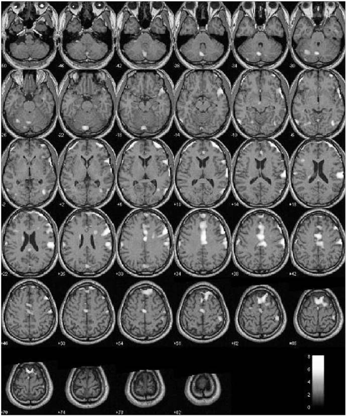
Fig. 1 Brain areas whose activation is related to erotic-visual stimuli in young healthy males.
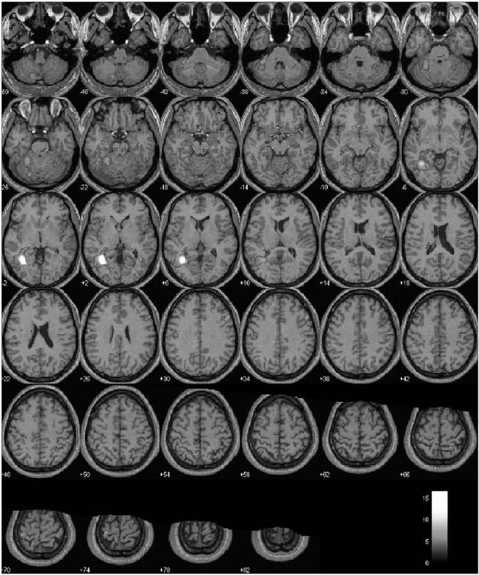
Fig. 2 Brain areas whose activation is related to erotic-visual stimuli in middle aged healthy males.
Reference
-
1. Park K, Seo JJ, Kang HK, Ryu SB, Kim HJ, Jeong GW. A new potential of blood oxygenation level dependent (BOLD) functional MRI for evaluating cerebral centers of penile erection. Int J Impot Res. 2001. 13:73–81.2. Arnow BA, Desmond JE, Banner LL, Glover GH, Solomon A, Polan ML, et al. Brain activation and sexual arousal in healthy, heterosexual males. Brain. 2002. 125:1014–1023.3. Karama S, Lecours AR, Leroux JM, Bourgouin P, Beaudoin G, Joubert S, et al. Areas of brain activation in males and females during viewing of erotic film excerpts. Hum Brain Mapp. 2002. 16:1–13.4. Braun M, Wassmer G, Klotz T, Reifenrath B, Mathers M, Engelmann U. Epidemiology of erectile dysfunction: results of the 'Cologne Male Survey'. Int J Impot Res. 2000. 12:305–311.5. O'Leary MP, Fowler FJ, Lenderking WR, Barber B, Sagnier PP, Guess HA, Barry MJ. A brief male sexual function inventory for urology. Urology. 1995. 46:697–706.6. Garrett AS, Maddock RJ. Time-course of the subjective emotional response to aversive pictures: relevance to fMRI studies. Psychiatry Res. 2001. 108:39–48.7. Redoute J, Stoleru S, Gregoire MC, Costes N, Cinotti L, Lavenne F, et al. Brain processing of visual sexual stimuli in human males. Hum Brain Mapp. 2000. 11:162–177.8. Talairach J, Tournoux P. Co-planar stereotaxic atlas of the human brain. 1988. Stuttgart: Thieme;2133–2136.9. Stoleru S, Gregoire MC, Gerard D, Decety J, Lafarge E, Cinotti L, et al. Neuroanatomical correlates of visually evoked sexual arousal in human males. Arch Sex Behav. 1999. 28:1–21.10. Reiman EM, Lane RD, Ahern GL, Schwartz GE, Davidson RJ, Friston KJ, et al. Neuroanatomical correlates of externally and internally generated human emotion. Am J Psychiatry. 1997. 154:918–925.11. Lane RD, Reiman EM, Ahern GL, Schwartz GE, Davidson RJ. Neuroanatomical correlates of happiness, sadness, and disgust. Am J Psychiatry. 1997. 154:926–933.12. Francis S, Rolls ET, Bowtell R, McGlone F, O'Doherty J, Browning A, et al. The representation of pleasant touch in the brain and its relationship with taste and olfactory areas. Neuroreport. 1999. 10:453–459.13. Llinas R, Ribary U, Contreras D, Pedroarena C. The neuronal basis for consciousness. Philos Trans R Soc Lond B Biol Sci. 1998. 353:1841–1849.
- Full Text Links
-
- Actions
-
Cited
- CITED
-
- Close
- Share
-
- Similar articles
-
- Effect of Sertraline on Current-Source Distribution of the High Beta Frequency Band: Analysis of Electroencephalography under Audiovisual Erotic Stimuli in Healthy, Right-Handed Males
- Time-Course Analysis of the Neuroanatomical Correlates of Sexual Arousal Evoked by Erotic Video Stimuli in Healthy Males
- Treatment with Selective Serotonin Reuptake Inhibitors and Mirtapazine Results in Differential Brain Activation by Visual Erotic Stimuli in Patients with Major Depressive Disorder
- Brain Activation in Response to Visually Evoked Sexual Arousal in Male-to-Female Transsexuals: 3.0 Tesla Functional Magnetic Resonance Imaging
- Functional Neuroanatomy in Depressed Patients with Sexual Dysfunction: Blood Oxygenation Level Dependent Functional MR Imaging
