Human Mesenchymal Stem Cell Transplantation Induces Sympathetic Nerve Sprouting and Reduces the Gap Junction With Potential Proarrhythmias in Dogs
- Affiliations
-
- 1Division of Cardiology, Department of Internal Medicine, Korea University College of Medicine, Seoul, Korea. hnpak@korea.ac.kr
- 2Department of Pathology, College of Medicine, Pochon CHA University, Pochon, Korea.
- 3Department of Hematology, Korea University, Seoul, Korea.
- 4Division of Cardiology, Utah Valley Medical Center, Provo, UT, USA.
- KMID: 2225718
- DOI: http://doi.org/10.4070/kcj.2008.38.10.536
Abstract
- BACKGROUND AND OBJECTIVES
Although human mesenchymal stem cell (hMSC) transplantation has been known to improve ventricular function, the potential proarrhythmic effects have not yet been studied. MATERIALS AND METHODS: We monitored the heart rhythm of 6 dogs for 4 weeks after transplantation of hMSC (1x10(7), epicardial injection) (hMSC group) and in 5 Sham dogs after the injection of the vehicle alone. Cardiac sympathetic nerve sprouting {nerve growth factor (NGF)-beta; tyrosine hydroxylase (TH)} and gap junction expression {connexin (Cx) 43} were evaluated in 10 dogs (5 hMSC and 5 Sham) that survived longer than 4 weeks. RESULTS: The hMSC group expressed higher levels of NGF-beta messenger ribonucleic acid (mRNA) (56.0+/-66.8 fold; p<0.01) with TH+ sympathetic nerves (0.51+/-0.40 vs. 0.15+/-0.13% area; p<0.03) than the Sham control. In contrast, the hMSC group expressed lower levels of Cx43 mRNA (0.59+/-0.29 fold, p<0.001) and Cx43+ (1.64+/-1.79 vs. 2.12+/-1.07% area, p<0.001) than the Sham control. The incidences of ventricular fibrillation were 33.3% and 0% in the hMSC group and Sham control, respectively. One of the dogs with ventricular fibrillation (VF) in the hMSC group died suddenly. CONCLUSION: hMSC transplantation may be proarrhythmic since NGF-beta expression increased with cardiac sympathetic hyperinnervation and the expression of Cx43 and the gap junction decreased.
MeSH Terms
-
Animals
Connexin 43
Dogs
Gap Junctions
Heart
Humans
Incidence
Mesenchymal Stem Cell Transplantation
Mesenchymal Stromal Cells
Nerve Growth Factor
RNA
RNA, Messenger
Salicylamides
Tachycardia, Ventricular
Transplants
Tyrosine 3-Monooxygenase
Ventricular Fibrillation
Ventricular Function
Connexin 43
Nerve Growth Factor
RNA
RNA, Messenger
Salicylamides
Tyrosine 3-Monooxygenase
Figure
-
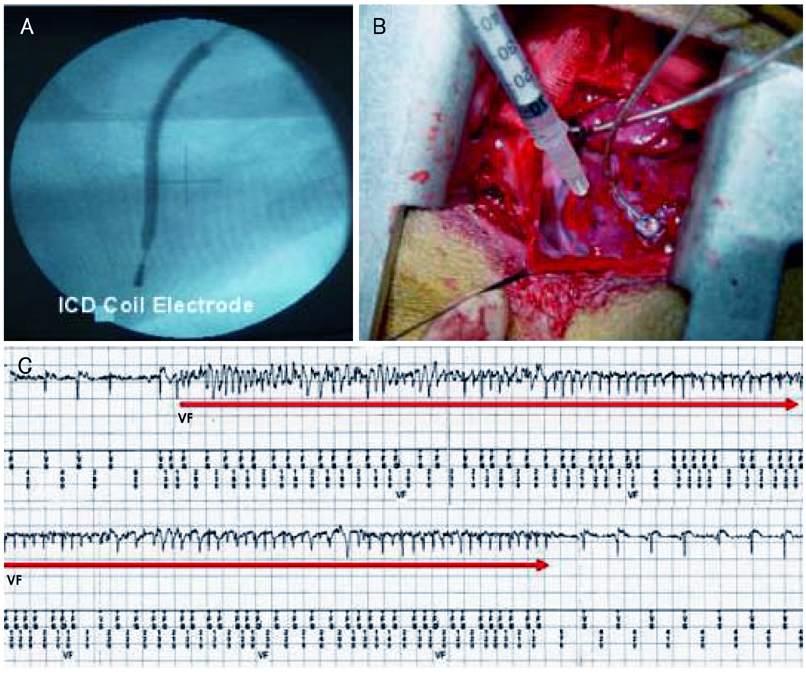
Fig. 1 Experimental procedures. A: the ICD lead was positioned using the left internal jugular venous approach, and the position of the ICD coil electrode was confirmed by fluoroscopic imaging. B: hMSC injection sites were marked with suture material, and the hMSCs were transplanted by direct epicardial injection. C: the ICD electrogram revealed an episode of spontaneously induced non-sustained VF. This dog survived for 4 weeks. ICD: implantable defibrillator, hMSC: human mesenchymal stem cell, VF: ventricular fibrillation.
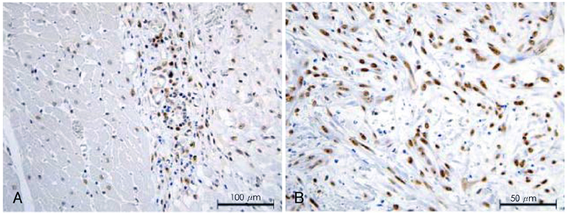
Fig. 2 Human nucleoline (hNC) immunostaining of ventricular tissue close to the area of hMSC injection. A: the brown colored nucleus represents hNC positive human cells (hMSC) at 200× magnification. B: the 400× magnification high power field shows no evidence of immune rejection (no lymphocytic infiltration, but polymorphonuclear cells due to surgical inflammation).
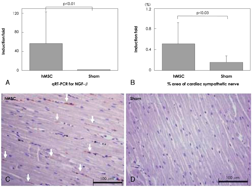
Fig. 3 A: mRNA expression levels of NGF-β was significantly higher in the hMSC group compared with Sham control group. B: calculated % area of TH positive (sympathetic) nerves was significantly higher in the hMSC transplanted tissues compared with the Sham group. C and D: tyrosine hydroxylase (TH) immunostaining of ventricular tissue close to the area of hMSC injection at 200× magnification. The hMSC group shows significant sympathetic hyperinnervation after 4 weeks of survival. By contrast, no significant increase in sympathetic nerves was observed in the Sham group. qRT-PCR: quantitative Reverse Transcription Polymerase chain Reaction, NGF-β: nerve growth factor-β, hMSC: human mesenchymal stem cell.
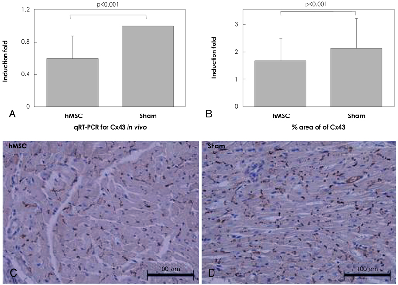
Fig. 4 mRNA expression levels. A: Cx43 was significantly lower in the hMSC group compared with Sham control group. B: calculated % area of Cx43 positive gap junction was significantly lower in the hMSC group compared with the Sham group. C and D: Cx43 immunostainings of ventricular tissue close to the area of hMSC injection at 200× magnification. The density of Cx43 positive gap junction is lower in hMSC transplanted tissue compared with the Sham after 4 weeks of survival. qRT-PCR: quantitative Reverse Transcription Polymerase chain Reaction, hMSC: human mesenchymal stem cell.
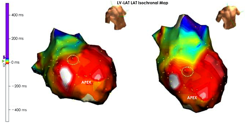
Fig. 5 Isochronal map of LV endocardium during high right atrial pacing (cycle length 500 ms) in right anterior oblique view and anterior posterior cranial view in animals that were transplanted with hMSC. As showed in the color scale bar, the impulse conducts from the white color to the purple color, and the earliest activation site is a septum. However, there is a significant conduction delay at the LV anterior wall (white dotted circle) where the hMSC was injected and Cx43 expression was reduced. LV: left ventricle, hMSC: human mesenchymal stem cell.
Reference
-
1. Schachinger V, Erbs S, Elsasser A, et al. Intracoronary bone marrow-derived progenitor cells in acute myocardial infarction. N Engl J Med. 2006. 355:1210–1221.2. Assmus B, Honold J, Schachinger V, et al. Transcoronary transplantation of progenitor cells after myocardial infarction. N Engl J Med. 2006. 355:1222–1232.3. Lim D. Stem cells for cardiovascular disease. Korean Circ J. 2004. 34:435–440.4. Tse WT, Pendleton JD, Beyer WM, Egalka MC, Guinan EC. Suppression of allogeneic T-cell proliferation by human marrow stromal cells: implications in transplantation. Transplantation. 2003. 75:389–397.5. Le Blanc K, Tammik C, Rosendahl K, Zetterberg E, Ringden O. HLA expression and immunologic properties of differentiated and undifferentiated mesenchymal stem cells. Exp Hematol. 2003. 31:890–896.6. Bartholomew A, Patil S, Mackay A, et al. Baboon mesenchymal stem cells can be genetically modified to secrete human erythropoietin in vivo. Hum Gene Ther. 2001. 12:1527–1541.7. Chang MG, Tung L, Sekar RB, et al. Proarrhythmic potential of mesenchymal stem cell transplantation revealed in an in vitro coculture model. Circulation. 2006. 113:1832–1841.8. Beeres SL, Atsma DE, van der Laarse A, et al. Human adult bone marrow mesenchymal stem cells repair experimental conduction block in rat cardiomyocyte cultures. J Am Coll Cardiol. 2005. 46:1943–1952.9. Valiunas V, Doronin S, Valiuniene L, et al. Human mesenchymal stem cells make cardiac connexins and form functional gap junctions. J Physiol. 2004. 555:617–626.10. Pak HN, Qayyum M, Kim DT, et al. Mesenchymal stem cell injection induces cardiac nerve sprouting and increased tenascin expression in a Swine model of myocardial infarction. J Cardiovasc Electrophysiol. 2003. 14:841–848.11. Cao JM, Chen LS, KenKnight BH, et al. Nerve sprouting and sudden cardiac death. Circ Res. 2000. 86:816–821.12. Cao JM, Fishbein MC, Han JB, et al. Relationship between regional cardiac hyperinnervation and ventricular arrhythmia. Circulation. 2000. 101:1960–1969.13. Zhou S, Chen LS, Miyauchi Y, et al. Mechanisms of cardiac nerve sprouting after myocardial infarction in dogs. Circ Res. 2004. 95:76–83.14. Liu YB, Wu CC, Lu LS, et al. Sympathetic nerve sprouting, electrical remodeling, and increased vulnerability to ventricular fibrillation in hypercholesterolemic rabbits. Circ Res. 2003. 92:1145–1152.15. Kessler PD, Byrne BJ. Myoblast cell grafting into heart muscle: cellular biology and potential applications. Ann Rev Physiol. 1999. 61:219–242.16. Liechty KW, MacKenzie TC, Shaaban AF, et al. Human mesenchymal stem cells engraft and demonstrate site-specific differentiation after in utero transplantation in sheep. Nat Med. 2000. 6:1282–1286.17. Saito T, Kuang JQ, Bittira B, Al-Khaldi A, Chiu RC. Xenotransplant cardiac chimera: immune tolerance of adult stem cells. Ann Thorac Surg. 2002. 74:19–24.18. Kang HJ, Kim HS, Zhang SY, et al. Effects of intracoronary infusion of peripheral blood stem-cells mobilised with granulocyte-colony stimulating factor on left ventricular systolic function and restenosis after coronary stenting in myocardial infarction. Lancet. 2004. 363:751–756.19. Lunde K, Solheim S, Aakhus S, et al. Intracoronary injection of mononuclear bone marrow cells in acute myocardial infarction. N Engl J Med. 2006. 355:1199–1209.20. Janssens S, Dubois C, Bogaert J, et al. Autologous bone marrowderived stem-cell transfer in patients with ST-segment elevation myocardial infarction: double-blind, randomised controlled trial. Lancet. 2006. 367:113–121.21. Meyer GP, Wollert KC, Lotz J, et al. Intracoronary bone marrow cell transfer after myocardial infarction: eighteen months' follow-up data from the randomized, controlled BOOST (BOne marrOw transfer to enhance ST-elevation infarct regeneration) trial. Circulation. 2006. 113:1287–1294.22. Menasche P, Hagege AA, Vilquin JT, et al. Autologous skeletal myoblast transplantation for severe postinfarction left ventricular dysfunction. J Am Coll Cardiol. 2003. 41:1078–1083.23. Abraham MR, Henrikson CA, Tung L, et al. Antiarrhythmic engineering of skeletal myoblasts for cardiac transplantation. Circ Res. 2005. 97:159–167.24. Fukushima S, Varela-Carver A, Coppen SR, et al. Direct intramyocardial but not intracoronary injection of bone marrow cells induces ventricular arrhythmias in a rat chronic ischemic heart failure model. Circulation. 2007. 115:2254–2261.25. Shake JG, Gruber PJ, Baumgartner WA, et al. Mesenchymal stem cell implantation in a swine myocardial infarct model: engraftment and functional effects. Ann Thorac Surg. 2002. 73:1919–1925.26. Kocher AA, Schuster MD, Szabolcs MJ, et al. Neovascularization of ischemic myocardium by human bone-marrow-derived angioblasts prevents cardiomyocyte apoptosis, reduces remodeling and improves cardiac function. Nat Med. 2001. 7:430–436.27. Orlic D, Kajstura J, Chimenti S, et al. Bone marrow cells regenerate infarcted myocardium. Nature. 2001. 410:701–705.28. Fuchs S, Baffour R, Zhou YF, et al. Transendocardial delivery of autologous bone marrow enhances collateral perfusion and regional function in pigs with chronic experimental myocardial ischemia. J Am Coll Cardiol. 2001. 37:1726–1732.29. Lim SY, Jeong MH, Ahn YK, et al. The effects of mesenchymal stem cells transduced with Ark in a porcine myocardial infarction model. Korean Circ J. 2005. 35:734–741.30. Piao H, Youn TJ, Kwon JS, et al. Cellular cardiomyoplasty using bone marrow derived mesenchymal stem cells transplantation in post myocardial infarction heart failure. Korean Circ J. 2004. 34:1113–1121.31. Yang KM, Park CS, Jang SW, et al. Effect of adult bone marrow stem cells on myocardial regeneration in doxorubicin-induced mouse cardiomyopathy. Korean Circ J. 2008. 38:110–118.32. Woodbury D, Schwarz EJ, Prockop DJ, Black IB. Adult rat and human bone marrow stromal cells differentiate into neurons. J Neurosci Res. 2000. 61:364–370.33. Weimann JM, Johansson CB, Trejo A, Blau HM. Stable reprogrammed heterokaryons form spontaneously in Purkinje neurons after bone marrow transplant. Nat Cell Biol. 2003. 5:959–966.34. Alvarez-Dolado M, Pardal R, Garcia-Verdugo JM, et al. Fusion of bone-marrow-derived cells with Purkinje neurons, cardiomyocytes and hepatocytes. Nature. 2003. 425:968–973.35. Rehman J, Li J, Orschell CM, March KL. Peripheral blood "endothelial progenitor cells" are derived from monocyte/macrophages and secrete angiogenic growth factors. Circulation. 2003. 107:1164–1169.36. Martins JB, Zipes DP. Effects of sympathetic and vagal nerves on recovery properties of the endocardium and epicardium of the canine left ventricle. Circ Res. 1980. 46:100–110.37. Opthof T, Misier AR, Coronel R, et al. Dispersion of refractoriness in canine ventricular myocardium: effects of sympathetic stimulation. Circ Res. 1991. 68:1204–1215.
- Full Text Links
-
- Actions
-
Cited
- CITED
-
- Close
- Share
-
- Similar articles
-
- Clinical Safety and Efficacy of Autologous Bone Marrow-Derived Mesenchymal Stem Cell Transplantation in Sensorineural Hearing Loss Patients
- Safety and outcomes of subconjunctival allogenic mesenchymal stem cell transplantation in canine experimental corneal defects
- Clinical utilization of cord blood over human health: experience of stem cell transplantation and cell therapy using cord blood in Korea
- Transplantation of adipose derived mesenchymal stem cells for acute thoracolumbar disc disease with no deep pain perception in dogs
- Mesenchymal Stem Cells for the Treatment of Liver Disease: Present and Perspectives
