Clinical Aspect and Prognosis of Staphylococcus Epidermidis Keratitis
- Affiliations
-
- 1Department of Ophthalmology, Chonbuk National University School of Medicine, Jeonju, Korea. you2ic@paran.com
- KMID: 2214132
- DOI: http://doi.org/10.3341/jkos.2011.52.1.14
Abstract
- PURPOSE
To investigate the predisposing factors, clinical manifestations, treatment results and risk factors for treatment failure in Staphylococcus epidermidis keratitis.
METHODS
Sixty-one eyes of 61 patients who were diagnosed with Staphylococcus epidermidis keratitis were included in the present study. The past history, location and size of ulceration, hypopyon, treatment results, and antibiotic susceptibility were reviewed retrospectively. A logistic regression analysis was performed to identify the main prognostic risk factors for treatment failure.
RESULTS
Twenty-six eyes (42.6%) had previous histories of corneal traumas. Polymicrobial infections were observed in 31 cases (50.8%), including 11 cases (35.5%) combined with the Fusarium species. Twenty-five eyes (41.0%) had lesions located at the corneal center. The average size of ulceration was 7.3 +/- 7.2 mm2. Thirteen eyes (21.3%) with lesions that progressed or occurred in the corneal perforation underwent evisceration, penetrating keratoplasty or scleral graft. Risk factors for treatment failure were a history of previous keratitis (P = 0.003) and an ulcer exceeding 5.0 mm2 in size (P = 0.018).
CONCLUSIONS
Staphylococcus epidermidis keratitis usually has a good prognosis, although a history of previous keratitis and a large ulcer size are risk factors for treatment failure.
MeSH Terms
Figure
-
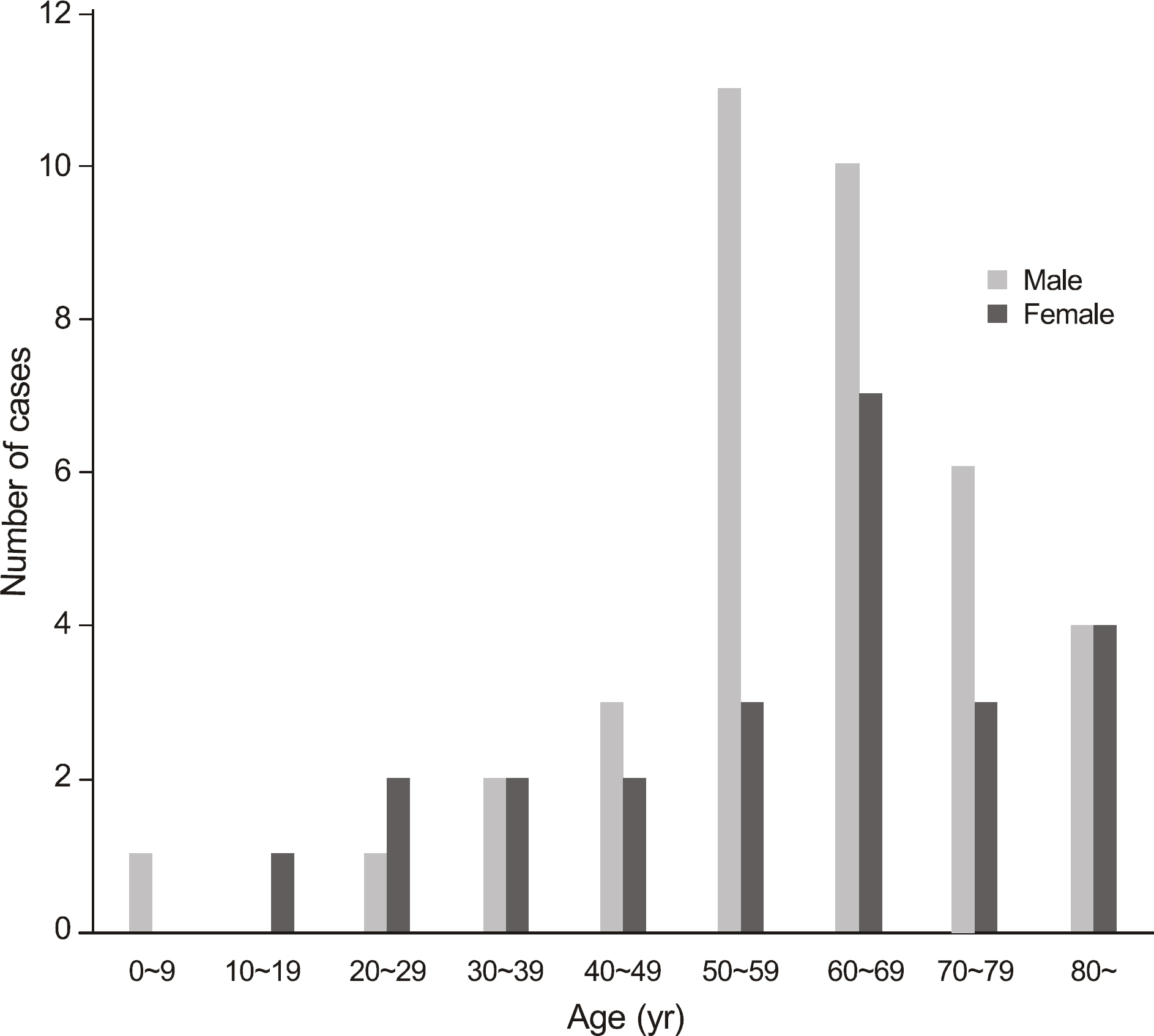
Figure 1. Age distribution of Staphylococcus epidermidis keratitis.
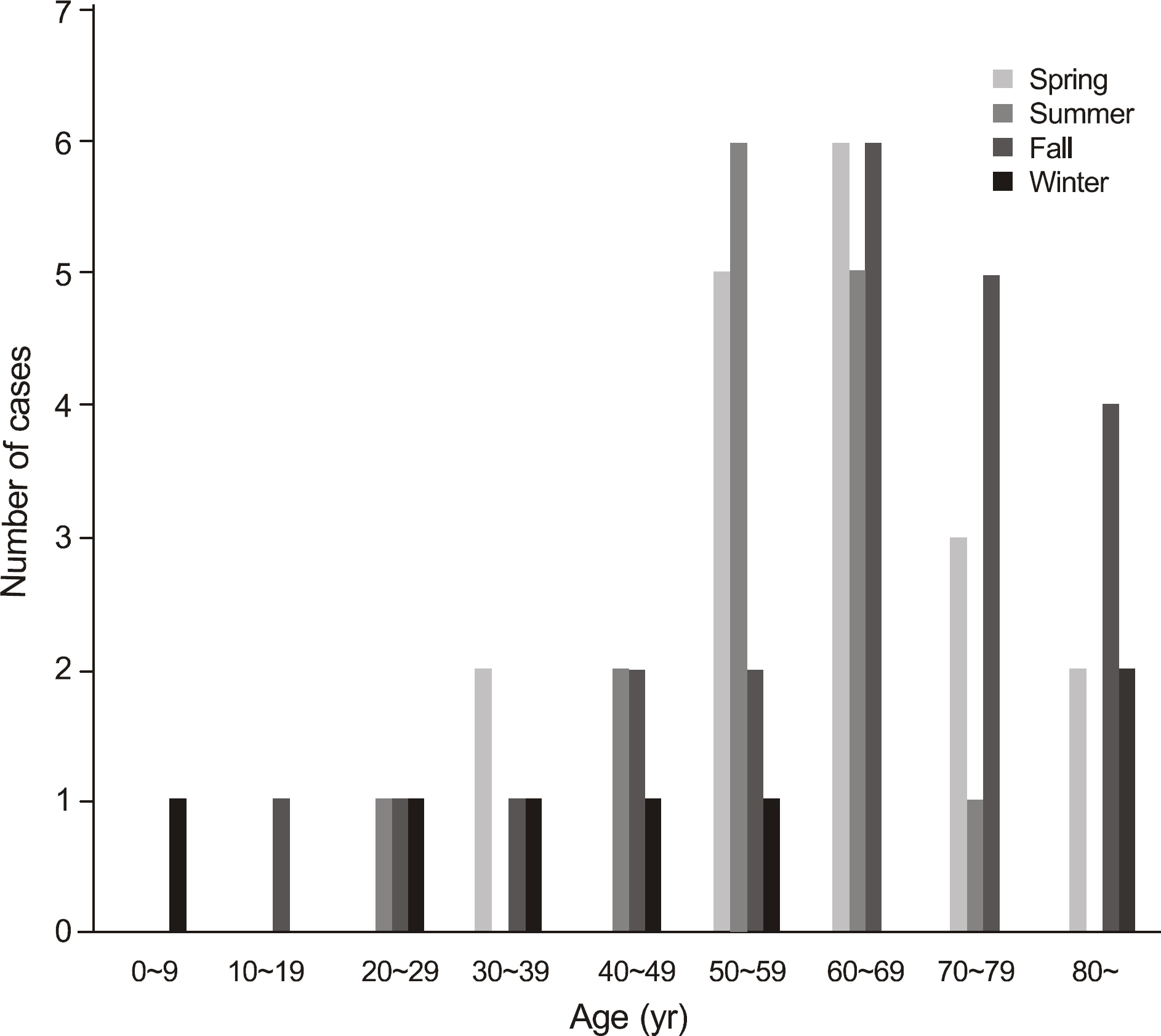
Figure 2. Seasonal variation of patients with Staphylococcus epidermidis keratitis.
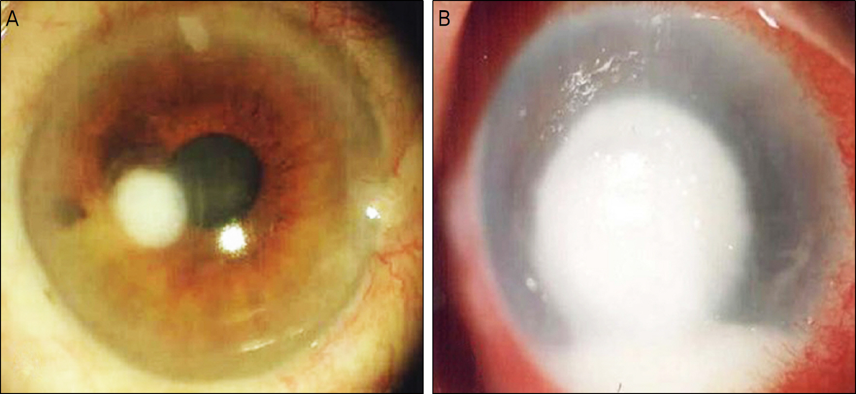
Figure 3. (A) Anterior segment photograph of a 46-year-old woman, who had a history of corneal trauma at the first visit. Mild conjunctival injection and corneal infiltration were observed. The lesion was completely healed in 2 weeks with antimicrobial therapy. (B) Anterior segment photograph of an 89-year-old woman, who had a history of keratitis at a first visit. Slit-lamp examination revealed severe conjunctival injection, corneal lysis with a size of approximately 4 mm, hypopyon, corneal edema, folding of the Descemet's membrane. Staphylococcus epidermidis and Fusarium were isolated by corneal scraping. Despite of antimicrobial and antifungal therapy, patient's ocular condition deteriorated and evisceration was ultimately performed.
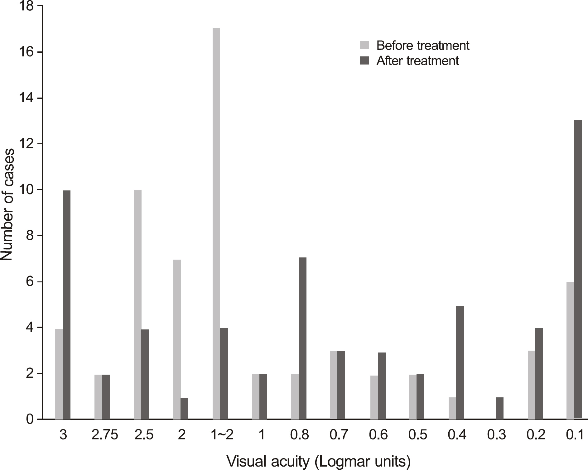
Figure 4. Visual acuities of the eyes with Staphylococcus epidermidis keratitis in the first and last medical examinations.
Reference
-
References
1. Lowy FD, Hammer SM. Staphylococcus epidermidis infections. Ann Intern Med. 1983; 99:834–9.
Article2. Tierno PM Jr, Stotzky G. Serologic typing of Staphylococcus epidermidis biotype 4. J Infect Dis. 1978; 137:514–23.
Article3. Fitzgerald RH Jr, Nolan DR, Ilstrup DM, et al. Deep wound sepsis following total hip arthroplasty. J Bone Joint Surg Am. 1977; 59:847–55.
Article4. Karchmer AW, Archer GL, Dismukes WE. Staphylococcus epidermidis causing prosthetic valve endocarditis: microbiologic and clinical observations as guides to therapy. Ann Intern Med. 1983; 98:447–55.
Article5. Rubin J, Rogers WA, Taylor HM, et al. Peritonitis during continuous ambulatory peritoneal dialysis. Ann Intern Med. 1980; 92:7–13.
Article6. Bannerman TL, Rhoden DL, Mcallister SK, et al. The source of co-agulase-negative staphylococci in the Endophthalmitis Vitrectomy Study. A comparison of eyelid and intraocular isolates using pulsed-field gel electrophoresis. Arch Ophthalmol. 1997; 115:357–61.7. Ormerod LD, Ho DD, Becker LE, et al. Endophthalmitis caused by the coagulase-negative staphylococci. Ophthalmology. 1993; 100:715–23.
Article8. Maske R, Hill JC, Oliver SP. Management of bacterial corneal ulcers. Br J Ophthalmol. 1986; 70:199–201.
Article9. Kunimoto DY, Sharma S, Garg P, et al. Corneal ulceration in the elderly in Hyderabad, south India. Br J Ophthalmol. 2000; 84:54–9.
Article10. Ammous MW, Noor Sunba MS. The nature of ulcerative keratitis in Kuwait (clinical and microbiological study). APMIS Suppl. 1988; 3:104–6.11. Jang YS, Hahn YH. Epidemiology of Staphylococcus epidermidis keratitis. J Korean Ophthalmol Soc. 2002; 43:665–71.12. Hahn YH, Hahn TW, Tchah HW, et al. Epidemiology of infectious keratitis (2): a multicenter study. J Korean Ophthalmol Soc. 2001; 42:247–65.13. Lee KH, Chae HJ, Yoon KC. Analysis of risk factors for treatment failure in fungal keratitis. J Korean Ophthalmol Soc. 2008; 49:737–42.
Article14. Mukerji N, Vajpayee RB, Sharma N. Technique of area measurement of epithelial defects. Cornea. 2003; 22:549–51.
Article15. Khosla PK, Prakash OM, Agarwal LP. Clinicopathological study of soframycin in conjunctival flora. Orient Arch Ophthalmol. 1963; 1:212–20.16. Thylefors JD, Harbarth S, Pittet D. Increasing bacteremia due to coagulase-negative staphylococci: fiction or reality? Infect Control Hosp Epidemiol. 1998; 19:581–9.
Article17. Tabbara KF, El-Sheikh HF, Aabed B. Extended wear contact lens related bacterial keratitis. Br J Ophthalmol. 2000; 84:327–8.
Article18. Vallas V, Stapleton F, Willcox MD. Bacterial invasion of corneal epithelial cells. Aust N Z J Ophthalmol. 1999; 27:228–30.19. Rupp ME, Archer GL. Hemagglutination and adherence to plastic by Staphylococcus epidermidis. Infect Immun. 1992; 60:4322–7.
Article20. Ormerod LD, Hertzmark E, Gomez DS, et al. Epidemiology of microbial keratitis in southern California. A multivariate analysis. Ophthalmology. 1987; 94:1322–33.21. Khan JA, Hoover D, Ide CH. Methicillin-resistant Staphylococcus epidermidis blepharitis. Am J Ophthalmol. 1984; 98:562–5.
Article22. Fleischer AB, Hoover DL, Khan JA, et al. Topical vancomycin formulation for methicillin-resistant Staphylococcus epidermidis blepharoconjunctivitis. Am J Ophthalmol. 1986; 101:283–7.
Article23. Sotozono C, Inagaki K, Fujita A, et al. Methicillin-resistant Staphylococcus aureus and methicillin-resistant Staphylococcus epidermidis infections in the cornea. Cornea. 2002; 21:S94–101.
Article24. Miedziak AI, Miller MR, Rapuano CJ, et al. Risk factors in microbial keratitis leading to penetrating keratoplasty. Ophthalmology. 1999; 106:1166–70.
Article
- Full Text Links
-
- Actions
-
Cited
- CITED
-
- Close
- Share
-
- Similar articles
-
- Epidemiology of Staphylococcus epidermidis Keratitis
- Comparison of Methicillin-Sensitive Staphylococcus Epidermidis (MSSE) Keratits and Methicillin-Resistant Staphylococcus Epidermidis (MRSE) Keratitis
- Clinical and Microbiological Analysis of Gram-Positive Bacterial Keratitis, a 15-Year Review
- Bilateral Staphylococcus Epidermidis Endophthalmitis After Cataract Extraction
- Age-related Clinical Analysis of Infectious Keratitis in Two Tertiary Centers
