Analysis of gene expression during mineralization of cultured human periodontal ligament cells
- Affiliations
-
- 1Department of Periodontology, Kyungpook National University School of Dentistry, Daegu, Korea. jysuh@knu.ac.kr
- 2Institute for Hard Tissue and Bio-Tooth Regeneration, Kyungpook National University School of Dentistry, Daegu, Korea.
- KMID: 2212133
- DOI: http://doi.org/10.5051/jpis.2011.41.1.30
Abstract
- PURPOSE
Under different culture conditions, periodontal ligament (PDL) stem cells are capable of differentiating into cementoblast-like cells, adipocytes, and collagen-forming cells. Several previous studies reported that because of the stem cells in the PDL, the PDL have a regenerative capacity which, when appropriately triggered, participates in restoring connective tissues and mineralized tissues. Therefore, this study analyzed the genes involved in mineralization during differentiation of human PDL (hPDL) cells, and searched for candidate genes possibly associated with the mineralization of hPDL cells.
METHODS
To analyze the gene expression pattern of hPDL cells during differentiation, the hPDL cells were cultured in two conditions, with or without osteogenic cocktails (beta-glycerophosphate, ascorbic acid and dexamethasone), and a DNA microarray analysis of the cells cultured on days 7 and 14 was performed. Reverse transcription-polymerase chain reaction was performed to validate the DNA microarray data.
RESULTS
The up-regulated genes on day 7 by hPDL cells cultured in osteogenic medium were thought to be associated with calcium/iron/metal ion binding or homeostasis (PDE1A, HFE and PCDH9) and cell viability (PCDH9), and the down-regulated genes were thought to be associated with proliferation (PHGDH and PSAT1). Also, the up-regulated genes on day 14 by hPDL cells cultured in osteogenic medium were thought to be associated with apoptosis, angiogenesis (ANGPTL4 and FOXO1A), and adipogenesis (ANGPTL4 and SEC14L2), and the down-regulated genes were thought to be associated with cell migration (SLC16A4).
CONCLUSIONS
This study suggests that when appropriately triggered, the stem cells in the hPDL differentiate into osteoblasts/cementoblasts, and the genes related to calcium binding (PDE1A and PCDH9), which were strongly expressed at the stage of matrix maturation, may be associated with differentiation of the hPDL cells into osteoblasts/cementoblasts.
MeSH Terms
Figure
-

Figure 1 Alizarin red S staining of the mineralized nodules cultured for 7 days (A) and 14 days (B) with osteogenic medium and non-osteogenic medium. Circular photographs show human periodontal ligament cells in the culture plate (24 wells). On day 7, mineralized nodules were not observed at all, but on day 14, mineralized nodules were only observed in the osteogenic group.
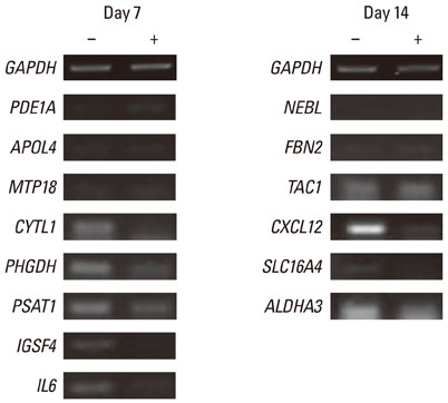
Figure 2 Expression on day 7 and 14 of selected genes by human periodontal ligament (hPDL) cells cultured in osteogenic or non-osteogeneic medium. The hPDL cells were cultured for 7 days in an osteogenic medium (+) or non-osteogenic medium (-). The 12 out of 14 genes were comparable with DNA microarray data. The expression patterns of tachykinin, precursor 1 were not significant, and nebulette were not expressed. Similar results were obtained in 2 separate experiments and representative data are shown. GAPDH: glyceraldehyde-3-phosphate dehydrogenase, PDE1A: phosphodiesterase 1A, calmodulin-dependent, APOL4: apolipoprotein L, 4, MTP18: mitochondrial protein 18 kDa, CYTL1: cytokine-like 1, PHGDH: phosphoglycerate dehydrogenase, PSAT1: phosphoserine aminotransferase 1, IGSF4: cell adhesion molecule 1, IL6: interleukin 6 (interferon, beta 2), NEBL: nebulette, FBN2: fibrillin 2, TAC1: tachykinin, precursor 1, CXCL12: chemokine (C-X-C motif ) ligand 12, SLC16A4: solute carrier family 16, member 4 (monocarboxylic acid transporter 5), ALDH1A3: aldehyde dehydrogenase 1 family, member A3.
Cited by 1 articles
-
Gene expression profile in mesenchymal stem cells derived from dental tissues and bone marrow
Su-Hwan Kim, Young-Sung Kim, Su-Yeon Lee, Kyoung-Hwa Kim, Yong-Moo Lee, Won-Kyung Kim, Young-Kyoo Lee
J Periodontal Implant Sci. 2011;41(4):192-200. doi: 10.5051/jpis.2011.41.4.192.
Reference
-
1. Lekic P, Rojas J, Birek C, Tenenbaum H, McCulloch CA. Phenotypic comparison of periodontal ligament cells in vivo and in vitro. J Periodontal Res. 2001. 36:71–79.
Article2. Ivanovski S, Haase HR, Bartold PM. Expression of bone matrix protein mRNAs by primary and cloned cultures of the regenerative phenotype of human periodontal fibroblasts. J Dent Res. 2001. 80:1665–1671.
Article3. Murakami Y, Kojima T, Nagasawa T, Kobayashi H, Ishikawa I. Novel isolation of alkaline phosphatase-positive subpopulation from periodontal ligament fibroblasts. J Periodontol. 2003. 74:780–786.
Article4. Wu L, Wei X, Ling J, Liu L, Liu S, Li M, et al. Early osteogenic differential protein profile detected by proteomic analysis in human periodontal ligament cells. J Periodontal Res. 2009. 44:645–656.
Article5. Seo BM, Miura M, Gronthos S, Bartold PM, Batouli S, Brahim J, et al. Investigation of multipotent postnatal stem cells from human periodontal ligament. Lancet. 2004. 364:149–155.
Article6. Flores MG, Yashiro R, Washio K, Yamato M, Okano T, Ishikawa I. Periodontal ligament cell sheet promotes periodontal regeneration in athymic rats. J Clin Periodontol. 2008. 35:1066–1072.
Article7. Iwata T, Yamato M, Tsuchioka H, Takagi R, Mukobata S, Washio K, et al. Periodontal regeneration with multi-layered periodontal ligament-derived cell sheets in a canine model. Biomaterials. 2009. 30:2716–2723.
Article8. Kulterer B, Friedl G, Jandrositz A, Sanchez-Cabo F, Prokesch A, Paar C, et al. Gene expression profiling of human mesenchymal stem cells derived from bone marrow during expansion and osteoblast differentiation. BMC Genomics. 2007. 8:70.
Article9. Yamada S, Ozawa Y, Tomoeda M, Matoba R, Matsubara K, Murakami S. Regulation of PLAP-1 expression in periodontal ligament cells. J Dent Res. 2006. 85:447–451.
Article10. Lallier TE, Spencer A. Use of microarrays to find novel regulators of periodontal ligament fibroblast differentiation. Cell Tissue Res. 2007. 327:93–109.
Article11. Shin JH, Park JW, Yeo SI, Noh WC, Kim MK, Kim JC, et al. Identification of matrix mineralization-related genes in human periodontal ligament cells using cDNA microarray. J Korean Acad Periodontol. 2007. 37:Suppl. 447–463.
Article12. Dahl LK. A simple and sensitive histochemical method for calcium. Proc Soc Exp Biol Med. 1952. 80:474–479.
Article13. Bartold PM, Shi S, Gronthos S. Stem cells and periodontal regeneration. Periodontol 2000. 2006. 40:164–172.
Article14. Nagatomo K, Komaki M, Sekiya I, Sakaguchi Y, Noguchi K, Oda S, et al. Stem cell properties of human periodontal ligament cells. J Periodontal Res. 2006. 41:303–310.
Article15. Kobayashi M, Takiguchi T, Suzuki R, Yamaguchi A, Deguchi K, Shionome M, et al. Recombinant human bone morphogenetic protein-2 stimulates osteoblastic differentiation in cells isolated from human periodontal ligament. J Dent Res. 1999. 78:1624–1633.
Article16. Kitagawa M, Tahara H, Kitagawa S, Oka H, Kudo Y, Sato S, et al. Characterization of established cementoblast-like cell lines from human cementum-lining cells in vitro and in vivo. Bone. 2006. 39:1035–1042.
Article17. Michibata H, Yanaka N, Kanoh Y, Okumura K, Omori K. Human Ca2+/calmodulin-dependent phosphodiesterase PDE1A: novel splice variants, their specific expression, genomic organization, and chromosomal localization. Biochim Biophys Acta. 2001. 1517:278–287.
Article18. Fidock M, Miller M, Lanfear J. Isolation and differential tissue distribution of two human cDNAs encoding PDE1 splice variants. Cell Signal. 2002. 14:53–60.
Article19. Zayzafoon M, Fulzele K, McDonald JM. Calmodulin and calmodulin-dependent kinase IIalpha regulate osteoblast differentiation by controlling c-fos expression. J Biol Chem. 2005. 280:7049–7059.
Article20. Etzrodt J, Krishna-K K, Redies C. Expression of classic cadherins and delta-protocadherins in the developing ferret retina. BMC Neurosci. 2009. 10:153.21. Gray MJ, Van Buren G, Dallas NA, Xia L, Wang X, Yang AD, et al. Therapeutic targeting of neuropilin-2 on colorectal carcinoma cells implanted in the murine liver. J Natl Cancer Inst. 2008. 100:109–120.
Article22. Hopwood B, Tsykin A, Findlay DM, Fazzalari NL. Gene expression profile of the bone microenvironment in human fragility fracture bone. Bone. 2009. 44:87–101.
Article23. Cho HM, Jun DY, Bae MA, Ahn JD, Kim YH. Nucleotide sequence and differential expression of the human 3-phosphoglycerate dehydrogenase gene. Gene. 2000. 245:193–201.
Article24. Pollari S, Käkönen SM, Edgren H, Wolf M, Kohonen P, Sara H, et al. Enhanced serine production by bone metastatic breast cancer cells stimulates osteoclastogenesis. Breast Cancer Res Treat. 2011. 125:421–430.
Article25. Gallagher SM, Castorino JJ, Philp NJ. Interaction of monocarboxylate transporter 4 with beta1-integrin and its role in cell migration. Am J Physiol Cell Physiol. 2009. 296:C414–C421.
- Full Text Links
-
- Actions
-
Cited
- CITED
-
- Close
- Share
-
- Similar articles
-
- Indirect Co-Culture of Stem Cells from Human Exfoliated Deciduous Teeth and Oral Cells in a Microfluidic Platform
- Effect of Extract of Seeds of Carthamus tinctorius L. on Mineralization in Periodontal Ligament Cells and Osteoblastic Cells
- Comparison of Gene Expression from Supernumerary Dental Pulp and Periodontal Ligament Stem Cells
- ULTRASTRUCTURAL INVESTIGATIONS OF THE INTERFACE BETWEEN CULTURED PERIODONTAL LIGAMENT CELLS AND TITANIUM
- Identification of Matrix Mineralization-Related Genes in Human Periodontal Ligament Cells Using cDNA Microarray
