J Korean Acad Conserv Dent.
2010 Sep;35(5):335-343. 10.5395/JKACD.2010.35.5.335.
Effect of moisture on sealing ability of root canal filling with different types of sealer through the glucose penetration model
- Affiliations
-
- 1Department of Conservative Dentistry, Chonbuk National University School of Dentistry, Jeonju, Korea. mkyou102@hanmail.net
- KMID: 2176431
- DOI: http://doi.org/10.5395/JKACD.2010.35.5.335
Abstract
OBJECTIVES
To compared the effect of different levels of moisture of root canal on the sealing ability after filling with four different types of sealer.
MATERIALS AND METHODS
Single-rooted teeth (n = 90) instrumented to and apical size of 0.06 / 45 were randomly assigned to 12 experimental groups (n = 7 per group), positive/negative control groups (n = 3 per group). The teeth of the experimental groups (a. DRY; b. PAPER POINT DRY; c. WET) were obturated with sealer (Group 1-3: Sealapex; Group 4-6: AH plus; Group 7-9: Tubuli-seal; Group 10-12: EndoRez) and warm vertical compaction method. After 7 days in 37degrees C, 100% humidity, the coronal-to-apical microleakage was evaluated quantitatively using a glucose leakage model. The leaked glucose concentration was measured with spectrophotometer at 1, 3, 7, 14, 21, and 30 days. Data were recorded ad mmol/L and statistically analysed with the two-way ANOVA and Duncan test (p = 0.05).
RESULTS
Throughout the experimental period Tubuli-seal/WET (Group 9) showed the highest mean cumulative glucose penetration (178.75 mmol/L), whereas AH plus/DRY (Group 4) had the least (20.78 mmol/L).
CONCLUSIONS
The results of this study demonstrated that the moisture condition of root canals at the time of obturation and the type of sealer that was used had a significant effect on leakage and sealing ability. Thus drying procedure according to sealer types is a critical step and should not be missed in endodontic treatment.
Keyword
MeSH Terms
Figure
-

Figure 1 Glucose leakage model.
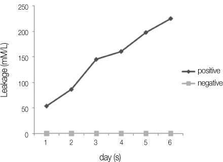
Figure 2 Leakage of positive and negative control groups.

Figure 3 Glucose microleakage of experimental groups. a, Sealapex groups; b, AH plus groups; c, Tubuliseal groups; d, EndoRez groups.
Cited by 1 articles
-
Evaluation of softening ability of Xylene & Endosolv-R on three different epoxy resin based sealers within 1 to 2 minutes - an
in vitro study
Pratima Ramakrishna Shenoi, Gautam Pyarelal Badole, Rajiv Tarachand Khode
Restor Dent Endod. 2014;39(1):17-23. doi: 10.5395/rde.2014.39.1.17.
Reference
-
1. Siqueira JF, Roças IN, Lopes HP, de Uzeda M. Coronal leakage of two root canal sealers containing calcium hydroxide after exposure to human saliva. J Endod. 1999. 25:14–16.
Article2. Glickman G, Gutmann J. Contemporary perspectives on canal obturation. Dent Clin North Am. 1992. 36:327–340.3. Ingle J, Taintor J. Endodontics. 1985. 3rd ed. Philadelphia: Lea & Febiger;223–225.4. Walton R, Torabinejad M. Principles and practice of endodontics. 1989. Philadelphia: WB Saunders;421.5. Limkangwalmongkol S, Burtscher P, Abbott PV, Sandler AB, Bishop BM. A comparative study of the apical leakage of four root canal sealers and laterally condensed gutta-percha. J Endod. 1991. 17:495–499.
Article6. Wu MK, De Gee AJ, Wesselink PR. Leakage of four root canal sealers at different thicknesses. Int Endod J. 1994. 27:304–308.
Article7. Himel VT, McSpadden JT, Goodis HE. Cohen S, Hargreaves KM, editors. Instruments, materials, and devices. Pathways of the Pulp. 2005. 9th edn. St Louis, MO: Mosby Elsevier;233–289.8. Madison S, Wilcox L. An evaluation of coronal microleakage in endodontically treated teeth. Part III. In vivo study. J Endod. 1988. 14:455–458.
Article9. Cohen S, Burns R. Pathways of the pulp. 1982. 5th ed. St. Louis: CV Mosby;207.10. Weine F. Endodontic therapy. 1982. 3rd ed. St. Louis: CV Mosby;341.11. Lee JK, Park SH, Choi GW. Time-dependent effects of EDTA application on removal of smear layer in the root canal system. J Korean Acad Conserv Dent. 2006. 31:169–178.
Article12. Xu Q, Fan MW, Fan B, Cheung GS, Hu HL. A new quantitative method using glucose for analysis of endodontic leakage. Oral Surg Oral Med Oral Pathol Oral Radiol Endod. 2005. 99:107–111.
Article13. Pommel L, Camps J. Effects of pressure and measurement time on the fluid filtration method in endodontics. J Endod. 2001. 27:256–258.
Article14. Yu YS, Kim TK, Lee KW, Yu MK. Evaluation of sealing ability of root fillings without smear layer by soft chelating irrigation. J Korean Acad Conserv Dent. 2009. 34:484–485.15. Shemesh H, Wu MK, Wesselink PR. Leakage along apical root fillings with and without smear layer using two different leakage models: a two-month longitudinal ex vivo study. Int Endod J. 2006. 39:968–976.
Article16. Wu MK, Wesselink PR. Endodontic leakage studies reconsidered. Part I. Methodology, Application and relevance. Int Endod J. 1993. 26:37–43.
Article17. Rao P, Pattabiraman TN. Reevaluation of the phenol-sulfuric acid reaction for the estimation of hexoses and pentoses. Anal Biochem. 1989. 181:18–22.
Article18. Horning TG, Kessler JR. A comparison of three different root canal sealers when used to obturate a moisture-contaminated root canal system. J Endod. 1995. 21:354–357.
Article19. Tagger M, Tagger E, Kfir A. Release of calcium and hydroxyl ions from set endodontic sealers containing calcium hydroxide. J Endod. 1988. 14:588–591.
Article20. Oguntebi BR, Shen C. Effect of different sealers on thermoplasticized gutta-percha root canal obturations. J Endod. 1992. 18:363–366.
Article21. Schäfer E, Zandbiglari T. Solubility of root-canal sealers in water and artificial saliva. Int Endod J. 2003. 36:660–669.
Article22. Roggendorf MJ, Ebert J, Petschelt A, Frankenberger R. Influence of moisture on the apical seal of root canal fillings with five different types of sealer. J Endod. 2007. 33:31–33.
Article23. Zmener O, Spielberg C, Lambergghini F, Rucci M. Sealing properties of a new epoxy resin-based root-canal sealer. Int Endod J. 1997. 30:332–334.
Article24. Ørstavik D, Nordahl I, Tibballs JE. Dimensional change following setting of root canal sealer materials. Dent Mater. 2001. 17:512–519.
Article25. Barnett F, Trope M, Rooney J, Tronstak L. In vivo sealing ability of calcium hydroxide containing root canal sealers. Endod Dent Traumatol. 1989. 5:23–26.
Article26. Zidan O, El Deeb M. The use of dental bonding agent as a root canal sealer. J Endod. 1985. 11:176–178.27. Markowitz K, Moynihan M, Liu M, Kim S. Biologic properties of eugenol and zinc oxide-eugenol. A clinically oriented review. Oral Surg Oral Med Oral Pathol. 1992. 73:729–737.28. Peters DD. Two-year in vitro solubility evaluation of four gutta-percha sealer obturation techniques. J Endod. 1986. 12:139–145.
Article29. Kuhre A, Kessler J. Effect of moisture on the apical seal of laterally condensed gutta percha. J Endod. 1993. 19:277–280.
Article30. Tay FR, Loushine RJ, Weller RN, et al. Ultrastructural evaluation of the apical seal in roots filled with a polycaprolactone-based root canal filling material. J Endod. 2005. 31:514–519.
Article31. Tay FR, Loushine RJ, Monticelli F, et al. Effectiveness of resin-coated gutta-percha cones and a dual-cured, hydrophilic methacrylate-based sealer in obturating root canals. J Endod. 2005. 31:659–664.
Article32. Zmener O, Pameijer CH, Serrano SA, Vidueira M, Macchi RI. Significance of moist root canal dentin with the use of methacrylate-based endodontic sealers: an in vitro coronal dye leakage study. J Endod. 2008. 34:76–79.
Article33. Wang Y, Spencer P. Continuity etching of an all-in-one adhesive in wet dentin tubules. J Dent Res. 2005. 84:350–354.
Article
- Full Text Links
-
- Actions
-
Cited
- CITED
-
- Close
- Share
-
- Similar articles
-
- Micro-computed tomographic evaluation of a new system for root canal filling using calcium silicatebased root canal sealers
- Effect of soft chelating irrigation on the sealing ability of GP/AH Plus root fillings
- Microleakage of resilon: Effects of several self-etching primer
- The effects of total-etch, wet-bonding, and light-curing of adhesive on the apical seal of a resin-based root canal filling system
- Efficacy of retreatment NiTi files for root canals filled with calcium silicate-based sealer
