J Korean Acad Conserv Dent.
2009 Nov;34(6):484-490. 10.5395/JKACD.2009.34.6.484.
Effect of soft chelating irrigation on the sealing ability of GP/AH Plus root fillings
- Affiliations
-
- 1Department of Conservative Dentistry, School of Dentistry, Chonbuk National University, Korea. mkyou102@hanmail.net
- KMID: 1986611
- DOI: http://doi.org/10.5395/JKACD.2009.34.6.484
Abstract
- The purpose of this study was to evaluate the effect of soft chelating irrigant on the sealing ability of root fillings by using a glucose leakage test. A total of 45 single-rooted teeth were selected for the study. The teeth were decoronated leaving a total length of 13mm. The root canals prepared using K3 NiTi rotary instruments to an apical dimension of size 45(0.06 taper). The specimens were then randomly divided into 3 experimental groups of 13 roots each and 2 control groups of 3 roots each. Specimen in each group were prepared with different irrigation protocols : group 1, 2.5% NaOCl; group 2, 2.5% NaOCl and 17% EDTA; group 3, 2.5% NaOCl and 15% HEBP. The root canals were filled with gutta-percha and AH Plus sealer using lateral condensation. After 7 days in 37degrees C, 100% humidity, the coronal-to-apical microleakage was evaluated quantitatively using a glucose leakage model. The leaked glucose concentration was measured with spectrophotometry at 1, 4, 7, 14, 21 and 28 days. There was a tendency of increase in leakage in all experimental groups during experimental period. HEBP-treated dentin showed no significant difference with EDTA-treated dentin during experimental period. From the 21th day onward, HEBP-treated dentin showed significantly lower leakage than smear-covered dentin. HEBP-treated dentin displayed a similar sealing pattern to EDTA-treated dentin and a better sealing ability than smear-covered dentin. Consequently, a soft chelator(HEBP) could be considered as the possible alternative to EDTA.
Keyword
MeSH Terms
Figure
-
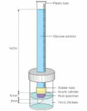
Figure 1 Glucose leakage model.
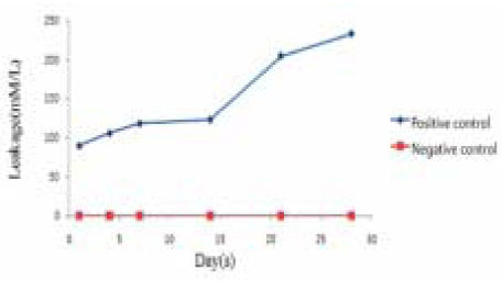
Figure 2 Leakage of positive and negative control groups.
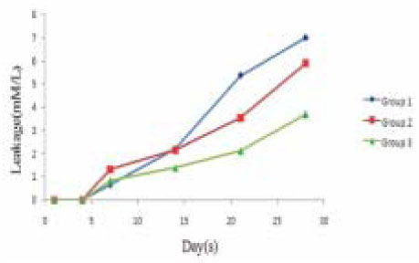
Figure 3 Glucose microleakage of experimental groups.
Reference
-
1. Czonstkowsky M, Wilson EG, Holstein FA. The smear layer in endodontics. Dent Clin North Am. 1990. 34:13–25.2. Pashley DH, Michelich V, Kehl TJ. Dentin permeability: effects of smear layer removal. J Prosthet Dent. 1981. 46:531–537.
Article3. Pashley DH. Smear layer: Physiological considerations. Oper Dent Suppl. 1984. 3:13–29.4. Foster KH, Kulild JC, Weller RN. Effect of smear layer removal on the diffusion of calcium hydroxide through radicular dentin. J Endod. 1993. 19:136–140.
Article5. White RR, Goldman M, Lin PS. The influence of the smeared layer upon dentinal tubule penetration by plastic filling materials. J Endod. 1984. 10:558–562.
Article6. Okşan T, Aktener BO, Sen BH, Tezel H. The penetration of root canal sealers into dentinal tubules. Int Endod J. 1993. 26:301–305.7. Economides N, Liolios E, Kolokuris I, Beltes P. Long-term evaluation of the influence of smear layer removal on the sealing ability of different sealers. J Endod. 1999. 25:123–125.
Article8. Lee JM, Park SH, Choi KW. The Effect of Smear Layer Treatment on the Microleakage. J Korean Acad Conserv Dent. 2006. 31:378–389.
Article9. Johnson WT, Gutmann JL. Cohen S, Hargreaves KM, editors. Obturation of the cleaned and shaped root canal system. Pathways of the Pulp. 2005. 9th edn. St Louis, MO: Mosby Elsevier;358–399.
Article10. Shahravan A, Haghdoost AA, Adl A, Rahimi H, Shadifar F. Effect of smear layer on sealing ability of canal obturation: a systematic review and meta-analysis. J Endod. 2007. 33:96–105.
Article11. Lester KS, Boyde A. Scanning electron microscopy of instrumented, irrigated and filled root canals. Br Dent J. 1977. 143:359–367.
Article12. Hulsmann N, Heckendorff M, Lennon A. Chelating agents in root canal treatment: mode of action and indications for their use. Int Endod J. 2003. 36:810–830.
Article13. Zehnder M, Schmidlin P, Sener B, Waltimo T. Chelation in root canal therapy reconsidered. J Endod. 2005. 31:817–820.
Article14. Grawehr M, Sener B, Waltimo T, Zehnder M. Interaction of ethylenediamine tetracetic acid with sodium hypochlorite in aqueous solutions. Int Endod J. 2003. 36:411–415.
Article15. Baumgartner JC, Marder CL. A scanning electron microscopic evaluation of four root canal irrigation regimens. J Endod. 1987. 13:147–157.
Article16. De-Deus G, Zehnder M, Reis C, Fidel S, Fidel RA, Galan J Jr, Paciornik S. Longitudinal co-site optical microscopy study on the chelating ability of etidronate and EDTA using a comparative single-tooth model. J Endod. 2008. 34:71–75.
Article17. Ari H, Erdemir A, Belli S. Evaluation of the effect of endodontic irrigation solution on the microhardness and the roughness of root canal dentin. J Endod. 2004. 30:792–795.
Article18. De-Deus G, Namen F, Galan J Jr, Zehnder M. Soft chelating irrigation protocol optimizes bonding quality of Resilon/Epiphany root fillings. J Endod. 2008. 34:703–705.
Article19. Russell RG, Gogers MJ. Bisphosphonates: from the laboratory to the clinic and back again. Bone. 1999. 25:97–106.
Article20. Xu Q, Fan MW, Fan B, Cheung GS, Hu HL. A new quantitative method using glucose for analysis of endodontic leakage. Oral Surg Oral Med Oral Pathol Oral Radiol Endod. 2005. 99:107–111.
Article21. Shemesh H, Wu MK, Wesselink PR. Leakage along apical root fillings with and without smear layer usting two different leakage models: a two-month longitudinal ex vivo study. Int Endod J. 2006. 39:968–976.
Article22. Wu MK, Wesselink PR. Endodontic leakage studies reconsidered. Part I. Methodology, Application and relevance. Int endod J. 1993. 26:37–43.
Article23. AliGhamdi A, Wennberg A. Testing of sealing ability of endodontic filling materials. Endod Dent Traumatol. 1994. 10:249–255.
Article24. Pommel L, Camps J. Effects of pressure and measurement time on the fluid filtration method in endodontics. J Endod. 2001. 27:256–258.
Article25. Rao P, Pattabiraman TN. Reevaluation of the phenol-sulfuric acid reaction for the estimation of hexoses and pentoses. Anal Biochem. 1989. 181:18–22.
Article26. Eldeniz AU, Erdemir A, Belli S. Shear bond strengths of three resin based sealers to dentin with and without the smear layer. J Endod. 2005. 31:293–296.
Article27. Behrend GD, Cutler CW, Gutmann JL. An in-vitro study of smear layer removal and microbial leakage along root-canal fiilings. Int Endod J. 1996. 29:99–107.
Article28. De-Deus G, Soares J, Leal F, Luna AS, Fidel S, Fidel RA. Similar glucose leakage pattern on smear-covered, EDTA-treated and BioPure MTAD-treated dentin. J Endod. 2008. 34:459–462.
Article29. Park DS. The effect of MTAD on the apical leakage of obturated root canals: an electrochemical study. J Korean Acad Conserv Dent. 2006. 31:119–124.
Article30. Shemesh H, Wu M-K, Wesselink PR. Leakage along apical root fillings with and without smear layer using two different leakage models:a two-month longitudinal ex vivo study. Int Endod J. 2006. 39:968–976.
Article31. Calt S, Serper A. Smear layer removal by EGTA. J Endod. 2000. 26:459–461.
Article32. Eldeniz AU, Erdemir A, Belli S. Effect of EDTA and citric acid solutions on the microhardness and the roughness of human root canal dentin. J Endod. 2005. 31:107–110.
Article33. Schwartz R. Adhesive dentistry and endodontics. Part 2: Bonding in the root canal system-The promise and the problems: A review. J Endod. 2006. 32:1125–1134.
Article34. Pashley DH, Tay FR, Yiu C, Hashimoto M, Breschi L, Carvalho RM, Ito S. Collagen degradation by host-derived enzymes during aging. J Dent Res. 2004. 83:216–221.
Article35. Eldeniz AU, Erdemir A, Belli S. Shear bond strength of three resin based sealers to dentin with and without the smear layer. J Endod. 2005. 31:293–296.
Article36. Mamootil K, Messer HH. Penetration of dentinal tubules by endodontic sealer cements in extracted teeth and in vivo. Int endod J. 2007. 40:873–881.
Article37. Tay FR, Hosoya Y, Loushine RJ, Pashley DH, Weller RN. Ultrastructure of intraradicular dentin after irrigation with BioPure MTAD. II. The consequence of obturation with an epoxy resin-based sealer. J Endod. 2006. 32:473–477.
Article
- Full Text Links
-
- Actions
-
Cited
- CITED
-
- Close
- Share
-
- Similar articles
-
- Bacterial leakage and micro-computed tomography evaluation in round-shaped canals obturated with bioceramic cone and sealer using matched single cone technique
- Effect of moisture on sealing ability of root canal filling with different types of sealer through the glucose penetration model
- The use of auxiliary devices during irrigation to increase the cleaning ability of a chelating agent
- Micro-computed tomographic evaluation of single-cone obturation with three sealers
- Comparison of apical seal with or without the use of dentin adhesive system
