The effects of total-etch, wet-bonding, and light-curing of adhesive on the apical seal of a resin-based root canal filling system
- Affiliations
-
- 1Department of Dentistry, Seoul National University School of Dentistry, Seoul, Korea.
- 2Department of Conservative Dentistry, Seoul National University School of Dentistry and Dental Research Institute, Seoul, Korea. chobh@snu.ac.kr
- KMID: 2176576
- DOI: http://doi.org/10.5395/JKACD.2011.36.5.385
Abstract
OBJECTIVES
This study evaluated the effects of adhesion variables such as the priming concepts of canal wall and the curing modes of adhesives on the sealing ability of a resin-based root canal filling system.
MATERIALS AND METHODS
Apical microleakage of the Resilon-RealSeal systems filled with 3 different combinations of adhesion variables was compared with the conventional gutta-percha filling using a dye penetration method. Experimental groups were SEDC, Resilon (Resilon Research LLC) filling with self-etch RealSeal (SybronEndo) primer and dual-cure RealSeal sealer; NELC, Resilon filling with no etching, Scotchbond Multi-Purpose (3M ESPE) primer application and light-curing adhesive; and TELC, Resilon filling with Scotchbond Multi-Purpose primer and adhesive used under total etch / wet bonding and light-cure protocols. GPCS, gutta-percha filling with conventional AH26 plus sealer, was the control group.
RESULTS
The median longitudinal dye penetration length of TELC was significantly shorter than those of GPCS and SEDC (Kruskal-Wallis test, p < 0.05). In the cross-sectional microleakage scores, TELC showed significant differences from other groups at 2 to 5 mm from the apical foramen (Kruskal-Wallis test, p < 0.05).
CONCLUSIONS
When a resin-based root canal filling material was used, compared to the self-etching primer and the dual-cure sealer, the total etch/wet-bonding with primer and light-curing of adhesive showed improved apical sealing and was highly recommended.
Keyword
MeSH Terms
Figure
-
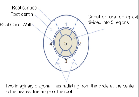
Figure 1 Schematic illustration of the segments for scoring microleakage from each section.
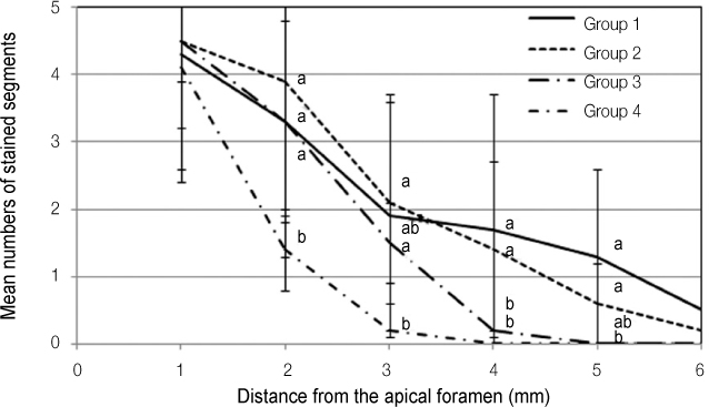
Figure 2 Cross-sectional microleakage scores of the experimental groups at each level of section. The cross-sectional microleakage scores decreased significantly along the distance from the apical foramen. Group 4 (TELC) showed significant differences in the cross-sectional microleakage scores from the other groups at 2 mm, from Groups 2 and 3 at 3 mm, and from Groups 1 and 2 at 4 mm and 5 mm from the apical foramen.
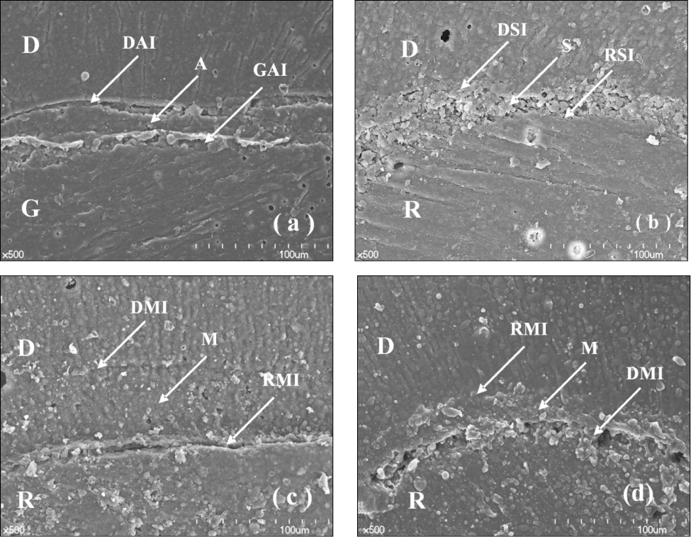
Figure 3 Scanning electron micrographs showing the interface between the root canal filling materials and the root canal dentin. a, Group 1 (GPCS), there were continuous gaps at the dentin side of the sealer; b, Group 2 (SEDC), the interface between the dentin wall and the sealer appeared tight, but the RealSeal sealer looked porous between the dentin wall and the Resilon filling material; c and d, Groups 3 (NELC) and 4 (TELC), the SMBP adhesive developed a thick adhesive layer between the dentin wall and the Resilon core filling material. D, root canal dentin; G, gutta-percha root canal filling material; R, Resilon root canal filling material; A, AH26 plus sealer; S, RealSeal sealer; M, Scotchbond Multi-Purpose adhesive; DAI, interface between the dentin and AH26 plus sealer; GAI, interface between the gutta-percha root canal filling material and AH26 plus sealer; DSI, interface between the dentin and the RealSeal sealer; RSI, interface between the Resilon root canal filling material and the RealSeal sealer; DMI, interface between the dentin and the Scotchbond Multi-Purpose adhesive; RMI, interface between the Resilon root canal filling material and the Scotchbond Multi-Purpose adhesive.
Reference
-
1. Baumgartner JC, Falkler WA Jr. Bacteria in the apical 5 mm of infected root canals. J Endod. 1991. 17:380–383.
Article2. Ricucci D, Siqueira JF Jr, Bate AL, Pitt Ford TR. Histologic investigation of root canal-treated teeth with apical periodontitis: a retrospective study from twenty-four patients. J Endod. 2009. 35:493–502.
Article3. Epley SR, Fleischman J, Hartwell G, Cicalese C. Completeness of root canal obturations: Epiphany techniques versus gutta-percha techniques. J Endod. 2006. 32:541–544.
Article4. Hammad M, Qualtrough A, Silikas N. Evaluation of root canal obturation: a three-dimensional in vitro study. J Endod. 2009. 35:541–544.5. Shipper G, Ørstavik D, Teixeira FB, Trope M. An evaluation of microbial leakage in roots filled with a thermoplastic synthetic polymer-based root canal filling material (Resilon). J Endod. 2004. 30:342–347.
Article6. Nagas E, Uyanik MO, Sahin C, Durmaz V, Cehreli ZC. Effects of different light-curing units and obturation techniques on the seal of the Resilon/Epiphany system. J Endod. 2008. 34:1230–1232.
Article7. Shipper G, Teixeira FB, Arnold RR, Trope M. Periapical inflammation after coronal microbial inoculation of dog roots filled with gutta-percha or resilon. J Endod. 2005. 31:91–96.
Article8. Pawińska M, Kierklo A, Marczuk-Kolada G. New technology in endodontics - the Resilon-Epiphany system for obturation of root canals. Adv Med Sci. 2006. 51:Suppl 1. 154–157.9. Oook S, Miyazaki M, Rikut A, Moore BK. Influence of polymerization mode of dual-polymerized resin direct core foundation systems on bond strengths to bovine dentin. J Prosthet Dent. 2004. 92:239–244.
Article10. Cekic-Nagas I, Ergun G, Vallittu PK, Lassila LV. Influence of polymerization mode on degree of conversion and micropush-out bond strength of resin core systems using different adhesive systems. Dent Mater J. 2008. 27:376–385.
Article11. Ariyoshi M, Nikaido T, Foxton RM, Tagami J. Influence of filling technique and curing mode on the bond strengths of composite cores to pulpal floor dentin. Dent Mater J. 2010. 29:562–569.
Article12. Cadenaro M, Antoniolli F, Sauro S, Tay FR, Di Lenarda R, Prati C, Biasotto M, Contardo L, Breschi L. Degree of conversion and permeability of dental adhesives. Eur J Oral Sci. 2005. 113:525–530.
Article13. Navarra CO, Cadenaro M, Codan B, Mazzoni A, Sergo V, De Stefano Dorigo E, Breschi L. Degree of conversion and interfacial nanoleakage expression of three one-step self-etch adhesives. Eur J Oral Sci. 2009. 117:463–469.
Article14. Ferracane JL, Greener EH. The effect of resin formulation on the degree of conversion and mechanical properties of dental restorative resins. J Biomed Mater Res. 1986. 20:121–131.
Article15. Dickens SH, Cho BH. Interpretation of bond failure through conversion and residual solvent measurements and Weibull analyses of flexural and microtensile bond strengths of bonding agents. Dent Mater. 2005. 21:354–364.
Article16. Suh BI, Feng L, Pashley DH, Tay FR. Factors contributing to the incompatibility between simplified-step adhesives and chemically-cured or dual-cured composites. Part III. Effect of acidic resin monomers. J Adhes Dent. 2003. 5:267–282.17. Xu Q, Fan MW, Fan B, Cheung GS, Hu HL. A new quantitative method using glucose for analysis of endodontic leakage. Oral Surg Oral Med Oral Pathol Oral Radiol Endod. 2005. 99:107–111.
Article18. Wu MK, Wesselink PR. Endodontic leakage studies reconsidered. Part I. Methodology, application and relevance. Int Endod J. 1993. 26:37–43.
Article19. Chailertvanitkul P, Saunders WP, Mackenzie D. An assessment of microbial coronal leakage in teeth root filled with gutta-percha and three different sealers. Int Endod J. 1996. 29:387–392.
Article20. Wedding JR, Brown CE, Legan JJ, Moore BK, Vail MM. An in vitro comparison of microleakage between Resilon and gutta-percha with a fluid filtration model. J Endod. 2007. 33:1447–1449.
Article21. Biggs SG, Knowles KI, Ibarrola JL, Pashley DH. An in vitro assessment of the sealing ability of Resilon/Epiphany using fluid filtration. J Endod. 2006. 32:759–761.
Article22. Shemesh H, Wu MK, Wesselink PR. Leakage along apical root fillings with and without smear layer using two different leakage models: a two-month longitudinal ex vivo study. Int Endod J. 2006. 39:968–976.
Article23. Paqué F, Sirtes G. Apical sealing ability of Resilon/Epiphany versus gutta-percha/AH Plus: immediate and 16-months leakage. Int Endod J. 2007. 40:722–729.
Article24. Onay EO, Ungor M, Unver S, Ari H, Belli S. An in vitro evaluation of the apical sealing ability of new polymeric endodontic filling systems. Oral Surg Oral Med Oral Pathol Oral Radiol Endod. 2009. 108:e49–e54.25. Tay FR, Loushine RJ, Lambrechts P, Weller RN, Pashley DH. Geometric factors affecting dentin bonding in root canals: a theoretical modeling approach. J Endod. 2005. 31:584–589.
Article26. Paqué F, Luder HU, Sener B, Zehnder M. Tubular sclerosis rather than the smear layer impedes dye penetration into the dentine of endodontically instrumented root canals. Int Endod J. 2006. 39:18–25.
Article27. Hiraishi N, Papacchini F, Loushine RJ, Weller RN, Ferrari M, Pashley DH, Tay FR. Shear bond strength of Resilon to a methacrylate-based root canal sealer. Int Endod J. 2005. 38:753–763.
Article
- Full Text Links
-
- Actions
-
Cited
- CITED
-
- Close
- Share
-
- Similar articles
-
- Effect of curing methods of resin cements on bond strength and adhesive interface of post
- The effect of different adhesive system applications on push-out bond strengths of glass fiber posts
- The effect of various bonding systems on the microtensile bond strength of immediate and delayed dentin sealing
- Microleakage and characteristics of resin-tooth tissues interface of a selfetch and an etch-and-rinse adhesive systems
- Shear bond strength and adhesive failure pattern in bracket bonding with plasma arc light
