Restor Dent Endod.
2020 Aug;45(3):e34. 10.5395/rde.2020.45.e34.
Micro-computed tomographic evaluation of a new system for root canal filling using calcium silicatebased root canal sealers
- Affiliations
-
- 1Department of Restorative Dentistry, São Paulo State University (UNESP), School of Dentistry, Araraquara, SP, Brazil
- KMID: 2512025
- DOI: http://doi.org/10.5395/rde.2020.45.e34
Abstract
Objectives
This study evaluated by using micro-computed tomography (micro-CT) the filling ability and sealer apical extrusion promoted by a new Sealer Injection System (SIS; Angelus) with side openings needle, in comparison with the conventional injection system, associated with a new ready-to-use calcium silicate-based sealer (Bio-C Sealer).
Materials and Methods
Acrylic resin models containing a main curved artificial canal and 3 simulated lateral canals in apical, middle and cervical thirds were used. The main root canals were prepared using a rotary system up to size 35.05. The canals were filled with Bio-C sealer by using a single cone technique and the conventional delivery system or SIS. Samples were scanned in micro-CT. The percentage of voids throughout the entire extension of the main root canal and in each third of the lateral canals, besides the apical extrusion of the sealer was calculated. Data were submitted to t-test (p < 0.05).
Results
There was no difference between both systems in the main root canals filling. Although the volume percentage of voids was similar in the apical and middle thirds of lateral canals, SIS had the greatest filling ability of the cervical third lateral canal. Moreover, the conventional system showed the highest apical extrusion of the sealer.
Conclusions
The conventional and SIS obturation systems had an appropriate filling ability of the main root canal. SIS had the best filling of the cervical third of the lateral canals, besides lower sealer apical extrusion, suggesting its clinical indication.
Figure
-
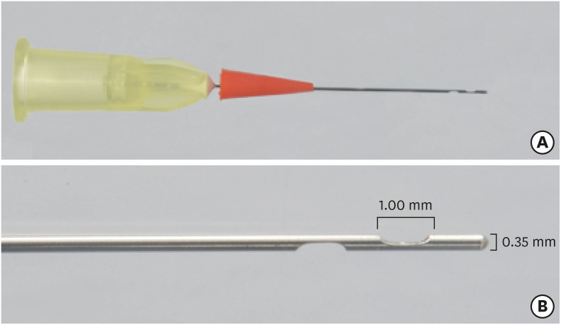
Figure 1 Representation of the new Sealer Injection System (SIS) with side openings that should be threaded to the Bio-C Sealer syringe for endodontic obturation (A), and the needle tip with more magnification (B).
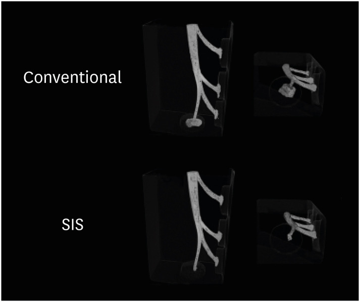
Figure 2 Three-dimensional models using CTVox software showing the artificial root canals filled with the conventional system or Sealer Injection System (SIS), and the apical extrusion of Bio-C Sealer after obturation by both systems. Voids are represented in black.
Reference
-
1. Yanpiset K, Banomyong D, Chotvorrarak K, Srisatjaluk RL. Bacterial leakage and micro-computed tomography evaluation in round-shaped canals obturated with bioceramic cone and sealer using matched single cone technique. Restor Dent Endod. 2018; 43:e30. PMID: 30135849.
Article2. Deniz Sungur D, Purali N, Coşgun E, Calt S. Push-out bond strength and dentinal tubule penetration of different root canal sealers used with coated core materials. Restor Dent Endod. 2016; 41:114–120. PMID: 27200279.
Article3. Oltra E, Cox TC, LaCourse MR, Johnson JD, Paranjpe A. Retreatability of two endodontic sealers, EndoSequence BC Sealer and AH Plus: a micro-computed tomographic comparison. Restor Dent Endod. 2017; 42:19–26. PMID: 28194360.
Article4. Zordan-Bronzel CL, Esteves Torres FF, Tanomaru-Filho M, Chávez-Andrade GM, Bosso-Martelo R, Guerreiro-Tanomaru JM. Evaluation of physicochemical properties of a new calcium silicate-based sealer, Bio-C Sealer. J Endod. 2019; 45:1248–1252. PMID: 31447172.
Article5. López-García S, Pecci-Lloret MR, Guerrero-Gironés J, Pecci-Lloret MP, Lozano A, Llena C, Rodríguez-Lozano FJ, Forner L. Comparative cytocompatibility and mineralization potential of Bio-C Sealer and TotalFill BC Sealer. Materials (Basel). 2019; 12:3087.
Article6. Shashirekha G, Jena A, Pattanaik S, Rath J. Assessment of pain and dissolution of apically extruded sealers and their effect on the periradicular tissues. J Conserv Dent. 2018; 21:546–550. PMID: 30294119.
Article7. Fonseca B, Coelho MS, Bueno CE, Fontana CE, Martin AS, Rocha DG. Assessment of extrusion and postoperative pain of a bioceramic and resin-based root canal sealer. Eur J Dent. 2019; 13:343–348. PMID: 31794999.
Article8. Chybowski EA, Glickman GN, Patel Y, Fleury A, Solomon E, He J. Clinical outcome of non-surgical root canal treatment using a single-cone technique with Endosequence Bioceramic Sealer: a retrospective analysis. J Endod. 2018; 44:941–945. PMID: 29606401.
Article9. Ho ES, Chang JW, Cheung GS. Quality of root canal fillings using three gutta-percha obturation techniques. Restor Dent Endod. 2016; 41:22–28. PMID: 26877987.
Article10. Tanomaru-FIlho M, Torres FF, Chávez-Andrade GM, Miano LM, Guerreiro-Tanomaru JM. Intermittent or continuous ultrasonically activated irrigation: micro-computed tomographic evaluation of root canal system cleaning. Clin Oral Investig. 2016; 20:1541–1546.
Article11. Yılmaz K, Uslu G, Özyürek T. In vitro comparison of the cyclic fatigue resistance of HyFlex EDM, One G, and ProGlider nickel titanium glide path instruments in single and double curvature canals. Restor Dent Endod. 2017; 42:282–289. PMID: 29142876.12. Tanomaru-Filho M, Torres FF, Chávez-Andrade GM, de Almeida M, Navarro LG, Steier L, Guerreiro-Tanomaru JM. Physicochemical properties and volumetric change of silicone/bioactive glass and calcium silicate-based endodontic sealers. J Endod. 2017; 43:2097–2101. PMID: 29032816.13. Silva EJ, Muniz BL, Pires F, Belladonna FG, Neves AA, Souza EM, De-Deus G. Comparison of canal transportation in simulated curved canals prepared with ProTaper universal and ProTaper gold systems. Restor Dent Endod. 2016; 41:1–5. PMID: 26877984.
Article14. Pinto JC, Pivoto-João MM, Espir CG, Ramos ML, Guerreiro-Tanomaru JM, Tanomaru-Filho M. Micro-CT evaluation of apical enlargement of molar root canals using rotary or reciprocating heat-treated NiTi instruments. J Appl Oral Sci. 2019; 27:e20180689. PMID: 31411264.
Article15. Song YS, Choi Y, Lim MJ, Yu MK, Hong CU, Lee KW, Min KS. In vitro evaluation of a newly produced resin-based endodontic sealer. Restor Dent Endod. 2016; 41:189–195. PMID: 27508160.
Article16. McMichen FR, Pearson G, Rahbaran S, Gulabivala K. A comparative study of selected physical properties of five root-canal sealers. Int Endod J. 2003; 36:629–635. PMID: 12950578.
Article17. Yeter KY, Evcil MS, Ayranci LB, Ersoy I. Weight of apically extruded debris following use of two canal instrumentation techniques and two designs of irrigation needles. Int Endod J. 2013; 46:795–799. PMID: 23441844.
Article18. Silva PB, Krolow AM, Pilownic KJ, Casarin RP, Lima RK, Leonardo RT, Pappen FG. Apical extrusion of debris and irrigants using different irrigation needles. Braz Dent J. 2016; 27:192–195. PMID: 27058383.
Article19. Ruparel NB, Ruparel SB, Chen PB, Ishikawa B, Diogenes A. Direct effect of endodontic sealers on trigeminal neuronal activity. J Endod. 2014; 40:683–687. PMID: 24767564.
Article20. Kim K, Kim DV, Kim SY, Yang S. A micro-computed tomographic study of remaining filling materials of two bioceramic sealers and epoxy resin sealer after retreatment. Restor Dent Endod. 2019; 44:e18. PMID: 31149616.
Article21. Kim J, Song YS, Min KS, Kim SH, Koh JT, Lee BN, Chang HS, Hwang IN, Oh WM, Hwang YC. Evaluation of reparative dentin formation of ProRoot MTA, Biodentine and BioAggregate using micro-CT and immunohistochemistry. Restor Dent Endod. 2016; 41:29–36. PMID: 26877988.
Article22. Torres FFE, Zordan-Bronzel CL, Guerreiro-Tanomaru JM, Chavez-Andrade GM, Pinto JC, Tanomaru-Filho M. Effect of immersion in distilled water or phosphate-buffered saline on the solubility, volumetric change and presence of voids within new calcium silicate-based root canal sealers. Int Endod J. 2020; 53:385–391. PMID: 31566768.
Article
- Full Text Links
-
- Actions
-
Cited
- CITED
-
- Close
- Share
-
- Similar articles
-
- A micro-computed tomographic study of remaining filling materials of two bioceramic sealers and epoxy resin sealer after retreatment
- Setting time of root canal sealers and root-end filling materials by different measuring methods
- A micro-computed tomographic evaluation of root canal filling with a single gutta-percha cone and calcium silicate sealer
- Effects of various root canal sealers on tooth discoloration and internal bleaching
- Calcium silicate-based root canal sealers: a literature review
