Pseudocirrhosis of Breast Cancer Metastases to the Liver Treated by Chemotherapy
- Affiliations
-
- 1Department of Radiology, Uijeongbu St. Mary's Hospital, The Catholic University of Korea College of Medicine, Uijeongbu, Korea.
- 2Department of Hospital Pathology, Uijeongbu St. Mary's Hospital, The Catholic University of Korea College of Medicine, Uijeongbu, Korea.
- 3Department of Nuclear Medicine, Uijeongbu St. Mary's Hospital, The Catholic University of Korea College of Medicine, Uijeongbu, Korea.
- 4Department of Surgery, Uijeongbu St. Mary's Hospital, The Catholic University of Korea College of Medicine, Uijeongbu, Korea.
- 5Department of Internal Medicine, Uijeongbu St. Mary's Hospital, The Catholic University of Korea College of Medicine, Uijeongbu, Korea. woncomet@catholic.ac.kr
Abstract
- Pseudocirrhosis refers to a condition that shows changes in hepatic contour that mimic cirrhosis radiographically in the absence of the typical histopathological findings of cirrhosis. This condition has been observed in patients with cancer metastatic to the liver, both in those who have undergone prior systemic chemotherapy and those who have not. Pseudocirrhosis may cause difficulty in interpretation of the response to chemotherapy and hepatic decompression and complication of portal hypertension have a negative effect on the prognosis. We report on a case of breast cancer with liver metastases that showed cirrhotic changes during disease progression. Progression of liver metastases was confirmed by F18 fluorodeoxyglucose positron emission tomography/computed tomography (PET-CT). We also performed ultrasound-guided liver biopsy and confirmed tumor infiltration with severe desmoplastic fibrosis. This case suggests the pathogenesis of pseudocirrhosis through histopathological findings and the role of PET-CT in evaluation of the response to chemotherapy in patients with pseudocirrhosis.
Keyword
MeSH Terms
Figure
-
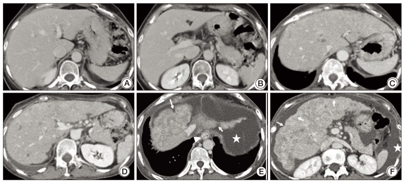
Fig. 1 Serial radiologic images of breast cancer with multiple liver metastases. (A, B) Contrast enhanced computed tomography (CT) scan (March 29, 2005) shows no evidence of liver metastasis or surface nodularity of the liver. (C, D) Contrast enhanced CT scan (September 22, 2011) shows multiple liver metastases in the liver, but no evidence of surface nodularity and esophageal varices. (E, F) Contrast enhanced CT scan (April 2, 2012) shows multiple liver metastases and a cirrhosis like appearance, such as surface nodularity (arrows), esophageal varices (arrowhead), and ascites (★).
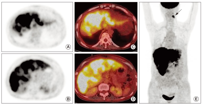
Fig. 2 F18 fluorodeoxyglucose positron emission tomography/computed tomography images taken on April 3, 2012. Axial positron emission tomography (A, C) and fusion (B, D) images show diffusely and heterogeneously increased activity in known hepatic metastases. Maximum intensity projection image (E) also shows extensive hepatic metastasis. Focally and incidentally increased activity (arrow) is seen in an abscess of the left mandible.
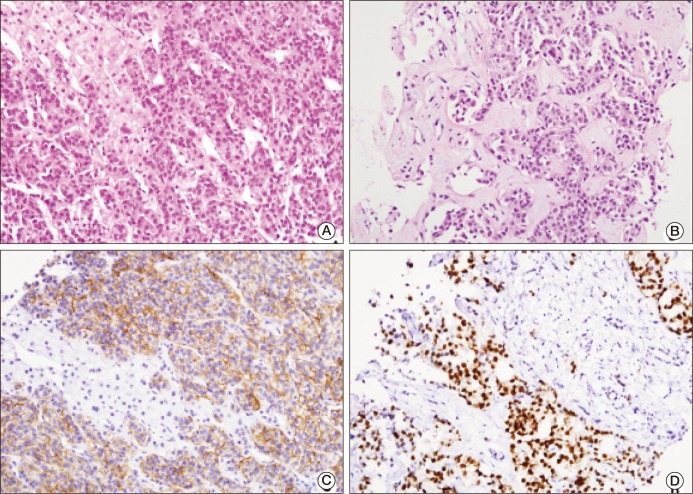
Fig. 3 (A, B) Diffusely infiltrated carcinoma cells with extensive fibrosis in liver parenchyma (H&E staining, ×200). Positive immunohistochemical staining for MOC 31 (C; ×200) and estrogen receptor (D; ×200).
Reference
-
1. Qayyum A, Lee GK, Yeh BM, Allen JN, Venook AP, Coakley FV. Frequency of hepatic contour abnormalities and signs of portal hypertension at CT in patients receiving chemotherapy for breast cancer metastatic to the liver. Clin Imaging. 2007; 31:6–10. PMID: 17189839.
Article2. Young ST, Paulson EK, Washington K, Gulliver DJ, Vredenburgh JJ, Baker ME. CT of the liver in patients with metastatic breast carcinoma treated by chemotherapy: findings simulating cirrhosis. AJR Am J Roentgenol. 1994; 163:1385–1388. PMID: 7992734.
Article3. Sonnenblick A, Appelbaum L, Peretz T. Liver failure on the background of pseudocirrhosis in patients with liver metastasis from breast cancer, who responded to treatment. Onkologie. 2011; 34:199–201. PMID: 21447980.
Article4. Sass DA, Clark K, Grzybicki D, Rabinovitz M, Shaw-Stiffel TA. Diffuse desmoplastic metastatic breast cancer simulating cirrhosis with severe portal hypertension: a case of "pseudocirrhosis". Dig Dis Sci. 2007; 52:749–752. PMID: 17265127.
Article5. Ma C, Brunt EM. Histopathologic evaluation of liver biopsy for cirrhosis. Adv Anat Pathol. 2012; 19:220–230. PMID: 22692285.
Article6. Jha P, Poder L, Wang ZJ, Westphalen AC, Yeh BM, Coakley FV. Radiologic mimics of cirrhosis. AJR Am J Roentgenol. 2010; 194:993–999. PMID: 20308502.
Article7. Kang SP, Taddei T, McLennan B, Lacy J. Pseudocirrhosis in a pancreatic cancer patient with liver metastases: a case report of complete resolution of pseudocirrhosis with an early recognition and management. World J Gastroenterol. 2008; 14:1622–1624. PMID: 18330959.
Article8. Kobashigawa C, Nakamoto M, Hokama A, Hirata T, Kinjo F, Fujita J. Pseudocirrhosis in metastatic esophageal cancer. South Med J. 2010; 103:488–489. PMID: 20375937.
Article9. Harry BL, Smith ML, Burton JR Jr, Dasari A, Eckhardt SG, Diamond JR. Medullary thyroid cancer and pseudocirrhosis: case report and literature review. Curr Oncol. 2012; 19:e36–e41. PMID: 22328846.
Article10. Liu CH, Chao TY. Education and imaging. Hepatobiliary and pancreatic: pseudocirrhosis after chemotherapy. J Gastroenterol Hepatol. 2011; 26:788. PMID: 21418309.
