Sclerosing Perivascular Epithelioid Cell Tumor of the Lung: A Case Report with Cytologic Findings
- Affiliations
-
- 1Department of Pathology, Korea University Guro Hospital, Seoul, Korea. sswords@naver.com
- KMID: 2164602
- DOI: http://doi.org/10.4132/jptm.2016.02.19
Abstract
- Benign perivascular epithelioid cell tumor (PEComa) of the lung is a rare benign neoplasm, a sclerosing variant of which is even rarer. We present a case of 51-year-old man who was diagnosed with benign sclerosing PEComa by percutaneous fine needle aspiration cytology and biopsy. The aspirate revealed a few cell clusters composed of bland-looking polygonal or spindle cells with fine granular or clear cytoplasm. Occasional fine vessel-like structures with surrounding hyalinized materials were seen. The patient later underwent wedge resection of the lung. The histopathological study of the resected specimen revealed sheets of polygonal cells with clear vacuolated cytoplasm, variably sized thin blood vessels, and densely hyalinized stroma. In immunohistochemical studies, reactivity of tumor cells for human melanoma black 45 and Melan-A further supported the diagnosis of benign sclerosing PEComa. To the best of our knowledge, this is the first case of benign sclerosing PEComa described in lung.
MeSH Terms
Figure
-
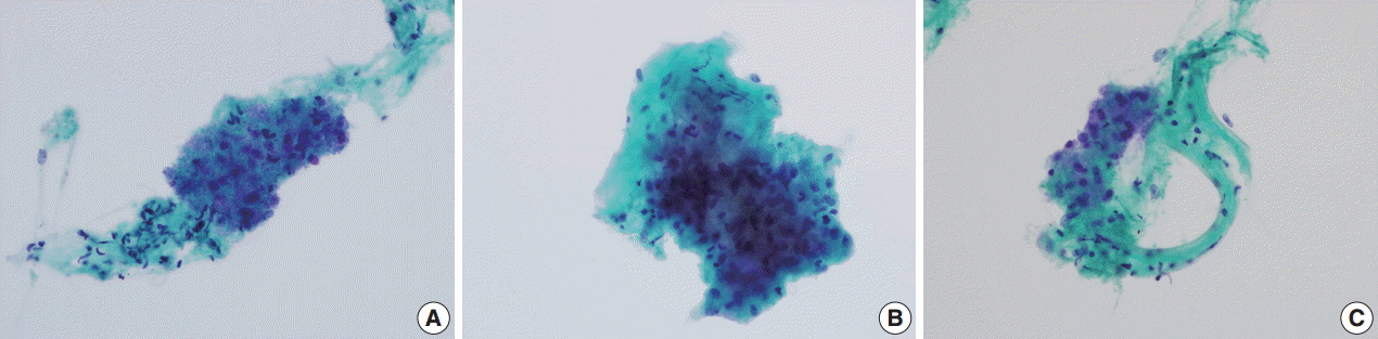
Fig. 1. Liquid-based aspiration cytology of benign sclerosing perivascular epithelioid cell tumor. (A) A few cohesive clusters of polygonal to spindle-shaped bland cells in a clean background (Papanicolaou staining). (B) The tumor cells show oval nuclei, small distinct nucleoli and abundant basophilic and granular cytoplasm. Most cells contain clear intracytoplasmic vacuoles (Papanicolaou staining). (C) Thin-walled blood vessels are occasionally seen. Semitranslucent hyalinizing material was noted around the vascular structure (Papanicolaou staining).
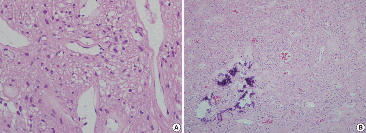
Fig. 2. Histologic section of benign sclerosing perivascular epithelioid tumor. (A) Dilated vessels surrounded by collagenous stroma and polygonal cells with clear cytoplasm. (B) Sheet-like arrangement of tumor cells around various-sized blood vessels and a focus of microcalcification.
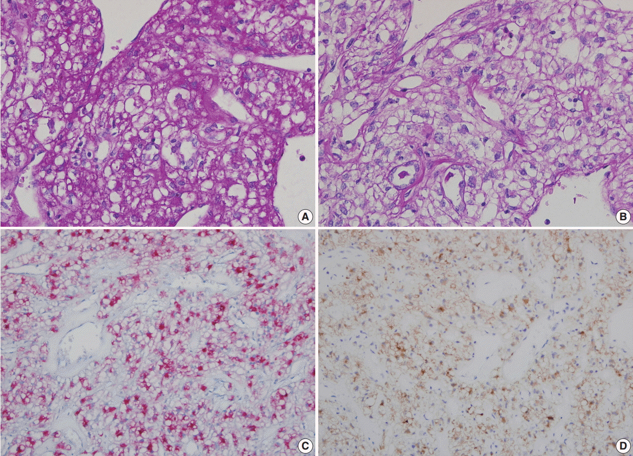
Fig. 3. Special stain and immunohistochemical stains (A, B) Periodic acid–Schiff (PAS) (A) stain and diastase-PAS (B) stain show positive reaction of the clear intracytoplasmic vacuoles in tumor cells. (C, D) Tumor cells show positive reaction to human melanoma black 45 (C) and to Melan-A (D).
Cited by 1 articles
-
Perivascular Epithelioid Cell Tumors (PEComas) of the Orbit
Panagiotis Paliogiannis, Giuseppe Palmieri, Francesco Tanda, Antonio Cossu
J Pathol Transl Med. 2017;51(1):7-8. doi: 10.4132/jptm.2016.10.26.
Reference
-
1. Liebow AA, Castleman B. Benign “clear cell tumors” of the lung. Am J Pathol. 1963; 43:13–4.2. Nguyen GK. Aspiration biopsy cytology of benign clear cell (“sugar”) tumor of the lung. Acta Cytol. 1989; 33:511–5.3. Policarpio-Nicolas ML, Covell J, Bregman S, Atkins K. Fine needle aspiration cytology of clear cell “sugar” tumor (PEComa) of the lung: report of a case. Diagn Cytopathol. 2008; 36:89–93.
Article4. Edelweiss M, Gupta N, Resetkova E. Preoperative diagnosis of clear cell “sugar” tumor of the lung by computed tomography-guided fine-needle biopsy and core-needle biopsy. Ann Diagn Pathol. 2007; 11:421–6.
Article5. Hornick JL, Fletcher CD. Sclerosing PEComa: clinicopathologic analysis of a distinctive variant with a predilection for the retroperitoneum. Am J Surg Pathol. 2008; 32:493–501.
Article6. Wang GX, Zhang D, Diao XW, Wen L. Clear cell tumor of the lung: a case report and literature review. World J Surg Oncol. 2013; 11:247.
Article7. Gora-Gebka M, Liberek A, Bako W, Szumera M, Korzon M, Jaskiewicz K. The “sugar” clear cell tumor of the lung-clinical presentation and diagnostic difficulties of an unusual lung tumor in youth. J Pediatr Surg. 2006; 41:e27–9.
Article8. Santana AN, Nunes FS, Ho N, Takagaki TY. A rare cause of hemoptysis: benign sugar (clear) cell tumor of the lung. Eur J Cardiothorac Surg. 2004; 25:652–4.
Article9. Sen S, Senturk E, Kuman NK, Pabuscu E, Kacar F. PEComa (clear cell “sugar” tumor) of the lung: a benign tumor that presented with thrombocytosis. Ann Thorac Surg. 2009; 88:2013–5.10. Kim WJ, Kim SR, Choe YH, et al. Clear cell “sugar” tumor of the lung: a well-enhanced mass with an early washout pattern on dynamic contrast-enhanced computed tomography. J Korean Med Sci. 2008; 23:1121–4.
Article11. Mizobuchi T, Masahiro N, Iwai N, Kohno H, Okada N, Nakada S. Clear cell tumor of the lung: surgical and immunohistochemical findings. Gen Thorac Cardiovasc Surg. 2010; 58:243–7.
Article12. Yamada Y, Yamamoto H, Ohishi Y, et al. Sclerosing variant of perivascular epithelioid cell tumor in the female genital organs. Pathol Int. 2011; 61:768–72.
Article13. Leão RR, Pereira BJ, Grenha V, Coelho H. Pararenal sclerosing PEComa. BMJ Case Rep. 2013; 2013:bcr2013009097.14. Wojcik EM, Sneige N, Lawrence DD, Ordóñez NG. Fine-needle aspiration cytology of sclerosing hemangioma of the lung: case report with immunohistochemical study. Diagn Cytopathol. 1993; 9:304–9.
Article
- Full Text Links
-
- Actions
-
Cited
- CITED
-
- Close
- Share
-
- Similar articles
-
- A case of perivascular epithelioid cell tumor of the uterus
- Primary Perivascular Epithelioid Cell Tumor (PEComa) of the Liver: A Case Report and Review of the Literature
- Uncommon of the Uncommon: Malignant Perivascular Epithelioid Cell Tumor of the Lung
- A Case of Primary Cutaneous Perivascular Epithelioid Cell Tumor
- Fine Needle Aspiration Cytology of Pulmonary Epithelioid Hemangioendothelioma with Prominent Hyaline Degeneration: A Case Report
