Uncommon of the Uncommon: Malignant Perivascular Epithelioid Cell Tumor of the Lung
- Affiliations
-
- 1Department of Radiology and Center for Imaging, Samsung Medical Center, Sungkyunkwan University School of Medicine, Seoul 135-710, Korea. hoyunlee96@gmail.com
- 2Department of Pathology, Samsung Medical Center, Sungkyunkwan University School of Medicine, Seoul 135-710, Korea.
- 3Department of Thoracic Surgery, Samsung Medical Center, Sungkyunkwan University School of Medicine, Seoul 135-710, Korea.
- KMID: 1715776
- DOI: http://doi.org/10.3348/kjr.2013.14.4.692
Abstract
- A perivascular epithelioid cell (PEC) tumor is a rare mesenchymal tumor characterized by abundant cytoplasmic Periodic acid-Schiff positive glycogen (also called sugar tumor or clear cell tumor of the lung for this characteristic) and is mostly benign. We report a case of a 63-year-old man who presented with an enlarging mass on chest radiograph. After a thorough workup, diagnosis of malignant pulmonary PEC tumor with lung to lung metastases was established. Herein, the difficulties of diagnosis and management we confronted are described.
MeSH Terms
Figure
-
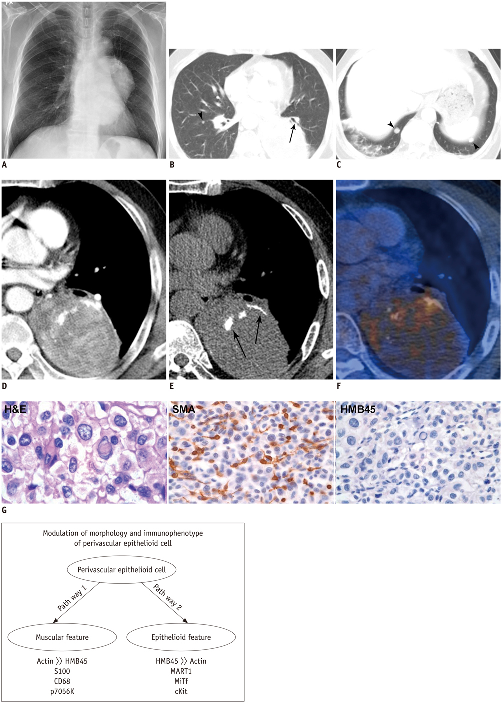
Fig. 1 Imaging and pathologic findings of 63-year-old man with malignant pulmonary PEComa (A-G) and modulation of PEC (H) Chest radiography revealing huge mass in left mid to lower lung field, abutting mediastinal structures (A). Computed tomography demonstrating mass attached to posteromedial aspect of left lower lobe with air-bronchogram (arrow) in peripheral portion and without displacement of bronchovascular bundle adjacent lung parenchyma (B), which indicates lung parenchymal lesion. Several metastatic nodules (arrowheads) in both lungs (B, C). Well circumscribed, heterogeneously enhancing mass (D) containing curvilinear calcifications (arrows, E). Positron emission tomography scan (F) showing hypermetabolic mass with SUVmax of 4.4. Specimen showing rounded or oval shaped tumor cells with abundant clear cytoplasm (histological examination), H&E × 400. Immunoreactivity for SMA and immunonegative for HMB45 of tumor cells (histological examination), × 200 (G). Concept of modulation of perivascular epithelioid cell in morphology and immunophenotype (H).
Reference
-
1. Liebow AA, Castleman B. Benign clear cell ("sugar") tumors of the lung. Yale J Biol Med. 1971; 43:213–222.2. Selvaggi F, Risio D, Claudi R, Cianci R, Angelucci D, Pulcini D, et al. Malignant PEComa: a case report with emphasis on clinical and morphological criteria. BMC Surg. 2011; 11:3.3. Martignoni G, Pea M, Reghellin D, Zamboni G, Bonetti F. PEComas: the past, the present and the future. Virchows Arch. 2008; 452:119–132.4. Ye T, Chen H, Hu H, Wang J, Shen L. Malignant clear cell sugar tumor of the lung: patient case report. J Clin Oncol. 2010; 28:e626–e628.5. Armah HB, Parwani AV. Perivascular epithelioid cell tumor. Arch Pathol Lab Med. 2009; 133:648–654.6. Kim WJ, Kim SR, Choe YH, Lee KY, Park SJ, Lee HB, et al. Clear cell "sugar" tumor of the lung: a well-enhanced mass with an early washout pattern on dynamic contrast-enhanced computed tomography. J Korean Med Sci. 2008; 23:1121–1124.7. Sale GE, Kulander BG. 'Benign' clear-cell tumor (sugar tumor) of the lung with hepatic metastases ten years after resection of pulmonary primary tumor. Arch Pathol Lab Med. 1988; 112:1177–1178.8. Parfitt JR, Keith JL, Megyesi JF, Ang LC. Metastatic PEComa to the brain. Acta Neuropathol. 2006; 112:349–351.9. Yan B, Yau EX, Petersson F. Clear cell 'sugar' tumour of the lung with malignant histological features and melanin pigmentation--the first reported case. Histopathology. 2011; 58:498–500.10. Zarbis N, Barth TF, Blumstein NM, Schelzig H. Pecoma of the lung: a benign tumor with extensive 18F-2-deoxy-D-glucose uptake. Interact Cardiovasc Thorac Surg. 2007; 6:676–678.11. Folpe AL, Mentzel T, Lehr HA, Fisher C, Balzer BL, Weiss SW. Perivascular epithelioid cell neoplasms of soft tissue and gynecologic origin: a clinicopathologic study of 26 cases and review of the literature. Am J Surg Pathol. 2005; 29:1558–1575.12. Wagner AJ, Malinowska-Kolodziej I, Morgan JA, Qin W, Fletcher CD, Vena N, et al. Clinical activity of mTOR inhibition with sirolimus in malignant perivascular epithelioid cell tumors: targeting the pathogenic activation of mTORC1 in tumors. J Clin Oncol. 2010; 28:835–840.
- Full Text Links
-
- Actions
-
Cited
- CITED
-
- Close
- Share
-
- Similar articles
-
- Primary Perivascular Epithelioid Cell Tumor (PEComa) of the Liver: A Case Report and Review of the Literature
- A case of perivascular epithelioid cell tumor of the uterus
- A Case of Malignant PEComa of the Uterus Associated with Intramural Leiomyoma and Endometrial Carcinoma
- Malignant Perivascular Epithelioid Cell Tumor (PEComa) Arising in the Omentum with Metastatic Peritoneal Seeding: A Case Report
- Primary Perivascular Epithelioid Cell Tumor of the Lung: A Case Report
