Unique Hippocampal Changes and Allodynia in a Model of Chronic Stress
- Affiliations
-
- 1Department of Internal Medicine, Inje University College of Medicine, Busan, Korea. junjan@paik.co.kr
- 2Department of Anatomy, Dongguk University College of Medicine, Gyeongju, Korea.
- 3Department of Anatomy, College of Oriental Medicine, Dongguk University, Gyeongju, Korea.
- KMID: 2158046
- DOI: http://doi.org/10.3346/jkms.2013.28.6.946
Abstract
- Sustained stress can have numerous pathologic effects. There have been several animal models for chronic stress. We tried to identify the changes of pain threshold and hippocampus in a model of chronic stress. Male Sprague-Dawley rats were kept in a cage filled with 23degrees C water to a height of 2.2 cm for 7 days. Nociceptive thresholds, expressed in grams, were measured with a Dynamic Plantar Aesthesiometer. Golgi staining was used to identify hippocampal changes. To demonstrate how long allodynia was lasting, behavioral test was repeated daily on another experiment. Compared to control group, chronic stress group showed bilateral mechanical hyper-responsiveness on days 5 (P = 0.047) and 7 (P = 0.032). In general, dendrite atrophic changes within hippocampus of chronic stress model were much more prominent in comparison with control. Compared to control, decreased spine number (P < 0.001) and spine length (P < 0.001) on Golgi staining were seen in the hippocampus of animals with chronic stress. Bilateral mechanical hyperresponsiveness was recovered on day 19 in animals with chronic stress. Chronic stress may bring about central sensitization and hippocampal changes in rats.
Keyword
MeSH Terms
Figure
-
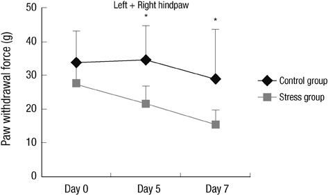
Fig. 1 Response to mechanical stimulation. Stress model showed bilateral mechanical hyper-responsiveness on day 5 and 7 compared to control. (Experiment I: n=5, group 1 and 2, respectively) The error bars indicate SD. Data from left and right hindpaws collapsed together for clarity of presentation. *P < 0.05.
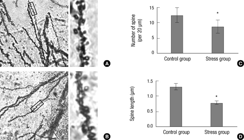
Fig. 2 Spine number and length changes on Golgi staining within the hippocampus of rats. (A) No significant changes of spine number and length within the hippocampus of control rats (×100). The rectangle of left is enlarged on the right. (B) Significantly decreased spine number and length within hippocampus of stress model (×100). The rectangle of left is enlarged on the right. (C) Comparison of spine number per 20 µm between control (12.5) and stress (8.7) group in the hippocampus. (D) Comparison of spine length between control (1.31 µm) and stress (0.78 µm) group in the hippocampus (Experiment I: n = 5, group 1 and 2, respectively). Bars show the mean and SD. *P < 0.001 vs control.

Fig. 3 Line graph representing the median mechanical threshold from stress and control animals according to observation day. Bilateral mechanical hyper-responsiveness was recovered on day 19 (Experiment II: n = 10, group 1 and 2, respectively). Data from left and right hindpaws collapsed together for clarity of presentation. *P < 0.05, †P < 0.01, ‡P < 0.001.
Reference
-
1. Ulrich-Lai YM, Herman JP. Neural regulation of endocrine and autonomic stress responses. Nat Rev Neurosci. 2009; 10:397–409.2. Sapolsky RM. Why stress is bad for your brain. Science. 1996; 273:749–750.3. Magariños AM, McEwen BS. Stress-induced atrophy of apical dendrites of hippocampal CA3c neurons: involvement of glucocorticoid secretion and excitatory amino acid receptors. Neuroscience. 1995; 69:89–98.4. Radley JJ, Rocher AB, Rodriguez A, Ehlenberger DB, Dammann M, McEwen BS, Morrison JH, Wearne SL, Hof PR. Repeated stress alters dendritic spine morphology in the rat medial prefrontal cortex. J Comp Neurol. 2008; 507:1141–1150.5. Tanaka M, Nakamura F, Mizokawa S, Matsumura A, Nozaki S, Watanabe Y. Establishment and assessment of a rat model of fatigue. Neurosci Lett. 2003; 352:159–162.6. Wu YW, Bi YP, Kou XX, Xu W, Ma LQ, Wang KW, Gan YH, Ma XC. 17-Beta-estradiol enhanced allodynia of inflammatory temporomandibular joint through upregulation of hippocampal TRPV1 in ovariectomized rats. J Neurosci. 2010; 30:8710–8719.7. Burke NN, Hayes E, Calpin P, Kerr DM, Moriarty O, Finn DP, Roche M. Enhanced nociceptive responding in two rat models of depression is associated with alterations in monoamine levels in discrete brain regions. Neuroscience. 2010; 171:1300–1313.8. Wood PB. Fibromyalgia syndrome: a central role for the hippocampus: a theoretical construct. J Musculoskelet Pain. 2004; 12:19–26.9. Emad Y, Ragab Y, Zeinhom F, El-Khouly G, Abou-Zeid A, Rasker JJ. Hippocampus dysfunction may explain symptoms of fibromyalgia syndrome: a study with single-voxel magnetic resonance spectroscopy. J Rheumatol. 2008; 35:1371–1377.10. Joëls M, Baram TZ. The neuro-symphony of stress. Nat Rev Neurosci. 2009; 10:459–466.11. McEwen BS, Chattarji S. Behavioral neurochemistry and neuroendocrinology. In : Lajtha A, editor. Handbook of Neurochemistry and Molecular Neurobiology. 3rd ed. New York: Springer;2007. p. 571–594.12. McKenna JE, Melzack R. Blocking NMDA receptors in the hippocampal dentate gyrus with AP5 produces analgesia in the formalin pain test. Exp Neurol. 2001; 172:92–99.13. Schuff N, Amend DL, Knowlton R, Norman D, Fein G, Weiner MW. Age-related metabolite changes and volume loss in the hippocampus by magnetic resonance spectroscopy and imaging. Neurobiol Aging. 1999; 20:279–285.14. Wood PB, Ledbetter CR, Glabus MF, Broadwell LK, Patterson JC 2nd. Hippocampal metabolite abnormalities in fibromyalgia: correlation with clinical features. J Pain. 2009; 10:47–52.15. Kundermann B, Lautenbacher S. Effects of impaired sleep quality and sleep deprivation on diurnal pain perception. In : Lavigne GS, Sessle BJ, Choinière M, Soja PJ, editors. Sleep and Pain. Seattle: IASP Press;2007. p. 137–152.16. Quintero L, Moreno M, Avila C, Arcaya J, Maixner W, Suarez-Roca H. Long-lasting delayed hyperalgesia after subchronic swim stress. Pharmacol Biochem Behav. 2000; 67:449–458.
- Full Text Links
-
- Actions
-
Cited
- CITED
-
- Close
- Share
-
- Similar articles
-
- Repeated restraint stress promotes hippocampal neuronal cell ciliogenesis and proliferation in mice
- The Changes of Metabotrophic Glutamate Receptor type 5 in Allodynia Induced by Nerve Ligation
- The effect of 4-Methylcatechol treatment in chronic constrictive injury of the rat sciatic nerve on the allodynia and spinal neurotrophic factors
- Hippocampal Volume and Memory Function in Patients with Posttraumatic Stress Disorder
- Effect of the Preoperative Intercostal Nerve Block in a Rat Model of Postthoracotomy Pain
